

Giant Cystic Pheochromocytoma with Low Risk of Malignancy: A Case Report and Literature Review
- PMID: 28396811
- PMCID: PMC5370478
- DOI: 10.1155/2017/4638608
Giant Cystic Pheochromocytoma with Low Risk of Malignancy: A Case Report and Literature Review
Abstract
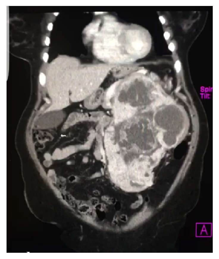
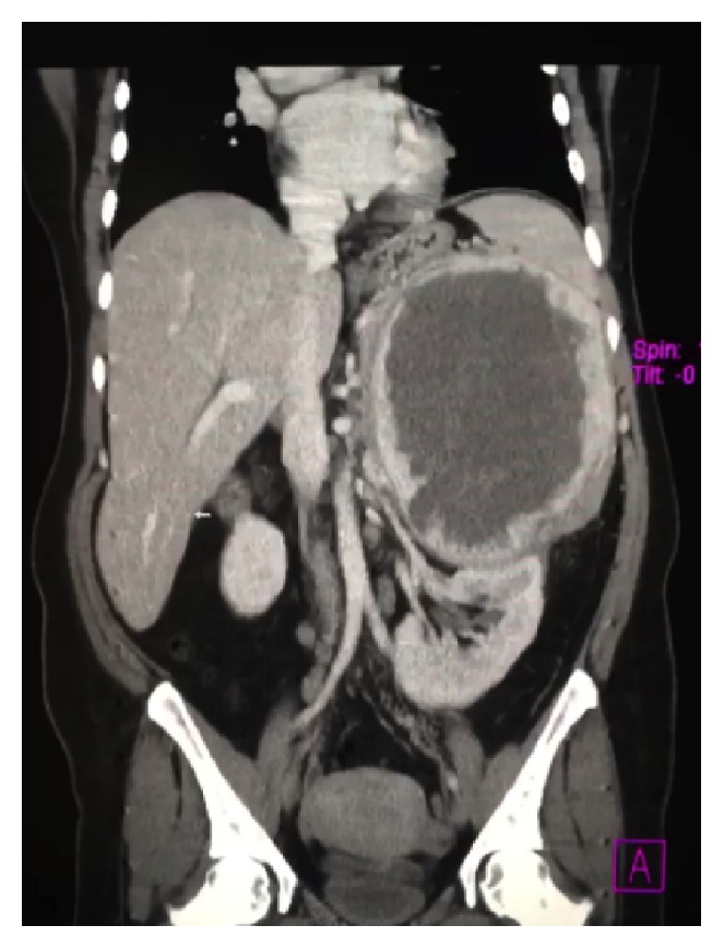
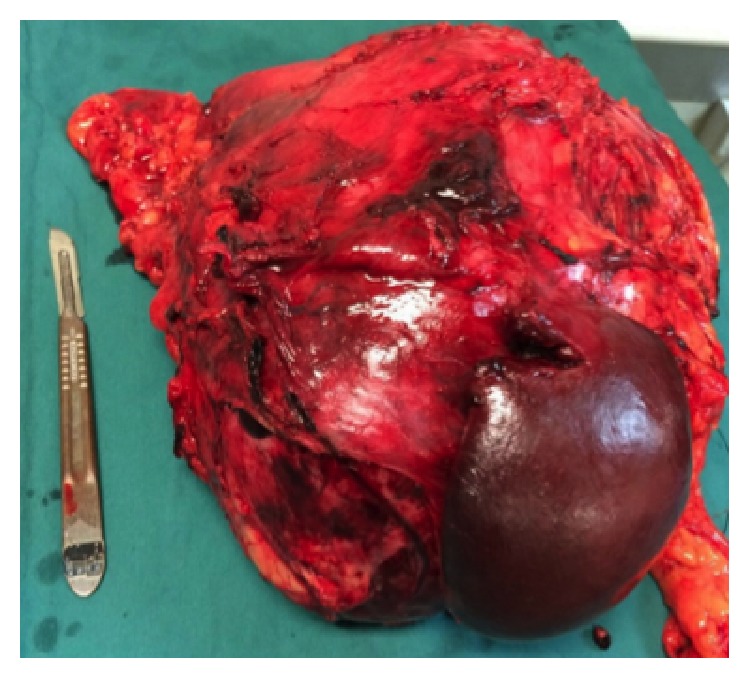
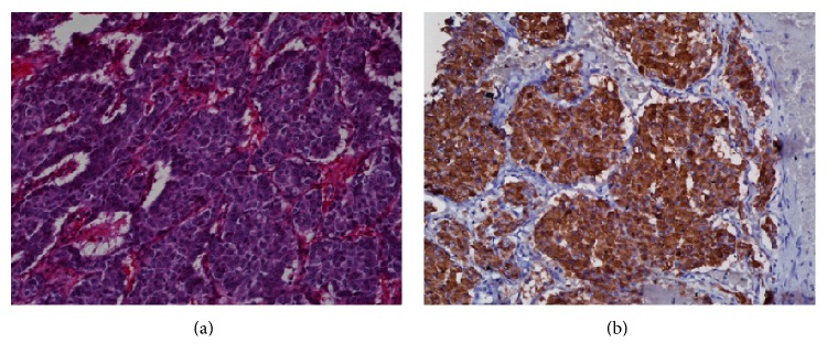
Giant pheochromocytomas are rare silent entities that do not present with the classical symptoms commonly seen in catecholamine-secreting tumors. In many cases they are accidentally discovered. The algorithm to diagnose a pheochromocytoma consists of biochemical evaluation and imaging of a retroperitoneal mass. The female patient in this case report presented with a palpable abdominal mass and was cured with surgical resection. She suffered no recurrence or complications on follow-up. The left retroperitoneal mass measured 27 × 18 × 12 cm and weighed 3,315 grams. Biochemical, radiological, and pathological examinations confirmed the diagnosis of a pheochromocytoma. In this paper, we report on our experience treating this patient and provide a summary of all giant pheochromocytomas greater than 10 cm reported to date in English language medical journals. Our patient's giant cystic pheochromocytoma was the fourth heaviest and fifth largest maximal diameter identified using our literature search criteria. Additionally, this tumor had the largest maximal diameter of all histologically confirmed benign/low metastatic risk pheochromocytomas. Giant cystic pheochromocytomas are rare entities requiring clinical suspicion coupled with strategic diagnostic evaluation to confirm the diagnosis.
Figures
References
-
- Nguyen-Martin M. A., Hammer G. D. Pheochromocytoma: an update on risk groups, diagnosis, and management. Hospital Physician. 2006;42(2):17–24.
Publication types
LinkOut - more resources
Full Text Sources
Other Literature Sources
