. 2017 Sep;13(3):279-292.
doi: 10.1007/s11302-017-9559-2. Epub 2017 Apr 11.

Potential role of P2X7R in esophageal squamous cell carcinoma proliferation

Affiliations

Potential role of P2X7R in esophageal squamous cell carcinoma proliferation

André A Santos Jr et al. Purinergic Signal. 2017 Sep.

Abstract

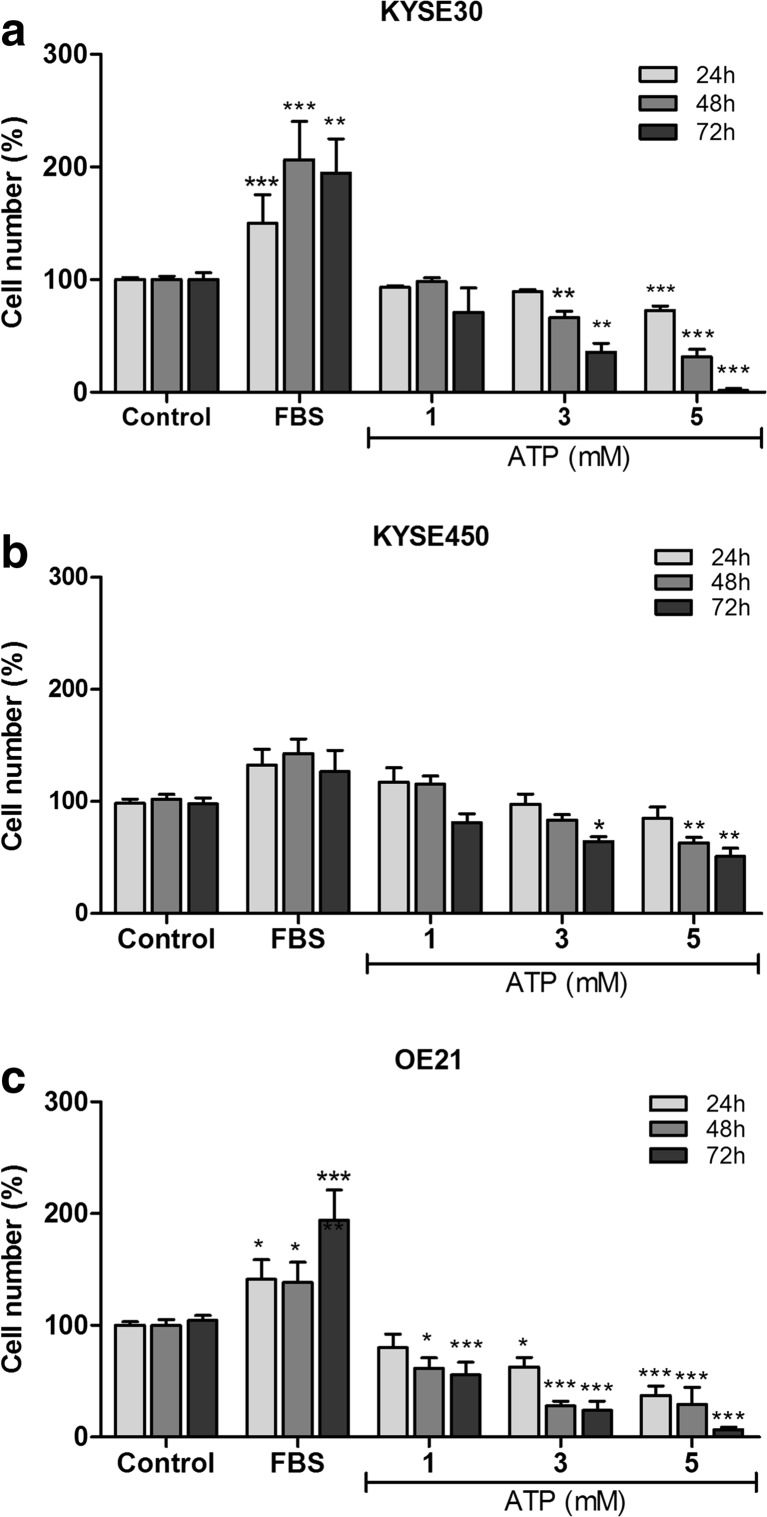
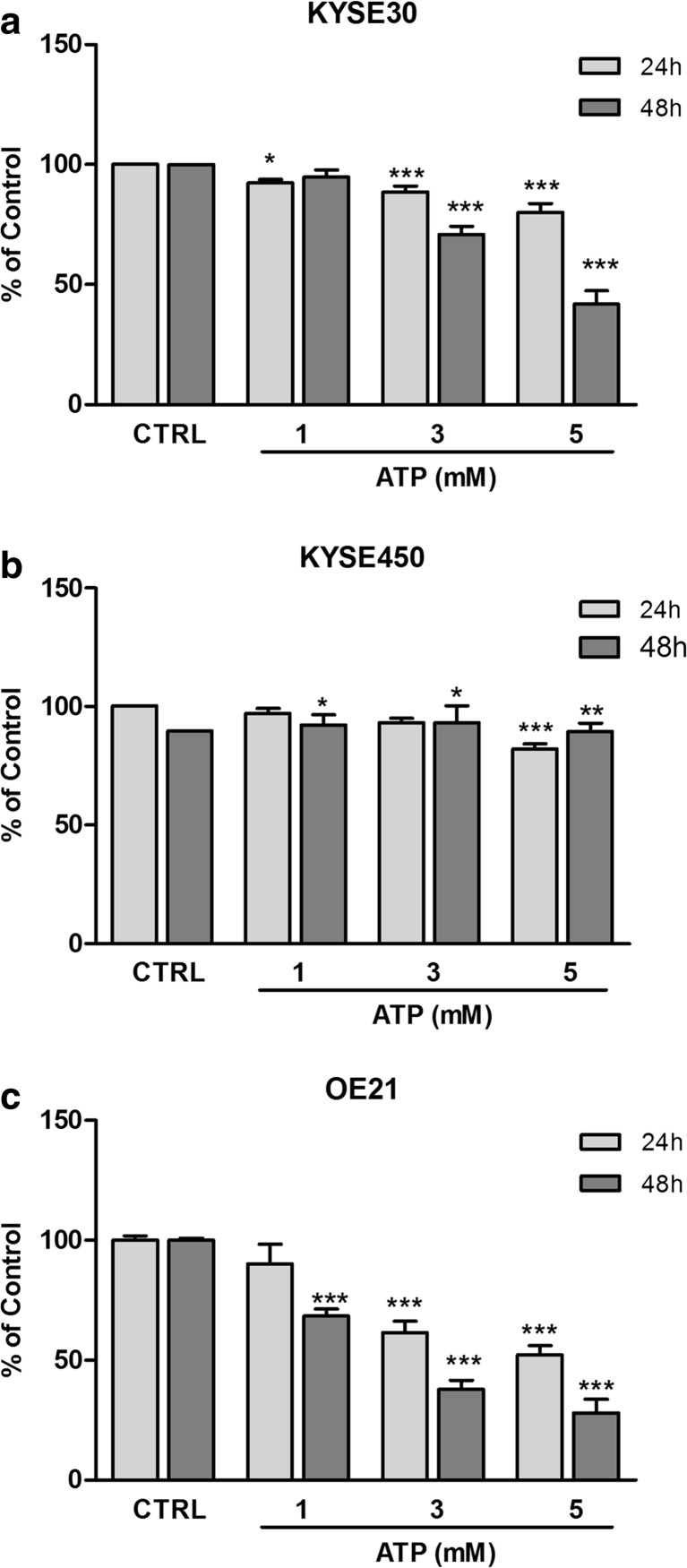
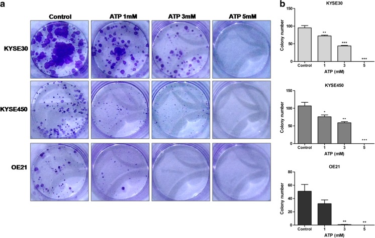
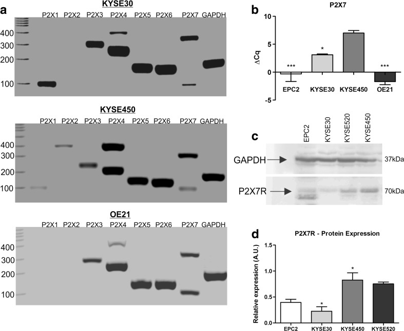
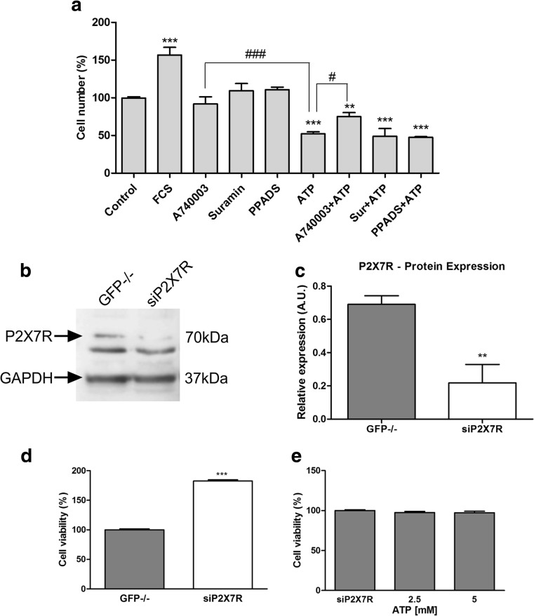
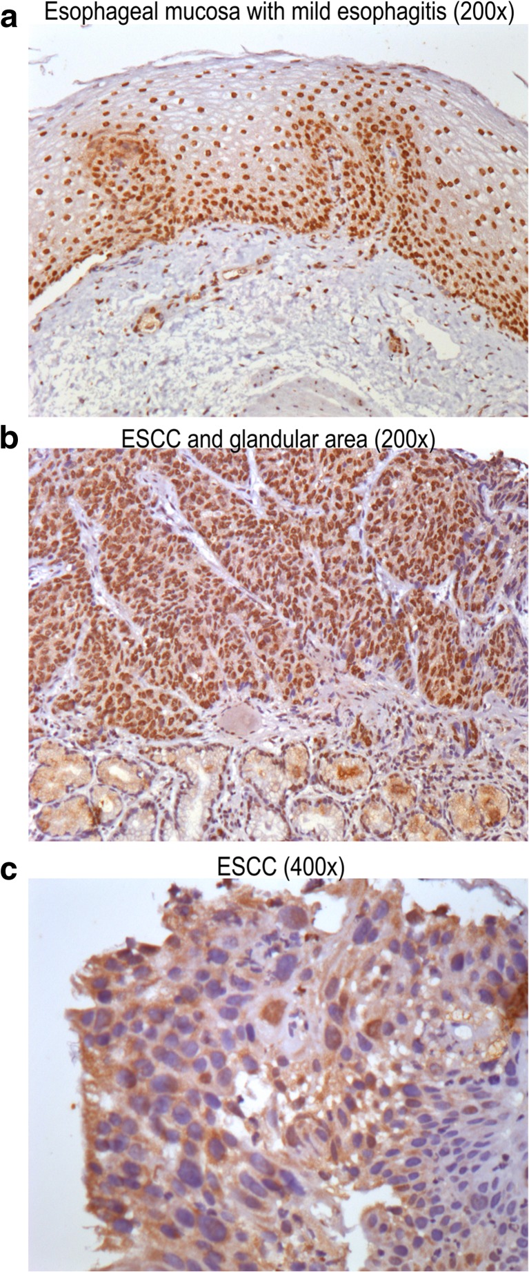
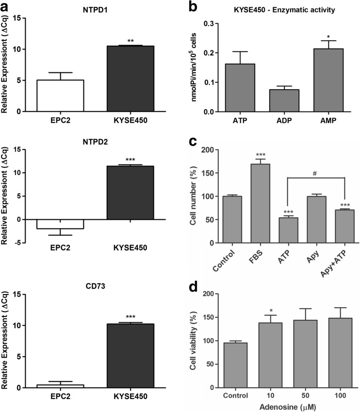
Esophageal cancer is an aggressive tumor and is the sixth leading cause of cancer death worldwide. ATP is well known to regulate cancer progression in a variety of models by different mechanisms, including P2X7R activation. This study aimed to evaluate the role of P2X7R in esophageal squamous cell carcinoma (ESCC) proliferation. Our results show that treatment with high ATP concentrations induced a decrease in cell number, cell viability, number of polyclonal colonies, and reduced migration of ESCC. The treatment with the selective P2X7R antagonist A740003 or siRNA for P2X7 reverted this effect in the KYSE450 cell line. In addition, results showed that P2X7R is highly expressed, at mRNA and protein levels, in KYSE450 lineage. Additionally, KYSE450, KYSE30, and OE21 cells express P2X3R, P2X4R, P2X5R, P2X6R, and P2X7R genes. P2X1R is expressed by KYSE30 and KYSE450, and only KYSE450 expresses the P2X2R gene. Furthermore, esophageal cancer cell line KYSE450 presented higher expression of E-NTPDases 1 and 2 and of Ecto-5'-NT/CD73 when compared to normal cells. This cell line also exhibits ATPase, ADPase, and AMPase activity, although in different levels, and the co-treatment of apyrase was able to revert the antiproliferative effects of ATP. Moreover, results showed high immunostaining for P2X7R in biopsies of patients with esophageal carcinoma, indicating the involvement of this receptor in the growth of this type of cancer. The results suggest that P2X7R may be a potential pharmacological target to treat ESCC and can lead us to further investigate the effect of this receptor in cancer cell progression.

Keywords: ATP; Ectonucleotidases; Esophageal cancer; P2X7R; Proliferation.


Conflict of interest statement

Conflicts of interest

André A Santos Jr declares that he has no conflict of interest.

Angélica R Cappellari declares that she has no conflict of interest.

Fernanda O de Marchi declares that she has no conflict of interest.

Marina P Gehring declares that she has no conflict of interest.

Aline Zaparte declares that she has no conflict of interest.

Caroline A Brandão declares that she has no conflict of interest.

Tiago Giuliani Lopes declares that he has no conflict of interest.

Luis Felipe Ribeiro Pinto that he has no conflict of interest.

Vinicius Duval da Silva declares that he has no conflict of interest.

Robson Coutinho-Silva declares that he has no conflict of interest.

Juliano D Paccez declares that she has no conflict of interest.

Luiz F Zerbini declares that he has no conflict of interest.

Fernanda B Morrone declares that she has no conflict of interest.

Ethical approval

Histological samples of human ESCC and esophageal tissue samples of patients with esophagitis were collected, between July and December 2015, from patients who underwent endoscopic procedures with biopsies and/or surgical resection at Pontificia Universidade Católica do Rio Grande do Sul (PUCRS, Porto Alegre, Brazil). The diagnosis was reviewed by two certified pathologists with at least 20-year experience in surgical pathology. Samples were obtained in accordance with approved ethical standards of the Institutional Research Ethics Committee (CAAE 4969 6115.0.0000.5336).

Figures

Fig. 1
Fig. 1
Evaluation of cell number after ATP treatment on human ESCC cell lines. The cell counting experiments were used as indicative of cell proliferation on KYSE30 (a), KYSE450 (b), and OE21 (c) cell lines. The cells were evaluated after 24, 48, and 72 h under treatment with ATP (1, 3, or 5 mM). The medium DMEM supplemented with 10% of FBS was used a positive control of cell proliferation. The experiments were performed three times in triplicate. Each column represents the mean ± SEM, *p < 0.05, **p < 0.01, and ***p < 0.001, and the results were established in relation to control cells
Fig. 2
Fig. 2
Evaluation of cell viability after ATP treatment on human ESCC cell lines. Cell viability was evaluated by MTT 24 and 48 h after treatment with ATP (1, 3, or 5 mM) in KYSE30 (a), KYSE450 (b), and OE21 (c) cell lines. Each column represents the mean ± SEM, *p < 0.05, **p < 0.01, and ***p < 0.001. The experiments were performed three times in triplicate to KYSE30 and OE21 and five times in triplicate to KYSE450. The results were established in relation to control cells (not treated and considered as 100% of cell viability)
Fig. 3
Fig. 3
Ability of ESCC cells to form new colonies after ATP treatment. The clonogenic assay was performed to evaluate the interference of ATP on cell proliferation after 10 days of treatment as described in “Material and Methods.” Evaluation of polyclonal cell population (a) and quantification of colony number (b). Each column represents the mean ± SEM, *p < 0.05, **p < 0.01, and ***p < 0.001, and the results were established in relation to control cells. The experiments were performed in triplicate
Fig. 4
Fig. 4
Analysis of P2X receptor expression profile in human ESCC cell lines. Expression of purinergic receptors P2X1–7 in KYSE30, KYSE450, and OE21 cells lines by RT-PCR (a). GAPDH expression was used as reference gene. RT-qPCR analysis of P2X7R relative expression was performed, and the values were shown as ΔCq relative expression in relation to GAPDH in EPC2, KYSE30, KYSE450, and OE21 cell lines (b) and the significance was described as *p < 0.05 and *** p < 0.001 indicating difference in relation to KYSE450. Representative Western blotting assays showing positivity to P2X7R in EPC2, KYSE30, KYSE450, and KYSE520 cells (c). Relative P2X7R levels obtained by analysis of protein bands detected by Western blot were compared to GAPDH expression levels (d) and the significance was described as *p < 0.05 and *** p < 0.001 indicating difference in relation to EPC2. Each column represents the mean ± SEM and the experiments were performed three times in triplicate
Fig. 5
Fig. 5
Modulation of P2X7R reduces the effect cytotoxic generated by ATP in high concentrations. a Pharmacological modulation of P2X7R was performed by cell counting after treatment with A740003 10 μM, a specific antagonist of P2X7R, suramin 30 μM, and PPADS 10 μM as unspecific antagonist of P2X receptors and ATP 5 mM. The treatment was performed after sequential reduction of FBS concentration, and the cell counting was performed after 48 h posttreatment. The difference in relation to control was determined as **p < 0.01 and ***p < 0.001. The difference of ATP 5 mM was determined as #p < 0.05 and ###p < 0,001. b Representative Western blotting assays showing positivity to P2X7R in KYSE450 GFP−/− and KYSE450 siP2X7R. c Relative P2X7R levels obtained by analysis of protein bands detected by Western blot were compared to GAPDH expression levels and the significance was described as **p < 0.01 and indicated difference in relation to GFP−/−. d Effect of P2X7R silencing on cell viability. After 48 h of silencing, the cells were plated and MTT assays were performed following 24 h. The significance was described as ***p < 0.001 and indicated difference in relation to GFP−/−. e Effect of ATP (2.5 and 5 mM) treatment on siP2X7R cell viability. The experiments were performed three times in triplicate
Fig. 6
Fig. 6
Immunostaining for P2X7 receptors in human esophageal samples. Representative images demonstrate the area of normal appearing esophageal mucosa with mild esophagitis (DAB, nuclear brown staining, ×200) (a), ESCC and glandular cells (b), and ESCC area (DAB, membrane brown staining, ×400) (c)
Fig. 7
Fig. 7
Evaluation of E-NTPDases and CD73 expression and activity in KYSE450 esophageal cancer cell line. a RT-qPCR analysis of NTPD1, NTPD2, and CD73 relative expression was performed and the values were showed as ΔCq relative expression in relation to GAPDH in EPC2 and KYSE450 cell lines. The significance was described as **p < 0.01 and ***p < 0.001 and indicated difference in relation to EPC2 cell line. b Evaluation of enzymatic activity as described in “Material and Methods” section. *P < 0.05 and indicated difference in relation to ADP hydrolysis. c Cell counting of KYSE450 after 48 h under treatment with apyrase (2 U/mL) and ATP 5 mM. ***P < 0.001 indicated difference of control and #p < 0.05 indicated difference of ATP treatment. d Cell viability assays performed after 24 h under treatment with adenosine and *p < 0.05 indicated difference of control. All experiments were performed three times in triplicate

Similar articles

Cited by

References

    1. Zhang Y, Pan T, Zhong X, Cheng C. Nicotine upregulates microRNA-21 and promotes TGF-beta-dependent epithelial-mesenchymal transition of esophageal cancer cells. Tumour Biol. 2014;35(7):7063–7072. - PubMed
    1. Instituto Nacional de Câncer José de Alencar Gomes da Silva (2015) Estimate (2016) - Cancer Incidence in Brazil. Rio de Janeiro, INCA
    1. Dawsey SM, Fagundes RB, Jacobson BC, Kresty LA, Mallery SR, Paski S, van den Brandt PA. Diet and esophageal disease. Ann N Y AcadSci. 2014;1325:127–137. - PMC - PubMed
    1. Morita M, Kumashiro R, Kubo N, Nakashima Y, Yoshida R, Yoshinaga K, Saeki H, Emi Y, KakejiY SY, Toh Y, Maehara Y. Alcohol drinking, cigarette smoking, and the development of squamous cell carcinoma of the esophagus: epidemiology, clinical findings, and prevention. Int J ClinOncol. 2010;15(2):126–134. - PubMed
    1. Verschuur EM, Siersema PD. Diagnostics and treatment of esophageal cancers. NedTijdschrTandheelkd. 2010;117(9):427–431. - PubMed

MeSH terms

LinkOut - more resources