Review
. 2017 Nov;143(4):418-431.
doi: 10.1111/jnc.14037. Epub 2017 May 14.

Brain aging and neurodegeneration: from a mitochondrial point of view

Affiliations
Review

Brain aging and neurodegeneration: from a mitochondrial point of view

Amandine Grimm et al. J Neurochem. 2017 Nov.

Abstract

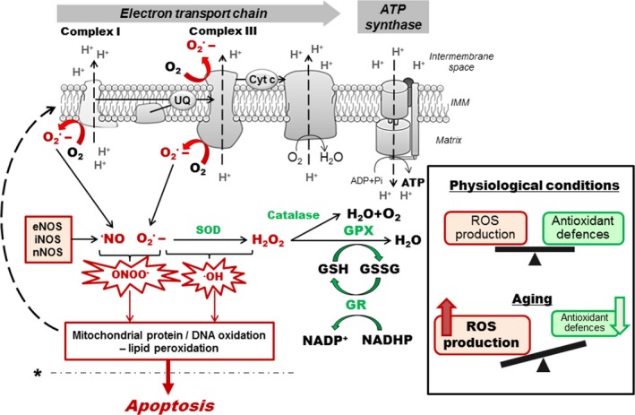
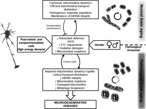
Aging is defined as a progressive time-related accumulation of changes responsible for or at least involved in the increased susceptibility to disease and death. The brain seems to be particularly sensitive to the aging process since the appearance of neurodegenerative diseases, including Alzheimer's disease, is exponential with the increasing age. Mitochondria were placed at the center of the 'free-radical theory of aging', because these paramount organelles are not only the main producers of energy in the cells, but also to main source of reactive oxygen species. Thus, in this review, we aim to look at brain aging processes from a mitochondrial point of view by asking: (i) What happens to brain mitochondrial bioenergetics and dynamics during aging? (ii) Why is the brain so sensitive to the age-related mitochondrial impairments? (iii) Is there a sex difference in the age-induced mitochondrial dysfunction? Understanding mitochondrial physiology in the context of brain aging may help identify therapeutic targets against neurodegeneration. This article is part of a series "Beyond Amyloid".

Keywords: Mitochondria; aging; bioenergetics; brain; mitochondrial dynamics.


Figures

Figure 1
Figure 1
Reactive oxygen species formation and detoxification. Superoxide anion radicals (O2˙‐) are mainly generated by the mitochondrial complexes I and III during electron transfer through the electron transport chain (ETC.). O2˙‐ can interact with •NO, produced by nitric oxide synthase (NOS), to generate peroxynitrite (ONOO‐) or can also react to form •OH (hydroxyl radical). Detoxification involves the enzymatic activity of superoxide dismutase (SOD) that converts O2˙‐ to H2O2 and may diffuse to the cytoplasmic compartment where glutathione peroxidase (GPX) and catalase convert H2O2 to H2O. In physiological conditions, reactive oxygen species (ROS) production and detoxification are in balance. In aging, increase in ROS production and/or defects in the antioxidant system may induce oxidative stress leading to protein and DNA oxidation as well as lipid peroxidation, which can in turn affect the ETC. and exacerbate ROS production (dashed arrow). This may trigger a vicious cycle of oxidative stress that lead to cell death by apoptosis when a pathological threshold is passed (dashed line marked by *). eNOS/iNOS/nNOS; endothelial/inducible/neuronal nitric oxide synthase, GSH/GSSG; reduced/oxidized glutathione, GPX; glutathione peroxidase, GR; glutathione reductase, IMM; inner mitochondrial membrane.
Figure 2
Figure 2
Schematic mechanisms of mitochondrial fusion, fission and mitophagy. Mitochondria cyclically shift between elongated (tubular) and fragmented state. The localization, as well as some interactions and modifications of the principal proteins involved in the two processes are shown. Once dephosphorylated, DRP1 (dynamin‐related protein 1) is recruited to the outer membrane by FIS1 (fission protein 1). The oligomerization of DRP1 is followed by constriction of the membrane and mitochondrial fission. Following the fission event, the mitochondrion can either be transported, or enter in fusion again. The pro‐fusion proteins mitofusin 1 and 2 (MFN1/2) on the outer membrane and optic atrophy 1 (OPA1) on the inner membrane) oligomerize to induce fusion of the membranes. Defective mitochondrion accumulates PINK1 kinase (PTEN‐induced putative kinase 1), recruiting the E3 ubiquitin ligase parkin, which ubiquitylates mitochondrial proteins and triggers mitophagy. The potential effects of aging on mitochondrial dynamics are marked by *. CAMK1α; Ca2+/calmodulin‐dependent protein kinase Iα, PKA: protein kinase A
Figure 3
Figure 3
Model of the influence of mitochondrial function on brain aging. Increased oxidative stress is a characteristic of brain aging with an increase in reactive oxygen species (ROS) production and/or defects in the antioxidant system. Mitochondrial dynamics protect the cells (especially highly differentiated cells such as neurons) against the accumulation of mitochondrial mutations. When the system is in balance, this leads to normal aging. However, when a pathological threshold is passed (dashed line), impaired mitochondrial dynamics may lead to the accumulation of defective organelle, triggering a cascade of event inducing neurodegeneration. The fact that neurons are post‐mitotic compartmentalized cells, as well as gender differences, brings additional points of complexity to the system. ETC.; electron transport chain, mtDNA; mitochondrial DNA.

References

    1. Alvarez‐Delgado C., Mendoza‐Rodriguez C. A., Picazo O. and Cerbon M. (2010) Different expression of alpha and beta mitochondrial estrogen receptors in the aging rat brain: interaction with respiratory complex V. Exp. Gerontol. 45, 580–585. - PubMed
    1. Amadoro G., Corsetti V., Florenzano F., Atlante A., Bobba A., Nicolin V., Nori S. L. and Calissano P. (2014) Morphological and bioenergetic demands underlying the mitophagy in post‐mitotic neurons: the pink‐parkin pathway. Front. Aging Neurosci. 6, 18. - PubMed
    1. Aon M. A., Cortassa S., Juhaszova M. and Sollott S. J. (2016) Mitochondrial health, the epigenome and healthspan. Clin. Sci. 130, 1285–1305. - PMC - PubMed
    1. Barja G. (1999) Mitochondrial oxygen radical generation and leak: sites of production in states 4 and 3, organ specificity, and relation to aging and longevity. J. Bioenerg. Biomembr. 31, 347–366. - PubMed
    1. Batlevi Y. and La Spada A. R. (2011) Mitochondrial autophagy in neural function, neurodegenerative disease, neuron cell death, and aging. Neurobiol. Dis. 43, 46–51. - PMC - PubMed

Publication types