. 2017 Aug;22(8):1085-1095.
doi: 10.1038/mp.2017.61. Epub 2017 Apr 11.

Neuronal plasticity and neurotrophic factors in drug responses

Affiliations

Neuronal plasticity and neurotrophic factors in drug responses

E Castrén et al. Mol Psychiatry. 2017 Aug.

Abstract

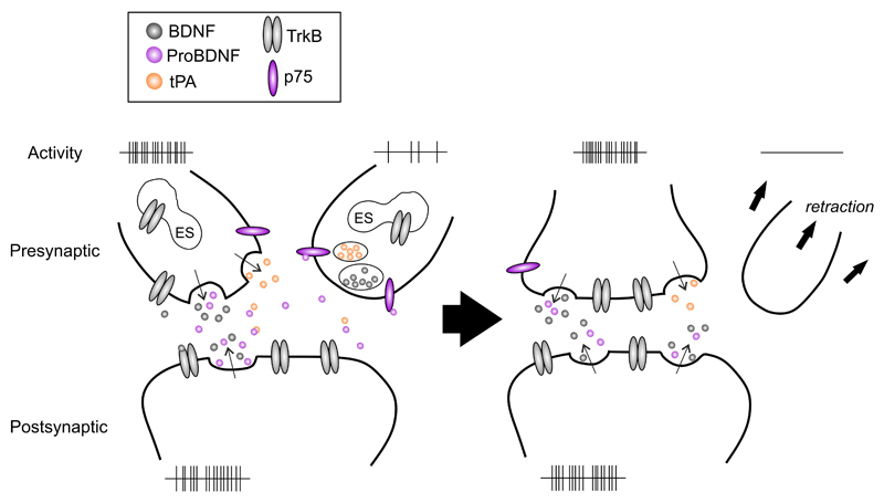
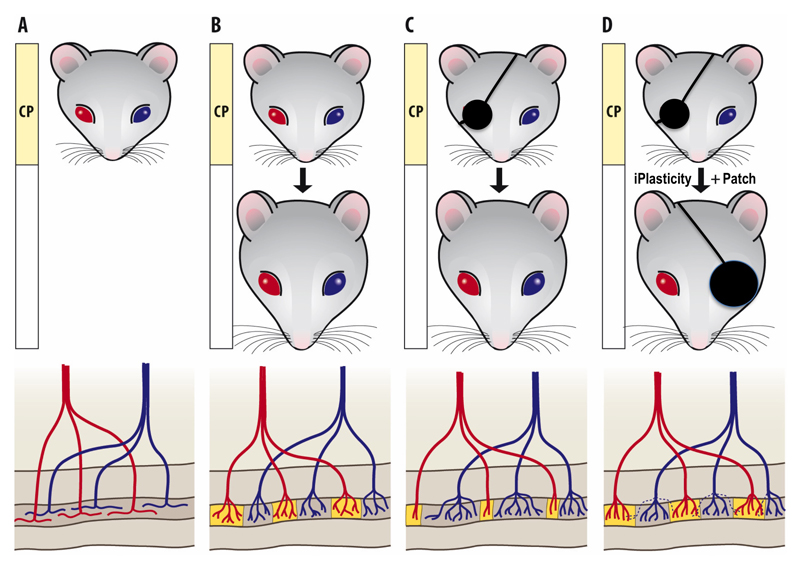
Neurotrophic factors, particularly brain-derived neurotrophic factor (BDNF) and other members of the neurotrophin family, are central mediators of the activity-dependent plasticity through which environmental experiences, such as sensory information are translated into the structure and function of neuronal networks. Synthesis, release and action of BDNF is regulated by neuronal activity and BDNF in turn leads to trophic effects such as formation, stabilization and potentiation of synapses through its high-affinity TrkB receptors. Several clinically available drugs activate neurotrophin signaling and neuronal plasticity. In particular, antidepressant drugs rapidly activate TrkB signaling and gradually increase BDNF expression, and the behavioral effects of antidepressants are mediated by and dependent on BDNF signaling through TrkB at least in rodents. These findings indicate that antidepressants, widely used drugs, effectively act as TrkB activators. They further imply that neuronal plasticity is a central mechanism in the action of antidepressant drugs. Indeed, it was recently discovered that antidepressants reactivate a state of plasticity in the adult cerebral cortex that closely resembles the enhanced plasticity normally observed during postnatal critical periods. This state of induced plasticity, known as iPlasticity, allows environmental stimuli to beneficially reorganize networks abnormally wired during early life. iPlasticity has been observed in cortical as well as subcortical networks and is induced by several pharmacological and non-pharmacological treatments. iPlasticity is a new pharmacological principle where drug treatment and rehabilitation cooperate; the drug acts permissively to enhance plasticity and rehabilitation provides activity to guide the appropriate wiring of the plastic network. Optimization of iPlastic drug treatment with novel means of rehabilitation may help improve the efficacy of available drug treatments and expand the use of currently existing drugs into new indications.


Conflict of interest statement

Conflict of interest statement: The authors declare no conflict of interest.

Figures

Figure 1
Figure 1. Neurotrophins as regulators of activity-dependent plasticity.
Neurotrophins and their pro-forms mediate synaptic strengthening and dendritic retraction, respectively. This dual action is regulated by neuronal activity that controls the expression and secretion of BDNF, cleavage of proBDNF and plasma membrane translocation of TrkB receptors. BDNF binds to TrkB receptors to mediate neuronal survival and stabilization whereas proBDNF binds with a higher affinity to p75NTRs that in the absence of Trk receptors promote apoptosis, dendritic retraction and synaptic depression, allowing differential responses in neurons depending on their activity state. When two axons are competing for the innervation of the same target neuron, the functional connection eventually forms between the two active neurons and the less active neuron retracts. Neuronal activity promotes the release of BDNF and proBDNF as well as tissue plasminogen activator (tPA) that promotes extracellular cleavage of proBDNF to BDNF. In addition, TrkB receptors that normally reside inside the cell are inserted to the plasma membrane in the active neurons. This allows BDNF to mediate survival promoting and synapse strengthening signals in the active neurons. In the less active neuron, TrkB receptors or tPA are not available, however, p75NTR is expressed and the proBDNF released from the active neighbor or the target cell promotes atrophic signals. These effects result in activity-dependent selection of neuronal connectivity. Abbreviations: BDNF, brain-derived neurotrophic factor; ES, endoplasmic system; p75, p75 neurotrophin receptor; proBDNF, pro-form of BDNF; tPA, tissue plasminogen activator; TrkB, tropomyosin receptor kinase.
Figure 2
Figure 2. iPlasticity in the adult visual cortex.
Development of the ocular dominance and its response to monocular development in the mammalian visual cortex is probably the best-characterized model of neuronal network development in the cerebral cortex. (A) During early life, thalamic inputs representing each eye diffusely innervate the entire visual cortex, but (B) during the critical period (CP) of early postnatal life, visual inputs from each eye segregate into alternating eye-specific regions in the primary visual cortex, called OD columns, such that each column becomes predominant innervated by one eye only. (C) If vision of one eye is blocked (monocular deprivation, MD) during the critical period, the more active inputs of the open eye take over the visual cortex through an activity-dependent competition involving BDNF signaling, and the closed eye loses its connectivity (see Fig. 1), thereby becoming poor in vision, or amblyopic. Vision of the amblyopic eye can be recovered if normal vision is restored during the critical period and the use of the weaker eye is encouraged by patching the better eye, but if MD extends beyond the end of the critical period, amblyopia becomes permanent and cannot be revised by patching. (D) However, vision of an amblyopic eye can be restored in adulthood if eye patching is combined with a treatment that induces iPlasticity, such as fluoxetine or environmental enrichment (see Table 1). iPlasticity promotes functional recovery also in other brain areas where abnormal environment has lead to miswiring of developing networks. Modified by permission from Elsevier Ltd. from.

References

    1. Katz LC, Shatz CJ. Synaptic activity and the construction of cortical circuits. Science. 1996;274:1133–1138. - PubMed
    1. Holtmaat A, Caroni P. Functional and structural underpinnings of neuronal assembly formation in learning. Nat Neurosci. 2016;19:1553–1562. - PubMed
    1. Castrén E, Hen R. Neuronal plasticity and antidepressant actions. Trends Neurosci. 2013;36:259–267. - PMC - PubMed
    1. Hensch TK. Critical period plasticity in local cortical circuits. Nat Rev Neurosci. 2005;6:877–888. - PubMed
    1. Maya Vetencourt JF, Sale A, Viegi A, Baroncelli L, De Pasquale R, O’leary OF, et al. The antidepressant fluoxetine restores plasticity in the adult visual cortex. Science. 2008;320:385–388. - PubMed

Publication types

MeSH terms