. 2017 Jun 15;26(12):2258-2276.
doi: 10.1093/hmg/ddx116.

Novel nesprin-1 mutations associated with dilated cardiomyopathy cause nuclear envelope disruption and defects in myogenesis

Affiliations

Novel nesprin-1 mutations associated with dilated cardiomyopathy cause nuclear envelope disruption and defects in myogenesis

Can Zhou et al. Hum Mol Genet. .

Abstract

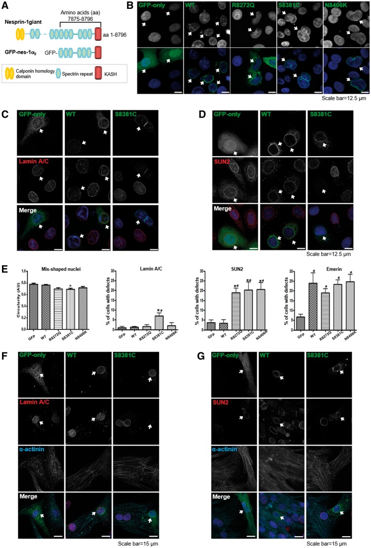
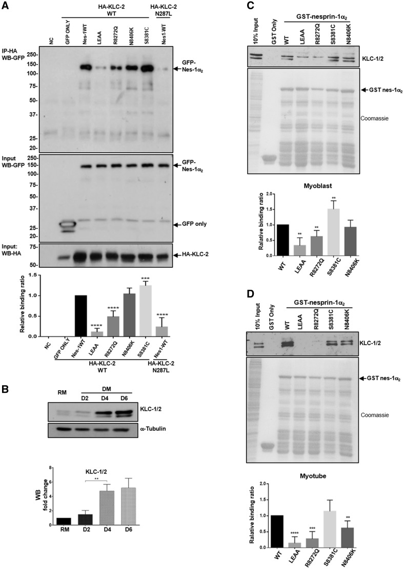
Nesprins-1 and -2 are highly expressed in skeletal and cardiac muscle and together with SUN (Sad1p/UNC84)-domain containing proteins and lamin A/C form the LInker of Nucleoskeleton-and-Cytoskeleton (LINC) bridging complex at the nuclear envelope (NE). Mutations in nesprin-1/2 have previously been found in patients with autosomal dominant Emery-Dreifuss muscular dystrophy (EDMD) as well as dilated cardiomyopathy (DCM). In this study, three novel rare variants (R8272Q, S8381C and N8406K) in the C-terminus of the SYNE1 gene (nesprin-1) were identified in seven DCM patients by mutation screening. Expression of these mutants caused nuclear morphology defects and reduced lamin A/C and SUN2 staining at the NE. GST pull-down indicated that nesprin-1/lamin/SUN interactions were disrupted. Nesprin-1 mutations were also associated with augmented activation of the ERK pathway in vitro and in hearts in vivo. During C2C12 muscle cell differentiation, nesprin-1 levels are increased concomitantly with kinesin light chain (KLC-1/2) and immunoprecipitation and GST pull-down showed that these proteins interacted via a recently identified LEWD domain in the C-terminus of nesprin-1. Expression of nesprin-1 mutants in C2C12 cells caused defects in myoblast differentiation and fusion associated with dysregulation of myogenic transcription factors and disruption of the nesprin-1 and KLC-1/2 interaction at the outer nuclear membrane. Expression of nesprin-1α2 WT and mutants in zebrafish embryos caused heart developmental defects that varied in severity. These findings support a role for nesprin-1 in myogenesis and muscle disease, and uncover a novel mechanism whereby disruption of the LINC complex may contribute to the pathogenesis of DCM.


Figures

Figure 1
Figure 1
Identification of nesprin-1 variants in DCM patients. Mutation screening in SYNE1 and 2 genes was performed in 218 DCM patients and 210 healthy controls, and identified seven patients harbouring three novel nesprin-1 mutations (R8272Q, S8381C, N8406K, shown in bold) in the C-terminus of nesprin-1 giant (A), equivalent to nesprin-1α, within an evolutionally conserved region containing the lamin and emerin binding domains (A, B). Previously identified nesprin-1 mutants in DCM (R8212H) and EDMD-CD patients (R8095H, V8387L and E8461K) were also shown (13,14). The KLC binding motif (LEWD) is shown in (B) (24,27).
Figure 2
Figure 2
Overexpression of nesprin-1 mutants caused abnormal nuclear morphology and reduced NE staining of lamin A/C and SUN2. GFP-tagged nesprin-1α2 WT and mutants are shown schematically (A). The full length of this isoform is 977 amino acids, equivalent to amino acids 7875–8796 in nesprin-1 Giant. U2OS cells were transfected with either GFP-nesprin-1α2 WT or mutants. IF staining showed overexpression of mutants, especially S8381C, led to abnormalities in nuclear morphology (B), reduced lamin A/C and SUN2 staining at the NE (C, D, arrowed). The mis-shapen nuclei were measured by circularity, and mislocalisation of lamin A/C, SUN2 and emerin in the transfected cells was quantified by comparing the NE staining in non-transfected cells on the same slide. Graphical representation of the frequency of misshaped nuclei and defects in lamin A/C, SUN2, and emerin at the NE caused by all three mutants (E). At least 100 transfected nuclei were counted in more than three individual experiments and the results are presented as mean ± SEM and also analysed by Student’s t-tests and one-way ANOVA. (*P < 0.05 compared with WT; #: P < 0.05 compared with GFP only). In addition, IF showed both lamin A/C and SUN2 staining at the NE was reduced in the transfected NRCs using GFP-tagged nesprin1α2 mutant S8381C compared with GFP-tagged WT nesprin-1α2 (F, G, arrowed).
Figure 3
Figure 3
Nesprin-1 mutants affect the interaction between nesprin-1α2 and lamin A/C or SUN2. GST-tagged WT nesprin-1α2 SR1-6 (equivalent to nesprin-1 giant SR 69–74) and mutants are generated and shown schematically (3A left panel, labelled with *), which constructs consisted of 837 amino acids, lacking KASH domain, equivalent to amino acids 7875–8662 of nespin-1 giant. GST pull-down using either GST-WT or mutant nesprin-1 beads showed all three mutants affected binding between nesprin-1 and lamin A/C or SUN2, but not emerin (A, B), which was confirmed by reverse GST pull down by transfecting either GFP-nesprin-1α2WT or each mutant and using either GST-lamin A (amino acids 356–665) (C) or GST-emerin (amino acids 1–176) beads (D). The binding for each mutant was quantified by densitometry with respect to the input material and expressed as a ratio of the value obtained for WT protein. Three independent experiments were performed shown as mean ± SEM, *P < 0.05 using one-way ANOVA analysis. Coomassie blue staining gel also showed equal amount of GST-nesprin 1α2, lamin or emerin beads used.
Figure 4
Figure 4
Nesprin-1 mutants cause aberrant activation of MAPKs. WB showed that aberrant activation of pERK was observed in human dermal fibroblasts from EDMD-DCM patients carrying nesprin-1 mutation (V8387L), lamin A/C (R249Q and R401C) and emerin (g.329del59) mutations (A), as well as nesprin-1 KASH KO mice heart collected at 17 and 81 weeks, respectively (B). WB also showed overexpression of all three nesprin-1 mutants and dominant negative-1KASH led to augmented pERK activity compared with WT nesprin-1 and GFP alone (C). GFP empty vector was used for negative control and a dominant negative-1KASH construct as a positive control. Three independent experiments were performed shown as mean ± SEM, *P < 0.05 using one-way ANOVA analysis.
Figure 5
Figure 5
Expression level of nesprin-1α increase during C2C12 myoblast differentiation. C2C12 myoblasts in regular medium (RM) were stimulated with a low serum differentiation medium (DM), leading to myotube formation during the process (A). WB showed endogenous expression levels of nesprin-1, myogenin and myosin/MHC increased during myotube formation (B, C). Nesprin-1α protein levels (detected by MANNES1E) were highest at day 6, whereas myogenin was highest at day 2, and MHC was detected from day 2 and increased until day 6.
Figure 6
Figure 6
Nesprin-1 mutants cause defects in myoblast differentiation. IF showed that exogenously expressed V5-tagged WT-1α2 or mutants and dominant genative-1KASH were localized at the NE. GFP was expressed as a reporter for retroviral infection (A). Upon differentiation, fewer multinucleated myotubes were observed in cells transduced with mutant R8272Q and dominant negative-1KASH compared with C2C12 cells transduced with eGFP alone (MIG only) or nesprin-1α2 WT (B). The fusion index was reduced in cells transduced with the mutant R8272Q and dominant negative-1KASH compared with 1α2 WT (C), more than 600 nuclei for each clone were counted by microscopy (63× objective) at day 6, three independent experiments were performed for each clone. Further analysis of the MHC positive multinucleated populations revealed that myotubes expressing nesprin-1 mutants contained fewer nuclei compared to the controls (D). qPCR and WB showed that nesprin-1 mutant R8272Q and dominant negative-1KASH caused significant reduction of myogenin (E, F) and MHC (E, G) levels at DM day 2 and day 6 respectively, qPCR also showed that dominant negative-1KASH caused significant reduction of MyoD (E) levels at DM day 2. All were normalized to GFP, three independent experiments were performed shown as mean ± SEM, *P < 0.05 using Student’s t-tests or two-way ANOVA analysis.
Figure 6
Figure 6
Nesprin-1 mutants cause defects in myoblast differentiation. IF showed that exogenously expressed V5-tagged WT-1α2 or mutants and dominant genative-1KASH were localized at the NE. GFP was expressed as a reporter for retroviral infection (A). Upon differentiation, fewer multinucleated myotubes were observed in cells transduced with mutant R8272Q and dominant negative-1KASH compared with C2C12 cells transduced with eGFP alone (MIG only) or nesprin-1α2 WT (B). The fusion index was reduced in cells transduced with the mutant R8272Q and dominant negative-1KASH compared with 1α2 WT (C), more than 600 nuclei for each clone were counted by microscopy (63× objective) at day 6, three independent experiments were performed for each clone. Further analysis of the MHC positive multinucleated populations revealed that myotubes expressing nesprin-1 mutants contained fewer nuclei compared to the controls (D). qPCR and WB showed that nesprin-1 mutant R8272Q and dominant negative-1KASH caused significant reduction of myogenin (E, F) and MHC (E, G) levels at DM day 2 and day 6 respectively, qPCR also showed that dominant negative-1KASH caused significant reduction of MyoD (E) levels at DM day 2. All were normalized to GFP, three independent experiments were performed shown as mean ± SEM, *P < 0.05 using Student’s t-tests or two-way ANOVA analysis.
Figure 7
Figure 7
Defects in nesprin-1 and KLC-1/2 interaction. Overexpression and IP showed the binding between nesprin-1WT/mutants and KLC WT/mutants in U2OS cells, HA-KLC2-N287L was co-expressed with GFP-nesprin-1α2 WT as a positive control, GFP empty vector with HA-KLC-2 WT as a negative control (A). WB showed endogenous expression levels of KLC-1/2 increased during myotube formation (B). GST pull-down showed the binding between nesprin-1 and KLC-1/2 using either GST-WT or mutant nesprin-1 beads in myoblasts (C) and myotubes (D), respectively. The binding for each mutant was quantified by densitometry and expressed as a ratio of the value obtained for WT protein. Three independent experiments were performed shown as mean ± SEM, *P < 0.05 using Student’s t-tests.
Figure 8
Figure 8
siRNA knockdown of KLC-1/2 cause defects in myoblast fusion and differentiation. WB showed the expression levels of both KLC-1 and -2 were reduced in myoblasts (A) and myotubes (B) upon KLC-1/2 depletion. The expression level of MHC (C) and the fusion index (D) were significantly reduced in myotubes especially upon KLC-2 depletion (using KLC-2A oligo), more than 800 nuclei for each clone were counted by microscopy (63× objective) at day 6, three independent experiments were performed for each clone. Further analysis of MHC positive multinucleated cells revealed that KLC-2 depletion resulted in fewer nuclei per myotube. In contrast, KLC-1 depletion (using KLC-1A oligo) led to significantly more clustered nuclei per myotube when compared with controls (E). Means and SEM were obtained from three independent experiments for each clone. *P < 0.05 using Student’s t-tests or two-way ANOVA analysis.
Figure 9
Figure 9
Human nesprin-1α2 WT induces heart development defects in zebrafish. Lateral views of zebrafish live embryos at 48 hpf. The pericardium (A, arrowed) and heart rate are shown for each corresponding mRNA injected (B), zebrafish embryos with nesprin1α2 WT mRNA showed slow heart rate and dilated atrial chambers. Whole-mount in situ hybridization (WISH) monitoring expression of the myl7 gene at 48 hpf (C), the numbers (left in brackets) indicate the percentage of embryos displaying the phenotype represented in the picture shown, the numbers (right in brackets) is the total numbers counted of observed embryos. The relative atrium area of embryos for the corresponding mRNA injection was measured and calculated by the area of myl7 expression using ImageJ (D), which was normalised to the atrium area of the embryos injected with GFP mRNA. Embryos are in ventral views with the anterior at the top. About 4–7 embryos for each injection were measured. Means and SEM were obtained from three independent experiments for each treatment. P < 0.001 using Student’s t-tests. a: atrium, v: ventricle.
Figure 10
Figure 10
Working model for the role nesprin-1. The schematic figure shows nesprin-1 plays multiple-functions at both INM and ONM, and how mutants can disrupt the NE-LINC complex, contributing to the pathogenesis of muscle disease. *Indicates where the mutants are.

Similar articles

Cited by

References

    1. Towbin J.A., Bowles N.E. (2002) The failing heart. Nature, 415, 227–233. - PubMed
    1. Hershberger R.E., Hedges D.J., Morales A. (2013) Dilated cardiomyopathy: the complexity of a diverse genetic architecture. Nat. Rev. Cardiol., 10, 531–547. - PubMed
    1. Capell B.C., Collins F.S. (2006) Human laminopathies: nuclei gone genetically awry. Nat. Rev. Genet., 7, 940–952. - PubMed
    1. Bonne G., Di Barletta M.R., Varnous S., Becane H.M., Hammouda E.H., Merlini L., Muntoni F., Greenberg C.R., Gary F., Urtizberea J.A., et al. (1999) Mutations in the gene encoding lamin A/C cause autosomal dominant Emery-Dreifuss muscular dystrophy. Nat. Genet., 21, 285–288. - PubMed
    1. Bione S., Maestrini E., Rivella S., Mancini M., Regis S., Romeo G., Toniolo D. (1994) Identification of a novel X-linked gene responsible for Emery-Dreifuss muscular dystrophy. Nat. Genet., 8, 323–327. - PubMed

MeSH terms