. 2017 May;187(5):599-611.
doi: 10.1667/RR14616.1. Epub 2017 Mar 6.

Cerebrovascular Remodeling and Neuroinflammation is a Late Effect of Radiation-Induced Brain Injury in Non-Human Primates

Affiliations

Cerebrovascular Remodeling and Neuroinflammation is a Late Effect of Radiation-Induced Brain Injury in Non-Human Primates

Rachel N Andrews et al. Radiat Res. 2017 May.

Abstract

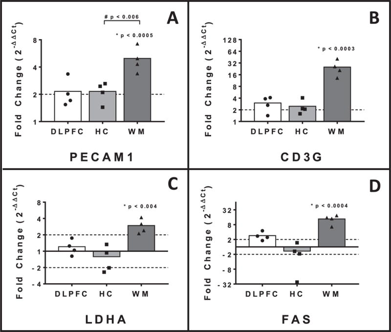
Fractionated whole-brain irradiation (fWBI) is a mainstay of treatment for patients with intracranial neoplasia; however late-delayed radiation-induced normal tissue injury remains a major adverse consequence of treatment, with deleterious effects on quality of life for affected patients. We hypothesize that cerebrovascular injury and remodeling after fWBI results in ischemic injury to dependent white matter, which contributes to the observed cognitive dysfunction. To evaluate molecular effectors of radiation-induced brain injury (RIBI), real-time quantitative polymerase chain reaction (RT-qPCR) was performed on the dorsolateral prefrontal cortex (DLPFC, Brodmann area 46), hippocampus and temporal white matter of 4 male Rhesus macaques (age 6-11 years), which had received 40 Gray (Gy) fWBI (8 fractions of 5 Gy each, twice per week), and 3 control comparators. All fWBI animals developed neurologic impairment; humane euthanasia was elected at a median of 6 months. Radiation-induced brain injury was confirmed histopathologically in all animals, characterized by white matter degeneration and necrosis, and multifocal cerebrovascular injury consisting of perivascular edema, abnormal angiogenesis and perivascular extracellular matrix deposition. Herein we demonstrate that RIBI is associated with white matter-specific up-regulation of hypoxia-associated lactate dehydrogenase A (LDHA) and that increased gene expression of fibronectin 1 (FN1), SERPINE1 and matrix metalloprotease 2 (MMP2) may contribute to cerebrovascular remodeling in late-delayed RIBI. Additionally, vascular stability and maturation associated tumor necrosis super family member 15 (TNFSF15) and vascular endothelial growth factor beta (VEGFB) mRNAs were increased within temporal white matter. We also demonstrate that radiation-induced brain injury is associated with decreases in white matter-specific expression of neurotransmitter receptors SYP, GRIN2A and GRIA4. We additionally provide evidence that macrophage/microglial mediated neuroinflammation may contribute to RIBI through increased gene expression of the macrophage chemoattractant CCL2 and macrophage/microglia associated CD68. Global patterns in cerebral gene expression varied significantly between regions examined (P < 0.0001, Friedman's test), with effects most prominent within cerebral white matter.


Figures

FIG. 1
FIG. 1
Cerebrovascular pathology in late-delayed radiation-induced brain injury. Histopathology showing the spectrum of vascular injury in animals with late-delayed radiation-induced brain injury. Panel A: Normal arteriole from an nonirradiated animal. Panel B: Perivascular accumulation of brightly eosinophilic protein and fibrin. Panel C: Disorganized angiogenesis characterized by haphazardly arranged, tortuous small caliber vessels, often lacking distinct lumina. Endothelial nuclei are enlarged with prominent nucleoli. Panel D: Accumulation of fibrous extracellular matrix surrounding arterioles. Reactive (gemistocytic) astrocytes are numerous within the surrounding parenchyma. Panel E: Pericapillary fibrous extracellular matrix, similar to that in seen in panel D.
FIG. 2
FIG. 2
Changes in gene expression associated with extracellular matrix deposition. Late-delayed radiation-induced brain injury was associated with up-regulated expression of MMP2 (35-fold), SERPINE1 (21-fold) and fibronectin1 (threefold) within white matter and MMP2 (sixfold) and fibronectin1 (threefold) within dorsolateral prefrontal cortex. Bars denote mean fold change, individual points represent gene expression values for each animal. Dashed line indicates twofold-expression change. DLPFC: Dorsolateral prefrontal cortex; HC: Hippocampus; WM: Temporal white matter. *P value indicates with the effect of radiation on relative-gene expression and #P value signifies differences in regional gene expression with respect to irradiation status
FIG. 3
FIG. 3
Up-regulated relative expression of factors associated with vascular maturation and stability. Expression of TNFSF15 and VEGFB mRNAs was up-regulated within temporal white matter in late-delayed radiation-induced brain injury, 44-fold and twofold, respectively. Bars denote mean fold change, individual points represent gene expression values for each animal. Dashed line indicates twofold-expression change. DLPFC: Dorsolateral prefrontal cortex, HC: Hippocampus, WM: Temporal white matter. *P value indicates with the effect of radiation on relative-gene expression and #P value signifies differences in regional gene expression with respect to irradiation status
FIG. 4
FIG. 4
Evidence of macrophage/microglial mediated neuroinflammation. Panel A: High-magnification image of focal white matter necrosis with axonal degeneration, mineralization and infiltration by macrophages/microglia. Panel B: Late-delayed radiation-induced brain injury was associated with increased gene expression of CD68 (28-fold) and CCL2 (49-fold) within temporal white matter. Expression of CCL2 was also increased within dorsolateral prefrontal cortex CCL2 (31-fold up-regulated). Bars denote mean fold change, individual points represent gene expression values for each animal. Dashed line indicates twofold-expression change. DLPFC: Dorsolateral prefrontal cortex, HC: Hippocampus, WM: Temporal white matter. *P value indicates with the effect of radiation on relative-gene expression and #P value signifies differences in regional gene expression with respect to irradiation status
FIG. 5
FIG. 5
Additional changes in gene expression. Panel A: Relative gene expression of endothelial inflammation associated PECAM1 was 4.8-fold increased within temporal white matter. Panel B: Expression of T-lymphocyte associated CD3G was 23.2 increased within white matter. Panel C: Hypoxia associated LDHA expression was 2.9-fold increased within temporal white matter. Panel D: Late-delayed radiation-induced brain injury was associated with a 12.7-fold increase in relative gene expression of cell surface death receptor FAS in temporal white matter. Bars denote mean fold change, individual points represent gene expression values for each animal. Dashed line indicates twofold-expression change. DLPFC: Dorsolateral prefrontal cortex, HC: Hippocampus, WM: Temporal white matter. *P value indicates with the effect of radiation on relative-gene expression and #P value signifies differences in regional gene expression with respect to irradiation status
FIG. 6
FIG. 6
Decreases in gene expression associated with neurotransmission. Late-delayed radiation-induced brain injury was associated with decreased expression of neurotransmission associated synaptophysin (−3.4-fold), and glutamatergic neurotransmitter receptor mRNAs for GRIA4 (−6.7-fold) and GRIN2A (−6.7-fold) within white matter. Bars denote mean fold change, individual points represent gene expression values for each animal. Dashed line indicates twofold-expression change. DLPFC: Dorsolateral prefrontal cortex, HC: Hippocampus, WM: Temporal white matter. *P value indicates with the effect of radiation on relative-gene expression and #P value signifies differences in regional gene expression with respect to irradiation status

References

    1. Maher EA, Mietz J, Arteaga CL, DePinho RA, Mohla S. Brain metastasis: opportunities in basic and translational research. Cancer Res. 2009;69(15):6015–20. - PubMed
    1. Meyers CA, Brown PD. Role and relevance of neurocognitive assessment in clinical trials of patients with CNS tumors. J Clin Oncol. 2006;24(8):1305–9. - PubMed
    1. Hsiao KY, Yeh SA, Chang CC, Tsai PC, Wu JM, Gau JS. Cognitive function before and after intensity-modulated radiation therapy in patients with nasopharyngeal carcinoma: a prospective study. Int J Radiat Oncol Biol Phys. 2010;77(3):722–6. - PubMed
    1. DeAngelis LM, Delattre JY, Posner JB. Radiation-induced dementia in patients cured of brain metastases. Neurology. 1989;39(6):789–96. - PubMed
    1. Yoritsune E, Furuse M, Kuwabara H, Miyata T, Nonoguchi N, Kawabata S, et al. Inflammation as well as angiogenesis may participate in the pathophysiology of brain radiation necrosis. J Radiat Res. 2014;55(4):803–11. - PMC - PubMed

Publication types

MeSH terms

Substances