Review
. 2017 Apr 10;41(1):10-22.
doi: 10.1016/j.devcel.2017.02.016.

Cleaning House: Selective Autophagy of Organelles

Affiliations
Review

Cleaning House: Selective Autophagy of Organelles

Allyson L Anding et al. Dev Cell. .

Abstract

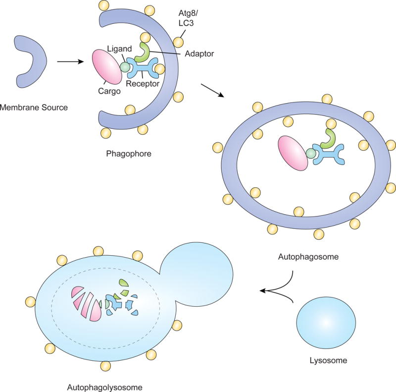
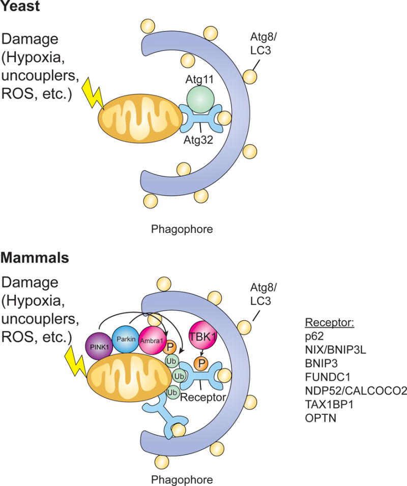
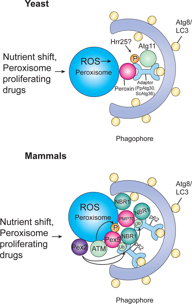
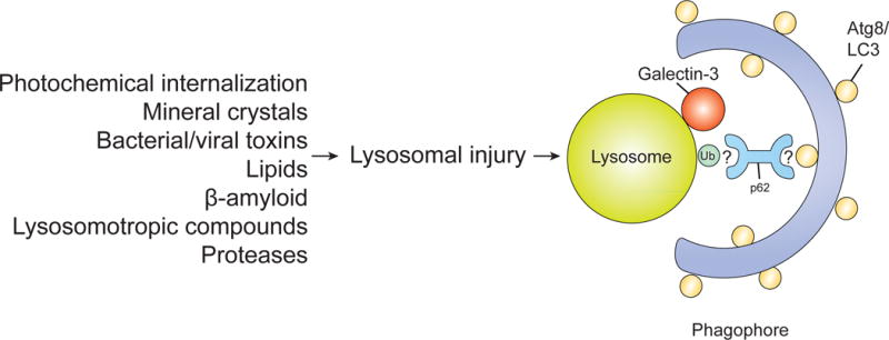
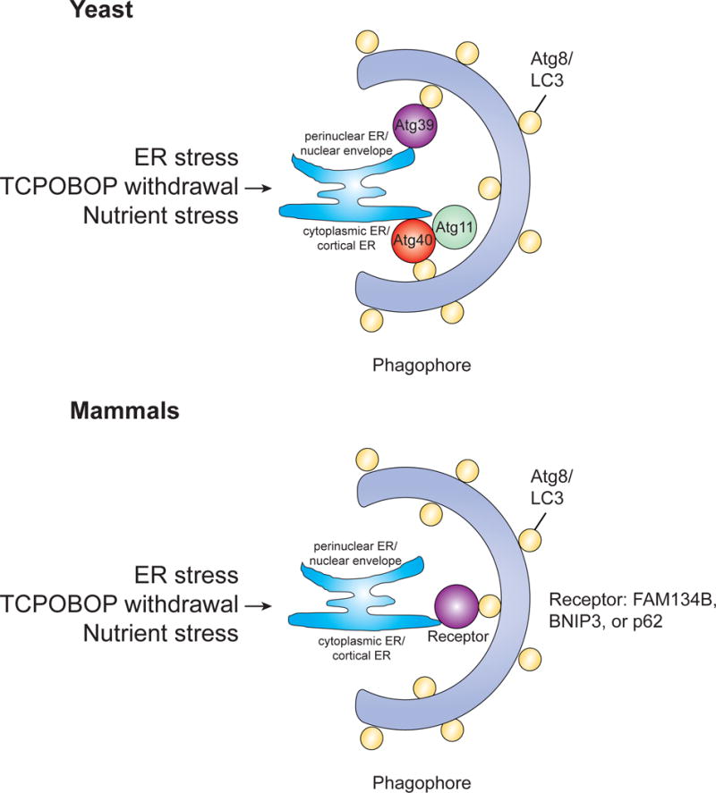
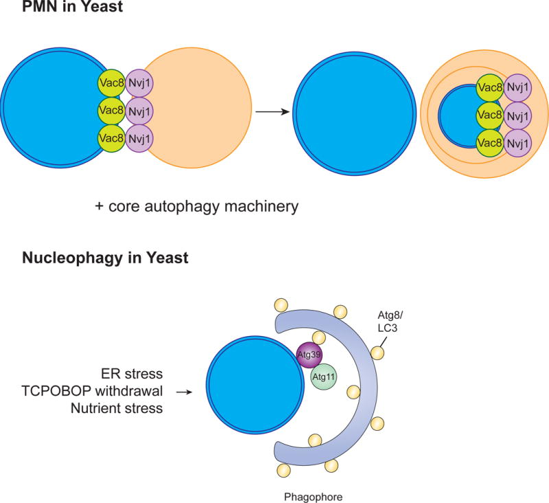
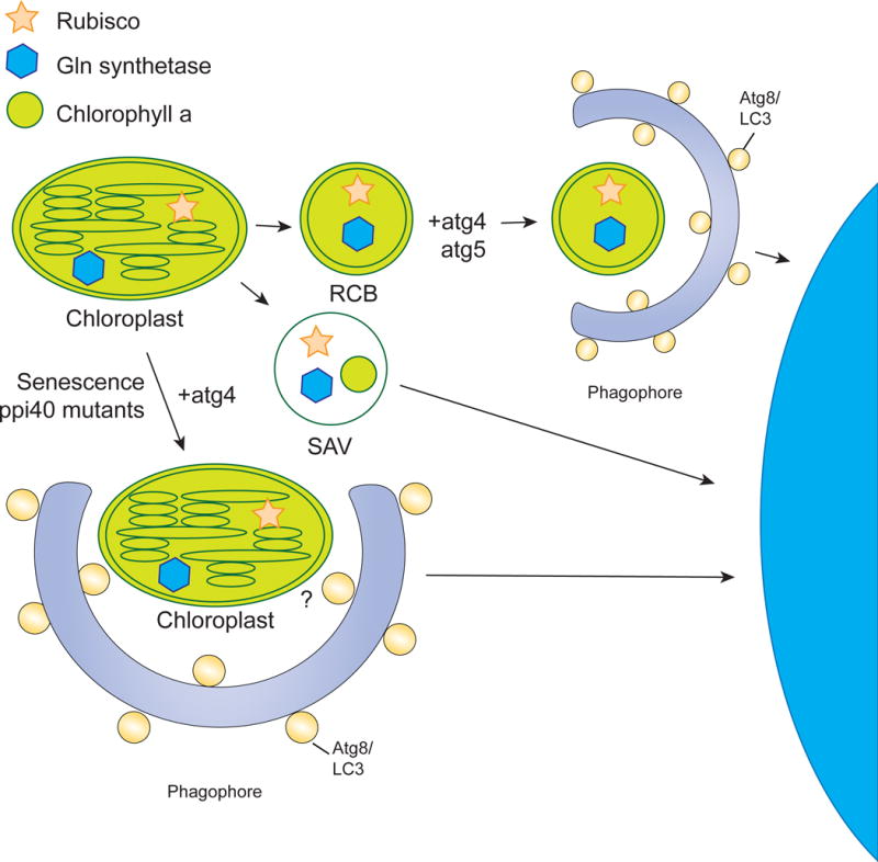
The selective clearance of organelles by autophagy is critical for the regulation of cellular homeostasis in organisms from yeast to humans. Removal of damaged organelles clears the cell of potentially toxic byproducts and enables reuse of organelle components for bioenergetics. Thus, defects in organelle clearance may be detrimental to the health of the cells, contributing to cancer, neurodegeneration, and inflammatory diseases. Organelle-specific autophagy can clear mitochondria, peroxisomes, lysosomes, ER, chloroplasts, and the nucleus. Here, we review our understanding of the mechanisms that regulate the clearance of organelles by autophagy and highlight gaps in our knowledge of these processes.

Keywords: ER-phagy; autophagy; chlorophagy; lysophagy; mitophagy; nucleophagy; pexophagy.


Figures

Figure 1
Figure 1. Selective autophagy
In selective autophagy, cargo, such as organelles, are degraded by the autophagolysosome. Cargo are often tagged with ligands such as ubiquitin, enabling them to interact with receptor proteins that can then interact with the autophagosomal membrane. This interaction occurs by binding to Atg proteins such as Atg8/LC3 or through interaction with adaptor proteins. After the cargo is brought to the phagophore, formation of the autophagosome occurs and this structure can then fuse with the lysosome to form the autophagolysosome, degrading the cargo.
Figure 2
Figure 2. Mitophagy
Mitophagy can be triggered by various stimuli, such as hypoxia, uncouplers, or ROS, that cause damage to mitochondria. In yeast, Atg32 acts as the mitophagy receptor, binding the adaptor protein Atg11 and interacting with Atg8 on the inner membrane of autophagosomes. In mammals, Pink1 phosphorylates various targets, including ubiquitin, and recruits Parkin which can then amplify the signal by ubiquitinating mitochondrial surface proteins. These ubiquitinated proteins can then be recognized by cargo receptor proteins that bring the mitochondria to forming autophagosomes for degradation. Mitochondrial surface receptor proteins can also recognize LC3 to facilitate recruitment to the phagophore. AMBRA1 also localizes to damaged mitochondria through LIR motif-dependent interactions with LC3, promoting both canonical PARKIN-dependent and –independent mitochondrial clearance. Additionally, TBK1 phosphorylates autophagy receptors to create a signal amplification loop in mitophagy.
Figure 3
Figure 3. Pexophagy
Pexophagy can be triggered by either a shift in nutrient conditions or peroxisome proliferating drugs. In yeast, Atg30 (P. pastoris) or Atg36 (S. cerevisiae) acts as the autophagy receptor, binding peroxisomes via their interaction with at least one peroxin and recruiting autophagic machinery by interacting with Atg proteins. Both Atg30 and Atg36 are phosphorylated (Atg36 by Hrr25), allowing them to interact with Atg11. In mammals, pexophagy has been shown to involve either NBR1 or p62. NBR1 binds peroxisomes through its JUBA domain which allows it to interact with phospholipids and ubiquitin. p62 supports and cooperates with NBR1 to bind ubiquitinated proteins. ATM kinase activates pexophagy in response to ROS by phosphorylating PEX5 leading to ubiquitination of PEX5 which can then be recognized by p62, targeting peroxisomes for pexophagy. PMP70 has also been implicatd in pexophagy and both PMP70 and Pex5 are ubiquitinated by Pex2.
Figure 4
Figure 4. Lysophagy
Lysophagy can be induced by a variety of stimuli that can lead to lysosome damage. Upon lysosomal injury, galectin-3 and LC3 are recruited to the injured lysosome. Lysosomal membranes are ubiquitinated and co-localize with p62, suggesting that ubiquitination and subsequent recruitment of p62 are involved in this process. However, the exact mechanisms of lysophagy regulation are still unclear.
Figure 5
Figure 5. ER-phagy/Reticulophagy
ER stress, TCPOBOP withdrawal, and nutrient stress are all stimuli that can lead to the induction of ER-phagy. In yeast, both Atg39 and Atg40 have been shown to mediate ER-phagy, localizing to distinct subdomains of the ER and interacting with Atg8. In mammals, the functional counterpart of Atg40 is FAM134B. Both BNIP3/Nix and p62 have also been implicated in ER-phagy.
Figure 6
Figure 6. Nucleophagy
The clearance of nuclear contents by autophagy using either PMN or the involvement of the cargo receptor Atg39. In yeast, PMN involves the formation of a junction between the nucleus and the vacuole generated by two key proteins, Vac8 and Nvi1. These junctions bulge into the vacuole, a piece of the nucleus buds off, and this vesicle is released into the vacuole where it is degraded by vacuolar hydrolases. This process involves much of the core autophagy machinery. The nucleophagy receptor Atg39 was also shown to localize to the yeast perinuclear ER/nuclear envelope and lead to degradation of nuclear components. Less is known about the mechanism behind nucleophagy in mammals.
Figure 7
Figure 7. Chlorophagy
The chloroplast can be degraded in a piecemeal fashion or through whole organelle autophagy. Piecemeal degradation occurs via RCBs and SAVs. RCBs are derived from the chloroplast envelope and contain rubisco and Gln synthetase. The RCB is surrounded by the isolation membrane in the cytoplasm. This process has been shown to be dependent on atg4 and atg5. SAVs are small lytic vacuoles that also degrade parts of the chloroplast and have been shown to contain rubisco, Gln synthetase, and chlorophyll a. However, they have not been shown to use the autophagy machinery. A decrease in the total number of chloroplasts during senescence has been shown to involve the degradation of the entire chloroplast, a process that is blocked in atg4 mutants. However, the cargo receptor for this process has not been identified.

References

    1. Aits S, Jäättelä M. Lysosomal cell death at a glance. J Cell Sci. 2013;126:1905–1912. - PubMed
    1. Akinduro O, Sully K, Patel A, Robinson DJ, Chikh A, McPhail G, Braun KM, Philpott MP, Harwood CA, Byrne C, et al. Constitutive Autophagy and Nucleophagy during Epidermal Differentiation. J Invest Dermatol. 2016;136:1460–1470. - PubMed
    1. Anding AL, Baehrecke EH. Autophagy in Cell Life and Cell Death. Curr Top Dev Biol. 2015;114:67–91. - PubMed
    1. Axe EL, Walker SA, Manifava M, Chandra P, Roderick HL, Habermann A, Griffiths G, Ktistakis NT. Autophagosome formation from membrane compartments enriched in phosphatidylinositol 3-phosphate and dynamically connected to the endoplasmic reticulum. J Cell Biol. 2008;182:685–701. - PMC - PubMed
    1. Bellot G, Garcia-Medina R, Gounon P, Chiche J, Roux D, Pouysségur J, Mazure NM. Hypoxia-induced autophagy is mediated through hypoxia-inducible factor induction of BNIP3 and BNIP3L via their BH3 domains. Mol Cell Biol. 2009;29:2570–2581. - PMC - PubMed