. 2017 Apr 10;31(4):501-515.e8.
doi: 10.1016/j.ccell.2017.03.005.

Eradication of Tumors through Simultaneous Ablation of CD276/B7-H3-Positive Tumor Cells and Tumor Vasculature

Affiliations

Eradication of Tumors through Simultaneous Ablation of CD276/B7-H3-Positive Tumor Cells and Tumor Vasculature

Steven Seaman et al. Cancer Cell. .

Abstract

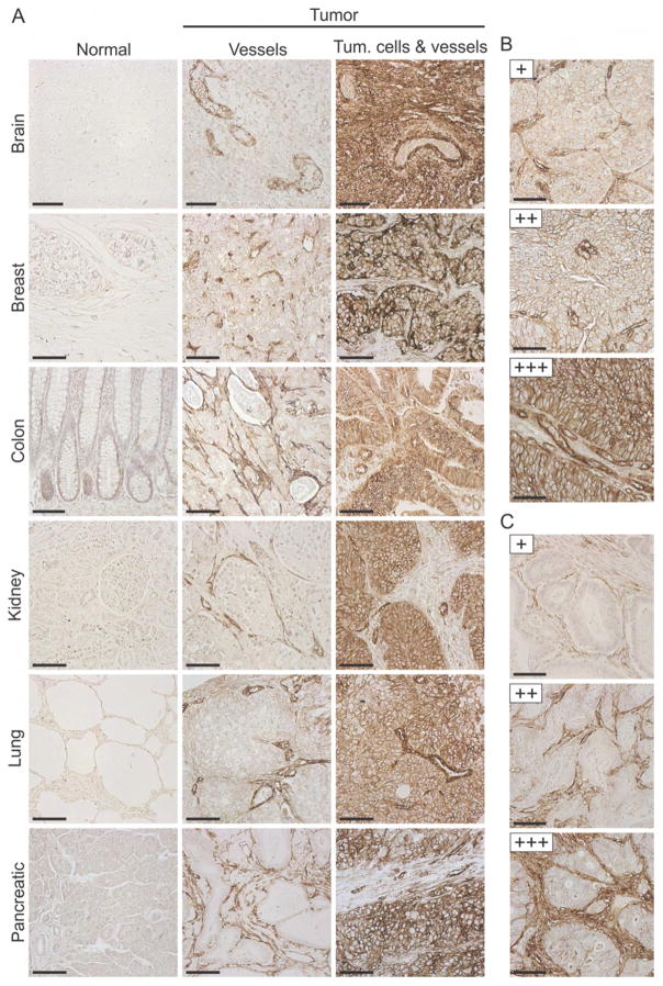
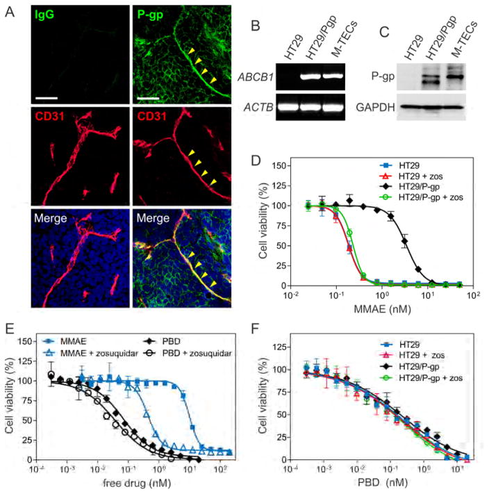
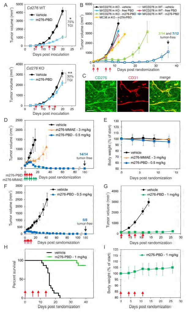
Targeting the tumor vasculature with antibody-drug conjugates (ADCs) is a promising anti-cancer strategy that in order to be realized must overcome several obstacles, including identification of suitable targets and optimal warheads. Here, we demonstrate that the cell-surface protein CD276/B7-H3 is broadly overexpressed by multiple tumor types on both cancer cells and tumor-infiltrating blood vessels, making it a potentially ideal dual-compartment therapeutic target. In preclinical studies CD276 ADCs armed with a conventional MMAE warhead destroyed CD276-positive cancer cells, but were ineffective against tumor vasculature. In contrast, pyrrolobenzodiazepine-conjugated CD276 ADCs killed both cancer cells and tumor vasculature, eradicating large established tumors and metastases, and improving long-term overall survival. CD276-targeted dual-compartment ablation could aid in the development of highly selective broad-acting anti-cancer therapies.

Keywords: ADC; Abcb1; B7H3; P-glycoprotein; P-gp; PBD; TEM; angiogenesis; cancer; endothelium.


Figures

Figure 1
Figure 1. CD276 is Overexpressed in Various Human Tumors
(A) IHC of FFPE tissues was used to evaluate CD276 expression in various human tumors or corresponding normal organs. (B) Tumor cell staining was ranked as low (+), moderate (++) or strong (+++), as shown in this renal cell cancer example. (C) Stromal cell staining was ranked as low (+), moderate (++) or strong (+++), as shown in this colon cancer example. Scale bar in (A) to (C): 100 μm. See also Figure S1.
Figure 2
Figure 2. CD276 is Overexpressed in Tumors but has Minimal Impact on Tumor Growth
(A) CD276 proteins were immunoprecipitated (IP) from MC38 parent cells or MC38 cells overexpressing full-length mouse (mCD276), rat (rCD276) or human (hCD276) CD276 and detected by immunoblotting. (B) Detection of human CD276 overexpressed in MC38 cells using flow cytometry. (C) CD276 IF staining (green) of an MDA-MB-231 breast cancer lung metastasis (yellow arrows indicate tumor margins). CD31 positive vessels are counterstained blue. (D) CD276 IF staining (green) of MC38 colon liver metastasis from Cd276 WT or KO mice. (E) vWF (top panel) or CD276 (bottom panel) IF staining of human colon cancer liver metastases. The two panels were taken from serial sections. Normal (N) / tumor (T) margins in (D) and (E) are indicated by a white dash. Scale bar in (C) to (E): 50μm. (F) CD276 IHC staining of human colon cancer lung metastases with co-opted vasculature. Tumor cells (T) have expanded inside the alveolar air space and are separated from stromal cells (S) by a thin layer of cytokeratin 7 (CK7) positive alveolar type I cells. Top and bottom panels represent serial sections, and arrows (inset) indicate the location of a large co-opted CD276+ blood-filled vessel. Staining is representative of 5 independent cases with vessel co-option. (G) CD276 IF staining of human colon cancer liver metastases with co-opted vasculature. A co-opted portal triad is found near the center of the section (inset), and is identified by large CD31+ vessels (red) next to bile ducts (arrows) that stain strongly for cytokeratin 19 (CK19; yellow). Surrounding colon tumor cells also express low levels of CK19. Arrowheads indicate the CD276 positive co-opted vessels. Staining is representative of 5 independent cases with vessel co-option. Scale bar in (F) to (H): 100μm. (H) CD276 IF staining of MC38 tumor vessels from Cd276 WT and Cd276 KO mice. Note that CD276 staining (green) was undetectable in desmin-positive vascular pericytes (red) of MC38 tumors (yellow arrowheads). CD31 staining (blue) was used to highlight the location of the endothelium in the KO. Scale bar: 10 μm. (I) Tumor cells were inoculated subcutaneously into athymic nude (DLD-1, HCT-116, and UACC melanoma) or C57BL/6 (MC38 colon) Cd276 WT or KO mice and tumor growth monitored. n = 10/ group (MC38), n = 12/group (DLD-1), n = 8 (KO) or 11 (WT) per group (HCT-116) and n = 9/group UACC. See also Figure S2.
Figure 3
Figure 3. m276-MMAE Elicits Potent Anti-Tumor Activity Against Various Human Tumor Xenografts Without Evidence of Toxicities
(A) The affinity of the monovalent m276 Fab for human, mouse, rat and monkey (cynomolgus) CD276 was determined using Biacore analysis. (B, C) Cell viability assays were used to measure the activity of m276-MMAE against HT29 (B) and OVCAR3 (C) cells. To verify specificity, competition with unlabeled m276 was performed. Error bars in (B) and (C) = SD. (D–H) Subcutaneous growth of colon HCT-116 (D), KM12 (E), HT29 (F), ovarian OVCAR3 (G), or orthotopic breast MDA-MB-231 (H) tumors. Treatments with vehicle, MMAE, m276, or m276-MMAE were initiated when tumors reached an average size of ~100mm3 and were administered on the days shown (green arrows). n = 10/group (DH). Error bars in (D) to (H) = SEM. (I) Six OVCAR3 tumors that had relapsed following treatment with 3 mg/kg of m276-MMAE (biwk x 3.5) were retreated with 3 mg/kg (biwk x 3) starting on day 55 and then 10 mg/kg (biwk x 5) starting on day 76. Individual tumor measurements are shown. See also Figures S3–S5.
Figure 4
Figure 4. CD276 ADC Activity against CD276+ Tumor Endothelium
(A) CD276 (green) and CD31 (red) IF staining of MC38 tumors 48 hr post treatment with vehicle (control), m276-MMAE (3 mg/kg) or m276-PBD (1 mg/kg). Scale bar: 100μm. (B) Mean vessel area (% of field) in MC38 tumors 48 hr post treatment with vehicle (control), m276-MMAE or m276-PBD. N.S.; non-significant. n = 25 fields/group, *p<0.001. (C) Flow cytometry was used to measure CD276 or CD31 levels in bEnd.3 Normal ECs (B-NECs) or MC38 Tumor ECs (M-TECs). (D) Cell viability of B-NECs or M-TECs following treatment with m276-MMAE and m276-MMAE. Error bars in (B) and (D) = SD. See also Figure S6.
Figure 5
Figure 5. m276-PBD helps Circumvent P-gp Mediated Resistance
(A) Co-immunofluorescence staining using anti-P-gp (green) and anti-CD31 (red) antibodies was performed on DMS-273 tumors. The yellow arrowheads mark a P-gp positive vessel. Non-specific rabbit IgG was used as a specificity control. Scale bar: 50 μm. (B) RT-PCR was used to evaluate ABCB1 mRNA expression in HT29, HT29/P-gp and M-TECs. The β-actin (ACTB) mRNA was used as a loading control. (C) Immunoblotting was used to evaluate P-gp protein expression in HT29, HT29/P-gp and M-TECs. GAPDH was used as a loading control. (D) Cell viability assays were used to measure the activity of the cell permeable MMAE free drug against HT29 and HT29/P-gp cells. P-gp activity was inhibited by treating cells with 1 μM zosuquidar (zos). (E) Cell viability assays were used to measure the activity of the cell permeable MMAE and PBD free drugs against M-TECs. P-gp activity was inhibited by treating cells with 1 μM zosuquidar. (F) Cell viability assays were used to measure the activity of the cell permeable PBD free drug against HT29 and HT29/P-gp cells. P-gp activity was inhibited by treating cells with 1 μM zosuquidar (zos). Error bars in (D) to (F) = SD.
Figure 6
Figure 6. m276-PBD Eradicates Common Tumor Types and is Well Tolerated
(A) Subcutaneous MC38 tumor growth in Cd276 WT or KO mice. Treatments with vehicle (control) or 1 mg/kg m276-PBD (red arrows) were initiated once tumors reached an average size of 60mm3. TGI; Tumor growth inhibition, n = 15/group, *p<0.0001, **p=0.02 (B) Growth of subcutaneous MC38 or MC38/CD276-c3 (M/CD276) tumors in Cd276 WT or KO mice. Treatments with vehicle (control), free PBD, or 1mg/kg m276-PBD (red arrows) began when tumors reached an average size of 100mm3. n = 10 to 14 mice/group. (C) CD276 (green) and CD31 (red) IF staining of MC38/CD276-c3 tumors from Cd276 WT mice. Note both tumor cells and tumor vasculature are CD276 positive. Scale bar: 50μm. (D) Growth of subcutaneous human DMS-273 lung tumors in athymic nude mice treated with PBS (vehicle), m276-MMAE (green arrows), or m276-PBD (red arrows) starting when tumors reached an average size of 140mm3. (E) Body weights of the DMS-273 tumor-bearing mice from (D). (F) Growth of subcutaneous human HCT-116 colon tumors in athymic nude mice treated with PBS (vehicle), or m276-PBD (red arrows) once tumors reached an average size of ~140mm3. (G) Growth of Py230 breast tumors inoculated orthotopically into syngeneic C57BL6 mice and treated with PBS (vehicle) or 1mg/kg of m276-PBD (biwk x 2 - red arrows) starting when tumors reached an average size of ~100 mm3. Error bars in (A), (B), (D) (F) and (G) = SEM. (H) Kaplan-Meier survival analysis from the Py230 study shown in (G). Log-rank analysis: p<0.0001 m276-PBD vs. vehicle. n=15/group (I) Body weights of the Py230 tumor-bearing mice from the study in (G). Error bars in (E) and (I) = SD. See also Figure S7.
Figure 7
Figure 7. m276-PBD Blocks Metastasis and Extends Overall Survival
(A) BLI of liver metastases following intrasplenic injection of HCT-116-luc colon cancer cells. 8 days post-inoculation (dpi) mice were sorted into two groups of equal average tumor burden and treatments initiated with PBS (vehicle) or m276-PBD (1 mg/kg) twice per week for two weeks (biwk x 2). Note the decrease in bioluminescence in the m276- PBD treated group at 35 dpi. (B) Quantification of tumor burden from the HCT-116 liver metastasis study shown in (A). Error bars = SEM. (C) Kaplan-Meier survival analysis of HCT-116 liver metastasis study shown in (A). Logrank analysis: p<0.0001 m276-PBD vs. vehicle. n=15–17/group. (D) Kaplan-Meier survival analysis of vehicle (PBS), m276 and m276-PBD in an experimental 4T1 breast cancer lung metastasis model. Treatments began 10 days after i.v. tumor cell inoculation and were administered twice per week (red arrows). Logrank analysis: p=0.0002 m276-PBD vs. untreated, p<0.0001 m276-PBD vs. m276. Nonsignificant: untreated vs. m276. n = 5 to 8/group. (E) CD276 IF staining in 4T1 breast cancer lung metastasis. Note the reduced number of CD31+ vessels within tumor nodules 48 hr post treatment with 1 mg/kg m276-PBD. Scale bar: 100μm. (F) Higher magnification of insets from (E). Arrowheads highlight some CD31+ tumor vessels that are also CD276+. Scale bar: 20μm. (G) Tumor vessel density in 4T1 lung metastases 48 hr post treatment with 1 mg/kg m276-PBD. n=30 metastases/group. Error bars = SD.

Comment in

References

    1. Abdollahi A, Folkman J. Evading tumor evasion: current concepts and perspectives of anti-angiogenic cancer therapy. Drug Resist Updat. 2010;13:16–28. - PubMed
    1. Ahmed M, Cheng M, Zhao Q, Goldgur Y, Cheal SM, Guo HF, Larson SM, Cheung NK. Humanized Affinity-matured Monoclonal Antibody 8H9 Has Potent Antitumor Activity and Binds to FG Loop of Tumor Antigen B7-H3. J Biol Chem. 2015;290:30018–30029. - PMC - PubMed
    1. Akiyama K, Ohga N, Hida Y, Kawamoto T, Sadamoto Y, Ishikawa S, Maishi N, Akino T, Kondoh M, Matsuda A, et al. Tumor endothelial cells acquire drug resistance by MDR1 up-regulation via VEGF signaling in tumor microenvironment. Am J Pathol. 2012;180:1283–1293. - PubMed
    1. Arigami T, Narita N, Mizuno R, Nguyen L, Ye X, Chung A, Giuliano AE, Hoon DS. B7-h3 ligand expression by primary breast cancer and associated with regional nodal metastasis. Ann Surg. 2010;252:1044–1051. - PubMed
    1. Baskin JM, Prescher JA, Laughlin ST, Agard NJ, Chang PV, Miller IA, Lo A, Codelli JA, Bertozzi CR. Copper-free click chemistry for dynamic in vivo imaging. Proc Natl Acad Sci U S A. 2007;104:16793–16797. - PMC - PubMed

Publication types

MeSH terms