Review
. 2017 May;23(5):465-481.
doi: 10.1016/j.molmed.2017.03.003. Epub 2017 Apr 8.

Cancer Manipulation of Host Physiology: Lessons from Pancreatic Cancer

Affiliations
Review

Cancer Manipulation of Host Physiology: Lessons from Pancreatic Cancer

Constantinos P Zambirinis et al. Trends Mol Med. 2017 May.

Abstract

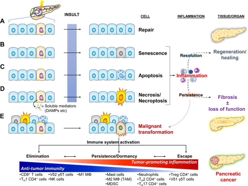
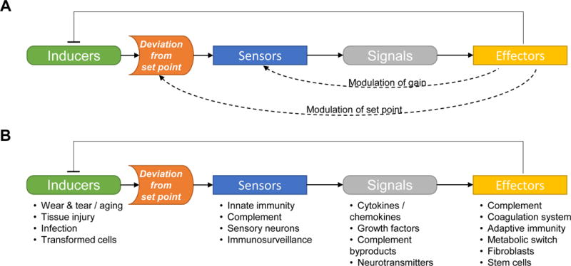
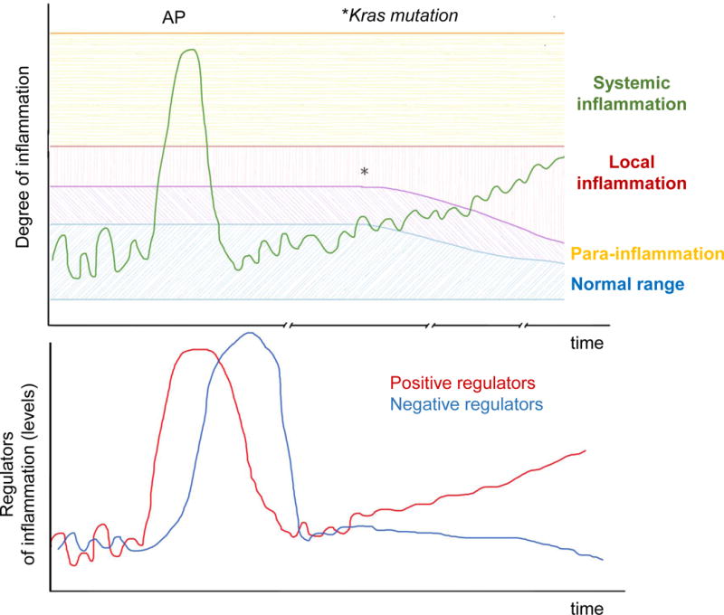
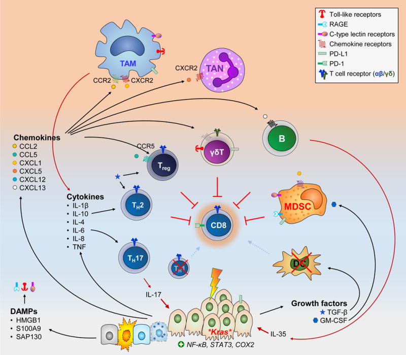
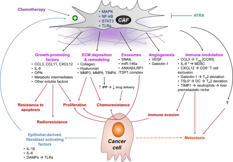
Homeostasis is a fundamental property of living organisms enabling the human body to withstand internal and external insults. In several chronic diseases, and especially in cancer, many homeostatic mechanisms are deranged. Pancreatic cancer in particular is notorious for its ability to invoke an intense fibroinflammatory stromal reaction facilitating its progression and resistance to treatment. In the past decade, several seminal discoveries have elucidated previously unrecognized modes of commandeering the host's defense systems. Here we review novel discoveries in pancreatic cancer immunobiology and attempt to integrate the notion of deranged homeostasis in the pathogenesis of this disease. We also highlight areas of controversy and obstacles that need to be overcome, hoping to further our mechanistic insight into this malignancy.

Keywords: cancer-associated fibroblasts; immunity; inflammation; pancreatic ductal adenocarcinoma; stroma.


Conflict of interest statement

Conflict of Interest

None declared.

Figures

Key Figure, Figure 1
Key Figure, Figure 1. Homeostatic Responses to Tissue Stress in the Context of Pancreatic Dysfunction and Malignancy
Cellular stress can be invoked by a great variety of internal (e.g. wear and tear/aging, reactive oxygen species, hypoxia, etc) and external causes (e.g. toxins, radiation, infectious agents, trauma, etc). Some can be mutagenic, potentially leading to cancer. Stressed/damaged cells activate defensive systems that attempt to repair the injury. If successful, the cell returns to its normal state and usually minimal or no inflammatory response is induced (a). (b–d) If the injury is extensive and irreparable, the cell may either go into a senescence – a “safe mode” characterized by cell cycle arrest and slowing of cellular functions [101] – or may activate programmed cell death by apoptosis or necroptosis. If the injury is sudden and overwhelming, it may induce cell rupture and resultant necrosis. Both senescence and the various modes of cell death are characterized by altered expression of cell surface molecules as well as release of soluble mediators that activate the immune system [101, 102]. The resultant inflammatory response attempts to clear off the debris and support tissue regeneration. If the normal homeostatic mechanisms malfunction, the inflammation may persist leading to a futile cycle of further tissue injury, fibrosis, and potentially carcinogenesis. (e) Malignant transformation usually arises de novo secondary to serial mutagenic insults to normal cells. The immune system usually recognizes single transformed cells and clears them off (elimination). Occasionally, some transformed cells may go unrecognized by the immune system and persist. As they accumulate additional mutations, they may initiate an inflammatory response that can clear them off. At the same time though, the inflammatory response places a selection pressure on the transformed cells which may enable the emergence of “resistant” clones and eventual escape from immunosurveillance mechanisms [103]. One of the ways the immune system escapes is by maintaining an immune infiltrate permissive to tumor growth through release of pro-tumorigenic mediators and simultaneous exclusion of cytotoxic cells (see Fig. 4). Finally, the sustained intratumoral inflammatory response may induce collateral damage to surrounding non-transformed epithelial cells. Activation of the responses to cell injury described above leads to release of additional pro-inflammatory mediators and further perpetuation of tumor-associated inflammation.
Figure 2
Figure 2. Simplified Homeostatic Circuit of the Inflammatory Response to Tissue Stress
(a) The general composition of a homeostatic circuit (examples include control of blood glucose levels, tissue oxygenation, and core body temperature). The set point may be fixed at a certain range (which may vary from between tissues or organs), or may be adjustable depending on the context (e.g. the body temperature set point is raised during the febrile response). The sensitivity (or gain) of the sensors can be fine-tuned, making them more or less responsive to deviations from the set point. (b) Inflammation has evolved to protect the organism from intrinsic and extrinsic challenges that, if not contained, can be detrimental to the involved tissue or even the entire body. It usually obeys the laws of homeostasis (although the system is in reality, more complex). Activators of the immune system (inducers, such as those described in Key Figure, Figure 1) are identified by the sensors as deviations from the set point, as a result of altered cell surface molecules and release of soluble mediators. The sensors initiate downstream cascades that attempt to alleviate the initial cause that deranged the system. In several disease states characterized by chronic inflammation (such as pancreatic cancer), the system is derailed secondary to imbalances between positive and negative regulators (e.g. additional activating signals elicited by cancer cells). These imbalances create new set points, modulate the threshold for sensor activation (gain) and alter the functions of effectors, such that instead of returning to baseline (i.e. homeostasis) the system is trapped in a self-perpetuating infinite loop that only benefits the cancer cells.
Figure 3
Figure 3. Examples of Regulated and Dysregulated Inflammatory Reactions in the Pancreas
The output of a homeostatic system (e.g. degree of inflammation in the pancreas) can be plotted in a graph against time (top). In healthy tissues, pro-inflammatory mediators and downstream immune responses fluctuate within a narrow “normal range” that are not sufficient to trigger inflammation at the steady state. Cellular stress or injury can trigger an inflammatory reaction of variable magnitude, which can range from a minimal reaction to tissue stress (para-inflammation); to a more pronounced inflammatory reaction to overt cellular injury, as in mild acute pancreatitis (AP), characterized by local inflammation; to a full-blown systemic inflammatory response as seen in severe AP. The inflammatory response is driven by positive regulators (e.g. IL-6), but is eventually tuned down by negative regulators of the system (e.g. IL-10; bottom graph). Therefore, inflammation eventually subsides and the system returns back to normal (homeostasis). When Kras is mutated, this balance is disturbed in two ways: (i) Kras mutant cells have a lower threshold for release of pro-inflammatory mediators; and (ii) the balance of positive and negative regulators of inflammation is tilted towards one that favors a sustained activation of immunosuppressive and pro-tumorigenic subsets. Besides pro-inflammatory signals from transformed cells, the tumor-enabling inflammation is further perpetuated by (i) soluble mediators released by stressed non-transformed epithelial cells within the tumor microenvironment; and (ii) systemic promoters of low-grade inflammatory states such as smoking, obesity, and microbial dysbiosis.
Figure 4
Figure 4. Mediators of Tumor-Enabling Inflammation within the Pancreatic Cancer Microenvironment
Cancer cells secrete a vast array of soluble factors that support an inflammatory infiltrate permissive of tumor growth. This is further fueled by similar mediators supplied by stressed non-transformed epithelial cells, particularly DAMPs that engage pattern recognition receptors (e.g. TLR-4, -7, and-9; CLR such as Mincle and Dectin-1; RAGE, etc) The effects of each mediator are far more complex than shown above. Several of them can be secreted by more than one source and act on multiple cell types. For example TGF-β and IL-10 can be secreted by both the cancer cells and regulatory T cells (Treg). TGF-β can supports Treg differentiation as well as TH2 skewing of CD4+ T cells and disabling of CD8+ CTLs. The combination and relative ratios of different soluble mediators will ultimately dictate the outcome. Furthermore, cell surface molecules expressed on tumor and stromal cells (such as PD-L1) can also disable CTLs. The end results of tumor-enabling inflammation can be conceptualized as (i) provision of cancer cells with growth signals (e.g. IL-6, IL-17); (ii) suppression of CTL anti-tumor activity; and (iii) activation of other non-immune stromal cells (shown in Figure 5). HMGB1, high mobility group box 1; SAP130, Sin3-associated protein 130; TAN, tumor-associated neutrophils.
Figure 5
Figure 5. Dysregulated Fibroblast Function in PDAC Leads to Desmoplasia where Cancer Cells Thrive
CAFs have a bidirectional communication with the epithelial compartment of the tumor. Epithelial-derived factors such as cytokines and chemokines, as well as DAMPs that ligate PRRs promote their activation via the MAPK, NF-κB, and STAT3 pathways [17, 18, 71, 104]. CAFs in turn provide, pro-proliferative and anti-apoptotic signals [18, 71, 105, 106]; promote angiogenesis [71, 104, 107]; modulate the ECM; and contribute to chemo- and radio-resistance [105, 106]. Importantly, CAFs also engage in crosstalk with immune cells and promote tumor tolerance: they recruit immunosuppressive cells such as Treg and MDSC [18, 104]; they skew helper T cells to TH2 deviation [25, 107, 108]; they sequester cytotoxic CD8+ T cells away from cancer cells [38, 51]; and recruit neutrophils to distant organs such as the liver, generating pre-metastatic niches [98, 99]. A newly appreciated mode of intercellular communication is exosomes, which are upregulated on CAFs under stressful conditions, such as hypoxia and exposure to chemotherapy [106, 109]. All the aforementioned mechanisms make the TME more hospitable to cancer cells, and contribute to tumor growth, immune escape, and metastasis. Agents such as all-trans retinoic acid (ATRA) may have a role in blocking their activation and disrupting their pro-tumorigenic function [110]. IFP, interstitial fluid pressure; MMP, matrix metalloprotease; OPN, osteoprotegerin; TSLP; thymic stromal lymphopoietin; VEGF, vascular endothelial growth factor.

References

    1. Greer JB, Whitcomb DC. Inflammation and pancreatic cancer: an evidence-based review. Curr Opin Pharmacol. 2009;9:411–418. - PubMed
    1. Siegel RL, et al. Cancer Statistics, 2017. CA Cancer J Clin. 2017;67:7–30. - PubMed
    1. Ricklin D, et al. Complement: a key system for immune surveillance and homeostasis. Nat Immunol. 2010;11:785–797. - PMC - PubMed
    1. Kotas ME, Medzhitov R. Homeostasis, inflammation, and disease susceptibility. Cell. 2015;160:816–827. - PMC - PubMed
    1. Chen GY, Nunez G. Sterile inflammation: sensing and reacting to damage. Nat Rev Immunol. 2010;10:826–837. - PMC - PubMed

Publication types

MeSH terms