Review
. 2017 Apr 15;144(8):1368-1381.
doi: 10.1242/dev.133108.

Stem cell therapies for retinal diseases: recapitulating development to replace degenerated cells

Affiliations
Review

Stem cell therapies for retinal diseases: recapitulating development to replace degenerated cells

Cuiping Zhao et al. Development. .

Abstract

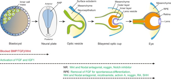
Retinal degenerative diseases are the leading causes of blindness worldwide. Replacing lost retinal cells via stem cell-based therapies is an exciting, rapidly advancing area of translational research that has already entered the clinic. Here, we review the status of these clinical efforts for several significant retinal diseases, describe the challenges involved and discuss how basic developmental studies have contributed to and are needed to advance clinical goals.

Keywords: Degenerative retinal diseases; Macular degeneration; Regenerative medicine; Retina pigment epithelial cells; Retinal development; Stem cell.


Conflict of interest statement

Competing interests

The authors declare no competing or financial interests.

Figures

Fig. 1.
Fig. 1.
Schematic of the key stages of retina development. Beginning with the blastocyst, which contains the pluripotent inner cell mass, gastrulation and neurulation lead to formation of the neural plate. The early eye field is located in the anterior neural plate (ANP) and develops into the optic vesicles. Blocking the activity of BMP, TGFβ and Wnt (red) promotes ANP development. Invagination of the optic vesicle leads to formation of the bilayered optic cup. The inner layer of the optic cup develops into the neural retina (NR) and the outer layer develops into the retinal pigment epithelium (RPE). Activation of FGF and IGF1 pathways (green) facilitates not only development of the ANP but also subsequent optic vesicle/cup formation and retina development. Wnt, FGF, BMP, Notch, SHH, RA and activin A signaling pathways (green) are involved in the specification of the RPE and NR. These factors have been used to promote NR and RPE production from stem cells in vitro, but the specific combinations and concentrations of each factor and the schedule of addition remain to be optimized for each lineage. It is also possible that additional factors not yet identified, and potentially specific to human retinal development, will aid neural retinal and RPE differentiation.

Similar articles

Cited by

References

    1. Abdelsalam A., Del Priore L. and Zarbin M. A. (1999). Drusen in age-related macular degeneration: pathogenesis, natural course, and laser photocoagulation-induced regression. Surv. Ophthalmol. 44, 1-29. 10.1016/S0039-6257(99)00072-7 - DOI - PubMed
    1. Adler R. and Canto-Soler M. V. (2007). Molecular mechanisms of optic vesicle development: complexities, ambiguities and controversies. Dev. Biol. 305, 1-13. 10.1016/j.ydbio.2007.01.045 - DOI - PMC - PubMed
    1. Ambati J. and Fowler B. J. (2012a). Mechanisms of age-related macular degeneration. Neuron 75, 26-39. 10.1016/j.neuron.2012.06.018 - DOI - PMC - PubMed
    1. Ambati J. and Fowler B. J. (2012b). Mechanisms of agerelated macular degeneration. Neuron 75, 26-39. 10.1016/j.neuron.2012.06.018 - DOI - PMC - PubMed
    1. Araki R., Uda M., Hoki Y., Sunayama M., Nakamura M., Ando S., Sugiura M., Ideno H., Shimada A., Nifuji A. et al. (2013). Negligible immunogenicity of terminally differentiated cells derived from induced pluripotent or embryonic stem cells. Nature 494, 100-104. 10.1038/nature11807 - DOI - PubMed

Publication types