

Effects of sintering temperature on surface morphology/microstructure, in vitro degradability, mineralization and osteoblast response to magnesium phosphate as biomedical material
- PMID: 28400583
- PMCID: PMC5429756
- DOI: 10.1038/s41598-017-00905-2
Effects of sintering temperature on surface morphology/microstructure, in vitro degradability, mineralization and osteoblast response to magnesium phosphate as biomedical material
Abstract
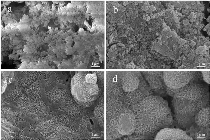
Magnesium phosphate (MP) was fabricated using a chemical precipitation method, and the biological performances of MP sintered at different temperatures as a biomedical material was investigated. The results indicated that the densification and crystallinity of MP increased as the sintering temperature increased. As the sintering temperature increased, the degradability of MP in PBS decreased, and the mineralization ability in SBF significantly increased. In addition, the MP sintered at 800 °C (MP8) possessed the lowest degradability and highest mineralization ability. Moreover, the positive response of MG63 cells to MP significantly increased as the sintering temperature increased, and MP8 significantly promoted the cell spreading, proliferation, differentiation and expressions of osteogenic differentiation-related genes. Faster degradation of MP0 resulted in higher pH environments and ion concentrations, which led to negative responses to osteoblasts. However, the appropriate degradation of MP8 resulted in suitable pH environments and ion concentrations, which led to positive responses to osteoblasts. This study demonstrated that the sintering temperature substantially affected the surface morphology/microstructure, degradability and mineralization, and osteoblasts response to magnesium phosphate.
Conflict of interest statement
The authors declare that they have no competing interests.
Figures


Similar articles
-
An injectable bioactive magnesium phosphate cement incorporating carboxymethyl chitosan for bone regeneration.Int J Biol Macromol. 2020 Oct 1;160:101-111. doi: 10.1016/j.ijbiomac.2020.05.161. Epub 2020 May 22. Int J Biol Macromol. 2020. PMID: 32450325
-
Magnesium phosphate ceramics incorporating a novel indene compound promote osteoblast differentiation in vitro and bone regeneration in vivo.Biomaterials. 2018 Mar;157:51-61. doi: 10.1016/j.biomaterials.2017.11.032. Epub 2017 Dec 7. Biomaterials. 2018. PMID: 29245051
-
Effect of strontium substitution on the material properties and osteogenic potential of 3D powder printed magnesium phosphate scaffolds.Mater Sci Eng C Mater Biol Appl. 2019 May;98:1145-1158. doi: 10.1016/j.msec.2019.01.053. Epub 2019 Jan 15. Mater Sci Eng C Mater Biol Appl. 2019. PMID: 30812998
-
In vitro Apatite Mineralization, Degradability, Cytocompatibility and in vivo New Bone Formation and Vascularization of Bioactive Scaffold of Polybutylene Succinate/Magnesium Phosphate/Wheat Protein Ternary Composite.Int J Nanomedicine. 2020 Sep 30;15:7279-7295. doi: 10.2147/IJN.S255477. eCollection 2020. Int J Nanomedicine. 2020. PMID: 33061381 Free PMC article.
-
Strength reliability and in vitro degradation of three-dimensional powder printed strontium-substituted magnesium phosphate scaffolds.Acta Biomater. 2016 Feb;31:401-411. doi: 10.1016/j.actbio.2015.11.050. Epub 2015 Nov 30. Acta Biomater. 2016. PMID: 26621692
Cited by
-
Osteoconductive Effect of a Nanocomposite Membrane Treated with UV Radiation.Polymers (Basel). 2022 Jan 11;14(2):289. doi: 10.3390/polym14020289. Polymers (Basel). 2022. PMID: 35054693 Free PMC article.
-
Fluorapatite and fluorohydroxyapatite apatite surfaces drive adipose-derived stem cells to an osteogenic lineage.J Mech Behav Biomed Mater. 2022 Jan;125:104950. doi: 10.1016/j.jmbbm.2021.104950. Epub 2021 Oct 29. J Mech Behav Biomed Mater. 2022. PMID: 34740011 Free PMC article.
-
3D plotting in the preparation of newberyite, struvite, and brushite porous scaffolds: using magnesium oxide as a starting material.J Mater Sci Mater Med. 2019 Jul 19;30(8):88. doi: 10.1007/s10856-019-6290-2. J Mater Sci Mater Med. 2019. PMID: 31325082
-
Rapid Fabrication of MgNH4PO4·H2O/SrHPO4 Porous Composite Scaffolds with Improved Radiopacity via 3D Printing Process.Biomedicines. 2021 Sep 2;9(9):1138. doi: 10.3390/biomedicines9091138. Biomedicines. 2021. PMID: 34572326 Free PMC article.
-
Natural bone-mimicking nanopore-incorporated hydroxyapatite scaffolds for enhanced bone tissue regeneration.Biomater Res. 2022 Feb 25;26(1):7. doi: 10.1186/s40824-022-00253-x. Biomater Res. 2022. PMID: 35216625 Free PMC article.
References
-
- Symietz C, et al. Fixation of bioactive calcium alkali phosphate on Ti6Al4V implant material with femtosecond laser pulses. Appl. Surf. Sci. 2011;257:5208–5212. doi: 10.1016/j.apsusc.2010.10.046. - DOI
-
- Neel EAA, et al. Bioactive functional materials: a perspective on phosphate-based glasses. J. Mater. Chem. 2009;19:690–701. doi: 10.1039/B810675D. - DOI
Publication types
MeSH terms
Substances
LinkOut - more resources
Full Text Sources
Other Literature Sources
Research Materials
