. 2017:2017:5629341.
doi: 10.1155/2017/5629341. Epub 2017 Mar 16.

Characterization of NO-Induced Nitrosative Status in Human Placenta from Pregnant Women with Gestational Diabetes Mellitus

Affiliations

Characterization of NO-Induced Nitrosative Status in Human Placenta from Pregnant Women with Gestational Diabetes Mellitus

Francisco Visiedo et al. Oxid Med Cell Longev. 2017.

Abstract

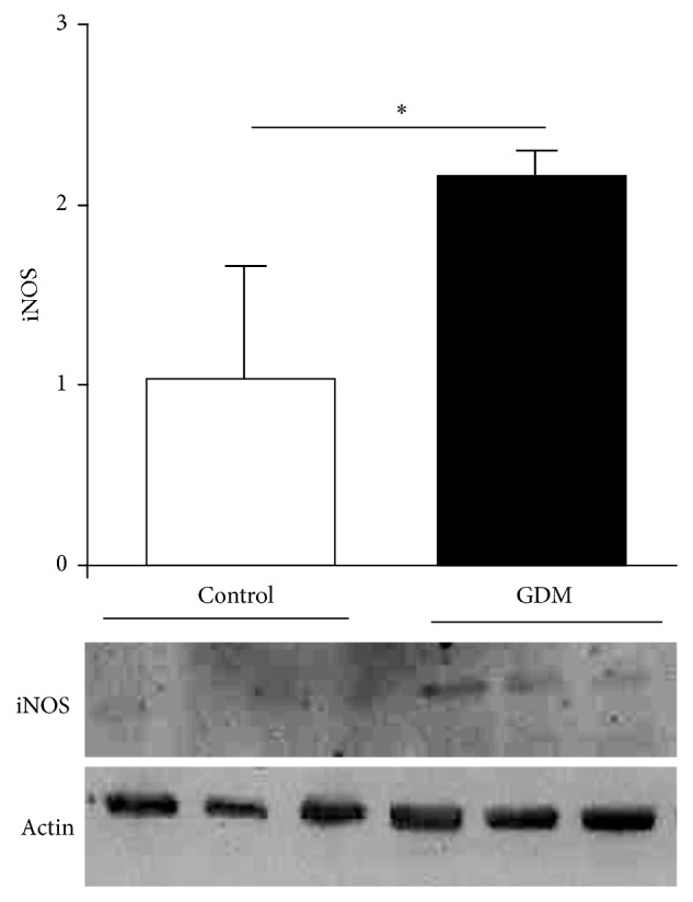
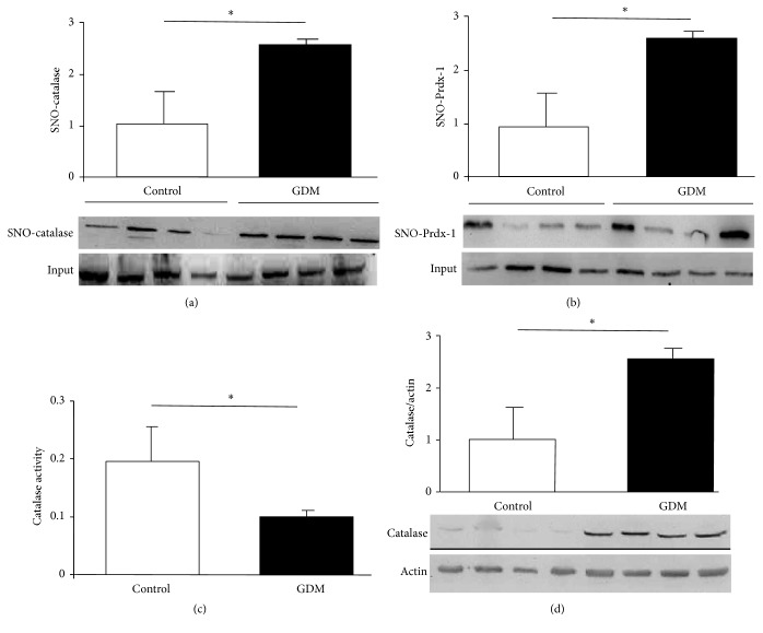
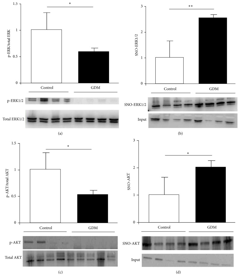
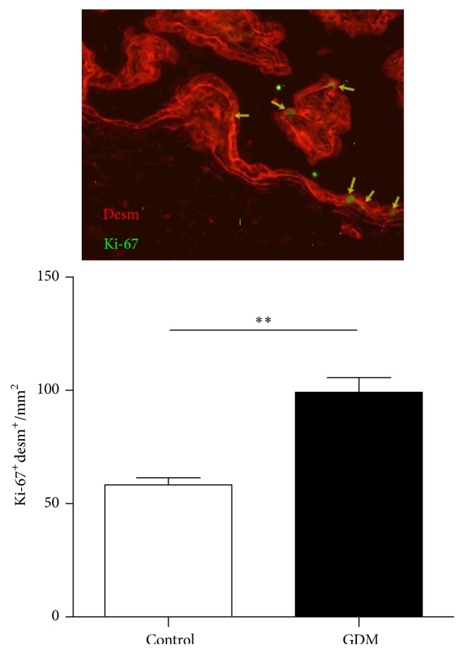
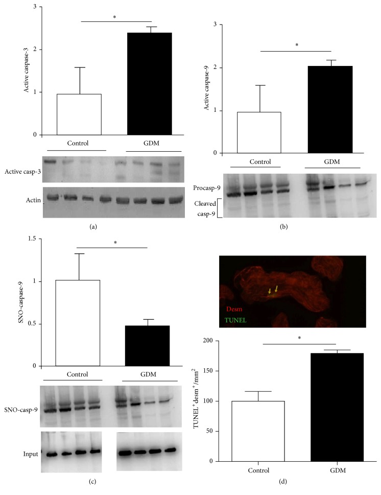
Dysregulation of NO production is implicated in pregnancy-related diseases, including gestational diabetes mellitus (GDM). The role of NO and its placental targets in GDM pregnancies has yet to be determined. S-Nitrosylation is the NO-derived posttranslational protein modification that can modulate biological functions by forming NO-derived complexes with longer half-life, termed S-nitrosothiol (SNO). Our aim was to examine the presence of endogenous S-nitrosylated proteins in cysteine residues in relation to antioxidant defense, apoptosis, and cellular signal transduction in placental tissue from control (n = 8) and GDM (n = 8) pregnancies. S-Nitrosylation was measured using the biotin-switch assay, while the expression and protein activity were assessed by immunoblotting and colorimetric methods, respectively. Results indicated that catalase and peroxiredoxin nitrosylation levels were greater in GDM placentas, and that was accompanied by reduced catalase activity. S-Nitrosylation of ERK1/2 and AKT was increased in GDM placentas, and their activities were inhibited. Activities of caspase-3 and caspase-9 were increased, with the latter also showing diminished nitrosylation levels. These findings suggest that S-nitrosylation is a little-known, but critical, mechanism by which NO directly modulates key placental proteins in women with GDM and, as a consequence, maternal and fetal anomalies during pregnancy can occur.


Figures

Figure 1
Figure 1
iNOS expression in GDM. Notes. iNOS expression quantified by Western blot of placental tissue obtained from control and GDM pregnancies. Results are expressed as means ± SEM of iNOS to actin ratio measured by densitometry (n = 3). Lower panel is a representative image from an experiment. Units on bar chart y-axis are arbitrary. p < 0.05, GDM versus control.
Figure 2
Figure 2
S-Nitrosylation in antioxidant enzymes. Notes. ((a) and (b)) S-Nitrosylation measured in placental tissue obtained from control and GDM pregnancies. Methods used were biotin-switch technique for catalase (a) and peroxiredoxin-1 (b). Results in bar charts are mean ± SEM of SNO-protein (n = 6) relative to input (total quantity of studied protein present in each sample). Units on bar chart y-axes are arbitrary. Representative images of immunoblotted SNO-proteins are shown under each graph. Catalase activity (c) and expression (d) were quantified by spectrophotometric analysis and immunoblotting, respectively. Placental tissues were obtained from control and GDM pregnancies. Results reported as mean ± SEM of enzymatic activity (n = 8) and catalase to actin ratio measured by densitometry (n = 3); lower panel is a representative image of immunoblotted proteins. p < 0.05, GDM versus control.
Figure 3
Figure 3
S-Nitrosylation and activation level in AKT and ERK1/2. Notes. ((a) and (c)) ERK1/2 (a) and AKT (c) phosphorylation quantified by Western blotting in control and GDM placental tissue. Results are expressed as means ± SEM of phosphoprotein to total-protein ratio measured by densitometry (n = 6). Lower panels are representative images of immunoblotted proteins. ((b) and (d)) S-Nitrosylation measured in placental tissue obtained from control and GDM pregnancies. Methods used were biotin-switch technique in ERK1/2 (b) and AKT (d). Results are presented in bar charts as mean ± SEM of SNO-protein (n = 6). Lower panels are representative images of immunoblotted SNO-proteins. Units on y-axes of bar charts are arbitrary. p < 0.05, GDM versus control; ∗∗p < 0.01, GDM versus control.
Figure 4
Figure 4
Proliferation level in trophoblasts. Notes. Cytotrophoblast proliferation quantified by immunohistochemistry in control and GDM placental tissue. Results are expressed as means ± SEM of desmosome protein (desm) and Ki-67 positive cells per area (mm2) (n = 7). Upper panel is a representative image of desmosome protein (red) and Ki-67 (green) immunostaining. Units on y-axes of bar charts are arbitrary. ∗∗p < 0.01, GDM versus control.
Figure 5
Figure 5
S-Nitrosylation and activation level in caspases. Notes. ((a) and (b)) Active fragment of caspase-3 (a) and caspase-9 (b) determined in control and GDM placental tissue by Western blotting. Results are expressed as means ± SEM of active caspases to actin ratio measured by densitometry (n = 3). Lower panels are representative images of immunoblotted proteins. (c) Caspase-9 S-nitrosylation measured in both experimental groups by biotin-switch technique. Results are presented in bar charts as mean ± SEM of SNO-caspase-9 (n = 6). Lower panel is a representative image of experiment. (d) Cytotrophoblast apoptosis quantified by TUNEL technique in control and GDM placental tissue. Results are expressed as means ± SEM of desmosome protein (desm) and TUNEL-positive cells per area (mm2) (n = 7). Upper panel is a representative image of desmosome protein (red) and TUNEL (green) immunostaining. Units on graph y-axis are arbitrary. p < 0.05, GDM versus control.

Similar articles

Cited by

References

    1. Ben-Haroush A., Yogev Y., Hod M. Epidemiology of gestational diabetes mellitus and its association with Type 2 diabetes. Diabetic Medicine. 2004;21(2):103–113. doi: 10.1046/j.1464-5491.2003.00985.x. - DOI - PubMed
    1. Albareda M., Caballero A., Badell G., et al. Diabetes and abnormal glucose tolerance in women with previous gestational diabetes. Diabetes Care. 2003;26(4):1199–1205. doi: 10.2337/diacare.26.4.1199. - DOI - PubMed
    1. Moncada S., Palmer R. M. J., Higgs E. A. Nitric oxide: physiology, pathophysiology, and pharmacology. Pharmacological Reviews. 1991;43(2):109–142. - PubMed
    1. Circu M. L., Aw T. Y. Reactive oxygen species, cellular redox systems, and apoptosis. Free Radical Biology and Medicine. 2010;48(6):749–762. doi: 10.1016/j.freeradbiomed.2009.12.022. - DOI - PMC - PubMed
    1. Myatt L. Review: reactive oxygen and nitrogen species and functional adaptation of the placenta. Placenta. 2010;31(supplement):S66–S69. doi: 10.1016/j.placenta.2009.12.021. - DOI - PMC - PubMed

MeSH terms

LinkOut - more resources