

Genomic and proteomic characterization of ARID1A chromatin remodeller in ampullary tumors
- PMID: 28401006
- PMCID: PMC5385638
Genomic and proteomic characterization of ARID1A chromatin remodeller in ampullary tumors
Abstract
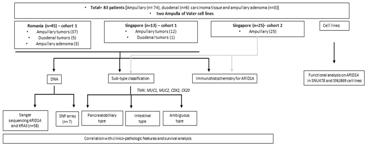
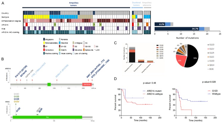
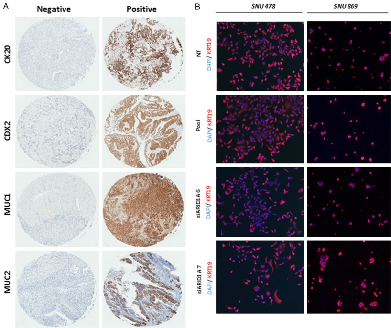
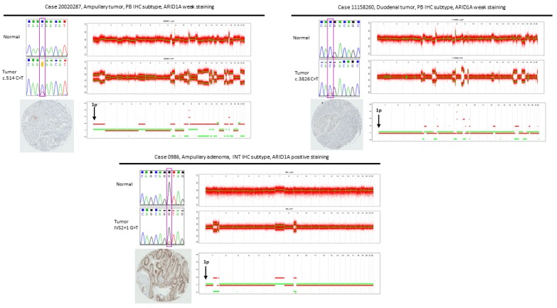
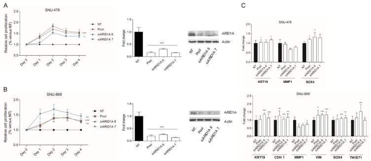
AT rich interactive domain 1A (ARID1A) is one of the most commonly mutated genes in a broad variety of tumors. The mechanisms that involve ARID1A in ampullary cancer progression remains elusive. Here, we evaluated the frequency of ARID1A and KRAS mutations in ampullary adenomas and adenocarcinomas and in duodenal adenocarcinomas from two cohorts of patients from Singapore and Romania, correlated with clinical and pathological tumor features, and assessed the functional role of ARID1A. In the ampullary adenocarcinomas, the frequency of KRAS and ARID1A mutations was 34.7% and 8.2% respectively, with a loss or reduction of ARID1A protein in 17.2% of the cases. ARID1A mutational status was significantly correlated with ARID1A protein expression level (P=0.023). There was a significant difference in frequency of ARID1A mutation between Romania and Singapore (2.7% versus 25%, P=0.04), suggestive of different etiologies. One somatic mutation was detected in the ampullary adenoma group. In vitro studies indicated the tumor suppressive role of ARID1A. Our results warrant further investigation of this chromatin remodeller as a potential early biomarker of the disease, as well as identification of therapeutic targets in ARID1A mutated ampullary cancers.
Keywords: ARID1A; Ampullary cancer; KRAS; Sanger sequencing.
Figures
References
-
- Demeure MJ, Craig DW, Sinari S, Moses TM, Christoforides A, Dinh J, Izatt T, Aldrich J, Decker A, Baker A, Cherni I, Watanabe A, Koep L, Lake D, Hostetter G, Trent JM, Von Hoff DD, Carpten JD. Cancer of the ampulla of vater: analysis of the whole genome sequence exposes a potential therapeutic vulnerability. Genome Med. 2012;4:56. - PMC - PubMed
-
- Chang DK, Jamieson NB, Johns AL, Scarlett CJ, Pajic M, Chou A, Pinese M, Humphris JL, Jones MD, Toon C, Nagrial AM, Chantrill LA, Chin VT, Pinho AV, Rooman I, Cowley MJ, Wu J, Mead RS, Colvin EK, Samra JS, Corbo V, Bassi C, Falconi M, Lawlor RT, Crippa S, Sperandio N, Bersani S, Dickson EJ, Mohamed MA, Oien KA, Foulis AK, Musgrove EA, Sutherland RL, Kench JG, Carter CR, Gill AJ, Scarpa A, McKay CJ, Biankin AV. Histomolecular phenotypes and outcome in adenocarcinoma of the ampulla of vater. J. Clin. Oncol. 2013;31:1348–1356. - PubMed
-
- Kimura W, Ohtsubo K. Incidence, sites of origin, and immunohistochemical and histochemical characteristics of atypical epithelium and minute carcinoma of the papilla of vater. Cancer. 1988;61:1394–1402. - PubMed
-
- Ang DC, Shia J, Tang LH, Katabi N, Klimstra DS. The utility of immunohistochemistry in subtyping adenocarcinoma of the ampulla of vater. Am J Surg Pathol. 2014;38:1371–1379. - PubMed
-
- Chung YE, Kim MJ, Park MS, Choi JY, Kim H, Kim SK, Lee M, Kim HJ, Choi JS, Song SY, Kim KW. Differential features of pancreatobiliary- and intestinal-type ampullary carcinomas at MR imaging. Radiology. 2010;257:384–393. - PubMed
Grants and funding
LinkOut - more resources
Full Text Sources
Miscellaneous