. 2017 Mar 1;7(3):531-542.
eCollection 2017.

Integrin β1 is a critical effector in promoting metastasis and chemo-resistance of esophageal squamous cell carcinoma

Affiliations

Integrin β1 is a critical effector in promoting metastasis and chemo-resistance of esophageal squamous cell carcinoma

Zhipeng Xu et al. Am J Cancer Res. .

Abstract

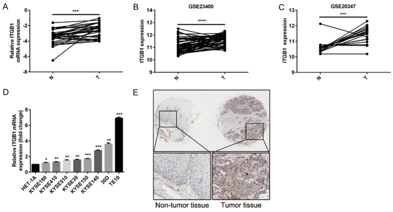
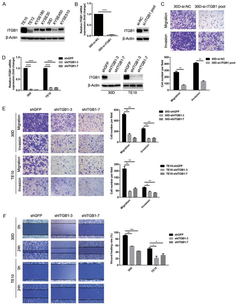
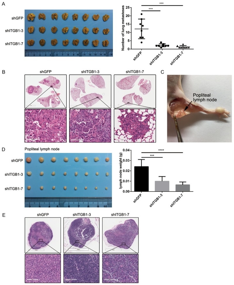
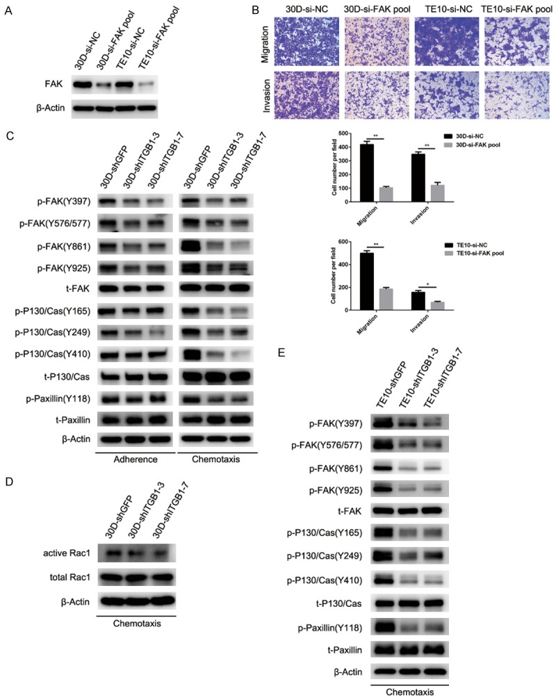
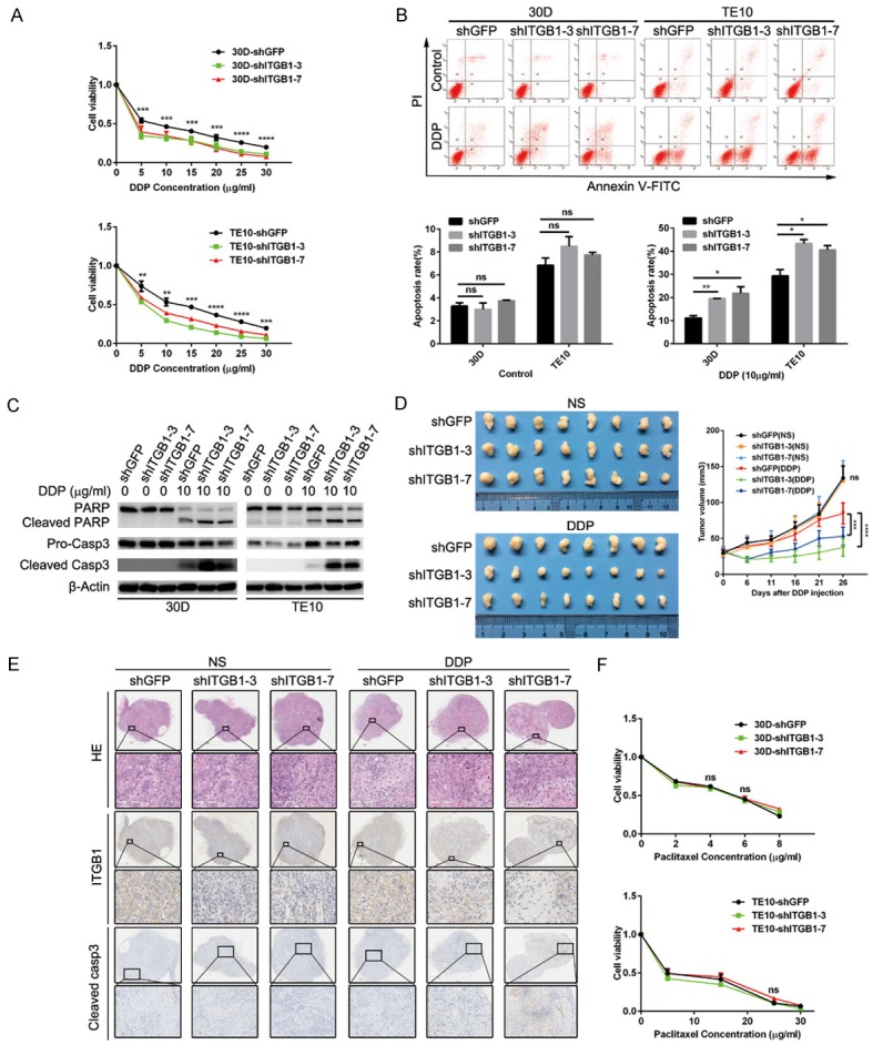
Metastasis of esophageal squamous cell carcinoma (ESCC) remains a challenge in clinical practice. In this study, we clarified that integrin β1 (ITGB1) plays critical roles in the metastasis of ESCC. By analyzing the expression of integrin β1 in ESCC specimens, we found that the expression of this integrin was higher in malignant than in normal tissues and that this increase was associated with lymph node metastasis. Moreover, in vitro functional experiments demonstrated that deletion of integrin β1 impaired the motility of ESCC cells, and we also showed that integrin β1 deletion significantly inhibited metastases formation in the lungs and lymph nodes of two murine models. Mechanistically, integrin β1 promoted cellular motility by regulating the FAK-Rac1 signaling pathway. Finally, we found that blocking integrin β1 significantly impaired the resistance of ESCC cells to cisplatin (DDP) treatment based on in vitro and in vivo experiments. Overall, our data suggest that integrin β1 promotes metastasis and confers DDP resistance to ESCC, which provides experimental evidence for targeting this protein to treat ESCC in the future.

Keywords: FAK; Integrin β1; chemo-resistance; esophageal cancer; metastasis.


Figures

Figure 1
Figure 1
Clinical properties of integrin β1 expression in human ESCC. A: Integrin β1 mRNA expression is higher in cancerous tissues than in adjacent normal tissues (N = 29). GAPDH was used as the internal control. The P-value was determined with the paired t-test. B and C: Mining two ESCC expression datasets (GSE23400 and GSE20347) in GEO showed significantly increased integrin β1 expression in malignant tissues compared with normal tissues. The P-values were determined with the paired t-test. D: The mRNA expression of integrin β1 was significantly lower in an immortalized esophageal epithelial cell line (HET-1A) than in a panel of ESCC cell lines. E: Integrin β1 staining in 88 paired ESCC tissues shows that this integrin was expressed in the membranes of epithelial cells and, importantly, significantly upregulated in cancerous tissues.
Figure 2
Figure 2
Knockdown of integrin β1 expression attenuates the motility of ESCC cells in vitro. (A) The protein level of integrin β1 was measured in a panel of ESCC cells using immunoblots. TE10 and 30D expressed higher levels of integrin β1 than the remaining tested cell lines. (B and C) siRNA knockdown of integrin β1 expression dramatically inhibited the migration and invasion of 30D cells in vitro. (D-F) The downregulation of integrin β1 expression in 30D or TE10 cells (shGFP and shITGB1-3/7 group) was verified using RT-qPCR and immunoblots (D). Stable knockdown of integrin β1 expression in 30D and TE10 cells significantly impeded cell motility compared with control cells based on Transwell assays (E) and wound healing assays (F). In all Transwell assays, five fields were randomly selected and the invading cells were counted. All scale bars in these images indicate 100 μm.
Figure 3
Figure 3
Blocking integrin β1 expression inhibits the metastasis of ESCC cells in vivo. (A and B) In the pulmonary metastasis model, β1-null 30D cells (shITGB1-3/7) formed far fewer metastases than control cells (shGFP) in the lungs of SCID/Beige mice (N = 8 for each group). The lungs were stained using 3% picric acid (A). H&E staining of fixed sections of these lungs shows the metastatic foci (B). The scale bars in the top three images indicate 4 mm, whereas those in the bottom three images indicate 100 μm (B). (C) Representative image showing the method used to harvest popliteal lymph nodes. (D and E) In the lymph node metastasis model, the popliteal lymph nodes of Balb/c nude mice in the shITGB1-3/7 group were smaller than those of the shGFP group (N = 8 for each group) (D). Representative images of the H&E-stained lymph nodes showing the metastatic lesions (E). The scale bars in the top three images indicate 1 mm or 2 mm, whereas those in the bottom three images indicate 100 μm (E).
Figure 4
Figure 4
Deletion of integrin β1 expression inhibits the motility of ESCC cells by suppressing FAK-Rac1 signaling. A: siRNA knockdown of FAK expression significantly reduced the FAK level in 30D cells or TE10 cells. B: Deletion of FAK in 30D or TE10 cells impeded their motility in vitro relative to the control cells. In the Transwell assays, five fields were randomly selected and the invading cells were counted. All scale bars in these images indicate 100 μm. C: In the stably β1-deleted 30D cells (30D-shITGB1-3/7), the levels of phosphorylated FAK (Y397/576/577/861/925), P130/Cas (Y16/249/410), and paxillin (Y118) were reduced compared with control cells (30D-shGFP) under adherent or chemotaxis conditions (2 h). D: Under chemotaxis conditions (2 h), 30D cells from the shITGB1-3/7 group expressed lower levels of Rac1-GTP than shGFP cells. E: Stable deletion of integrin β1 in TE10 cells (TE10-shITGB1-3/7) also reduced the level of phosphorylated FAK (Y397/576/577/861/925), P130/Cas (Y16/249/410), and paxillin (Y118) relative to the control ones (TE10-shGFP) under chemotaxis conditions for 2 h.
Figure 5
Figure 5
Reduced integrin β1 expression sensitizes ESCC cells to DDP administration. A: Integrin β1 deletion in 30D or TE10 cells impaired cellular viability when exposed to the indicated concentrations of DDP. B: The apoptosis rates were analyzed using flow cytometry, which showed that integrin β1 deletion promoted DDP-mediated apoptosis (10 μg/ml) in 30D or TE10 cells. C: Immunoblotting assays confirmed that DDP (10 μg/ml) treatment promoted apoptosis in integrin β1-null 30D or TE10 cells, as demonstrated by the dramatic increases in PARP and caspase-3. D: In vivo, the growth of xenografts formed by subcutaneously injected 30D cells was inhibited by 26 days of DDP treatment. Xenografts in the shITGB1-3/7 group were smaller than those in the shGFP group. E: Immunohistochemistry staining showed decreases in integrin β1 expression and increases in cleaved caspase-3 expression in the harvested xenografts. The scale bars indicate 1 mm or 2 mm in the original images and 100 μm or 200 μm in the magnified images. F: The deletion of integrin β1 did not affect the viability of 30D or TE10 cells when they were exposed to the indicated concentrations of paclitaxel.

Similar articles

Cited by

References

    1. Torre LA, Bray F, Siegel RL, Ferlay J, Lortet-Tieulent J, Jemal A. Global cancer statistics, 2012. CA Cancer J Clin. 2015;65:87–108. - PubMed
    1. Rustgi AK, El-Serag HB. Esophageal carcinoma. N Engl J Med. 2014;371:2499–2509. - PubMed
    1. Gupta GP, Massague J. Cancer metastasis: building a framework. Cell. 2006;127:679–695. - PubMed
    1. Hynes RO. Integrins: bidirectional, allosteric signaling machines. Cell. 2002;110:673–687. - PubMed
    1. Itou J, Tanaka S, Li W, Iida A, Sehara-Fujisawa A, Sato F, Toi M. The Sal-like 4-integrin alpha6beta1 network promotes cell migration for metastasis via activation of focal adhesion dynamics in basal-like breast cancer cells. Biochim Biophys Acta. 2017;1864:76–88. - PubMed

LinkOut - more resources