Review
. 2017 Aug;74(16):2999-3009.
doi: 10.1007/s00018-017-2511-3. Epub 2017 Apr 11.

Vinculin in cell-cell and cell-matrix adhesions

Affiliations
Review

Vinculin in cell-cell and cell-matrix adhesions

Jennifer L Bays et al. Cell Mol Life Sci. 2017 Aug.

Abstract

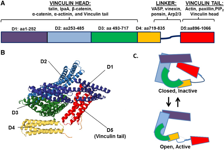
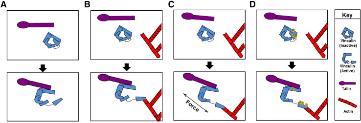
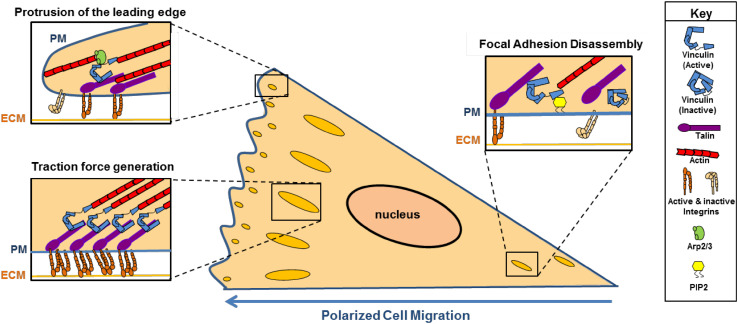
Vinculin was identified as a component of focal adhesions and adherens junctions nearly 40 years ago. Since that time, remarkable progress has been made in understanding its activation, regulation and function. Here we discuss the current understanding of the roles of vinculin in cell-cell and cell-matrix adhesions. Emphasis is placed on the how vinculin is recruited, activated and regulated. We also highlight the recent understanding of how vinculin responds to and transmits force at integrin- and cadherin-containing adhesion complexes to the cytoskeleton. Furthermore, we discuss roles of vinculin in binding to and rearranging the actin cytoskeleton.

Keywords: Cadherins; Cell adhesion; Cell migration; Force and mechanotransduction; Integrins; Vinculin.


Figures

Fig. 1
Fig. 1
Vinculin structure and binding partners. Vinculin is comprised of anti-parallel α-helical bundles organized into five distinct domains. a Domains 1–3 (D1D3) make up the vinculin head, while domain 5 (D5) encompasses the tail. The binding sites for many proteins interacting with vinculin have been mapped. b The ribbon diagram derived from the human full-length vinculin crystal structure shows vinculin resides in an inactive, closed conformation largely due to tight interactions between D1 and D5. The structure was derived from the PDB coordinates that were supplied by [72]. c A schematic of vinculin in closed, inactive conformation and open, active conformation
Fig. 2
Fig. 2
Models of vinculin activation. Vinculin exists in two conformations in the cell: an open, active form and a closed, auto-inhibited state in which the vinculin head domain interacts with the tail. Over the years, several models have been posed to explain how vinculin is opened and activated. a The helical bundle conversion model suggests that talin binding is sufficient to induce changes on the helical bundles in vinculin head to displace the head from vinculin tail, whereas others argue that two ligands—a head and a tail ligand—are required to separate vinculin head–tail interaction (b). Recent findings indicate that c force and d phosphorylation promote ligand binding and conformational changes within vinculin leading to activation
Fig. 3
Fig. 3
Role of vinculin in polarized cell migration. Vinculin is involved in many steps of cell migration. Its interaction with the Arp2/3 complex at nascent focal adhesions in the leading edge promotes protrusion of the membrane. During traction force generation, vinculin links integrins to the actin cytoskeleton and bears the forces exerted during motility. Lastly, vinculin interaction with PIP2 induces a conformation change that reduces vinculin interaction with actin, thereby promoting the disassembly of focal adhesions. PM plasma membrane, ECM extracellular matrix

References

    1. Geiger B. A 130 K protein from chicken gizzard: its localization at the termini of microfilament bundles in cultured chicken cells. Cell. 1979;18(1):193–205. doi: 10.1016/0092-8674(79)90368-4. - DOI - PubMed
    1. Burridge K, Feramisco JR. Microinjection and localization of a 130 K protein in living fibroblasts: a relationship to actin and fibronectin. Cell. 1980;19(3):587–595. doi: 10.1016/S0092-8674(80)80035-3. - DOI - PubMed
    1. Bakolitsa C, Cohen DM, Bankston LA, Bobkov AA, Cadwell GW, Jennings L, Critchley DR, Craig SW, Liddington RC. Structural basis for vinculin activation at sites of cell adhesion. Nature. 2004;430(6999):583–586. doi: 10.1038/nature02610. - DOI - PubMed
    1. Molony L, Burridge K. Molecular shape and self-association of vinculin and metavinculin. J Cell Biochem. 1985;29(1):31–36. doi: 10.1002/jcb.240290104. - DOI - PubMed
    1. Gimona M, Small JV, Moeremans M, Van Damme J, Puype M, Vandekerckhove J. Porcine vinculin and metavinculin differ by a 68-residue insert located close to the carboxy-terminal part of the molecule. EMBO J. 1988;7(8):2329–2334. - PMC - PubMed

Publication types

LinkOut - more resources