Review
. 2018 Mar;55(3):2547-2564.
doi: 10.1007/s12035-017-0503-9. Epub 2017 Apr 11.

Mitochondrial Quality Control and Disease: Insights into Ischemia-Reperfusion Injury

Affiliations
Review

Mitochondrial Quality Control and Disease: Insights into Ischemia-Reperfusion Injury

Anthony R Anzell et al. Mol Neurobiol. 2018 Mar.

Abstract

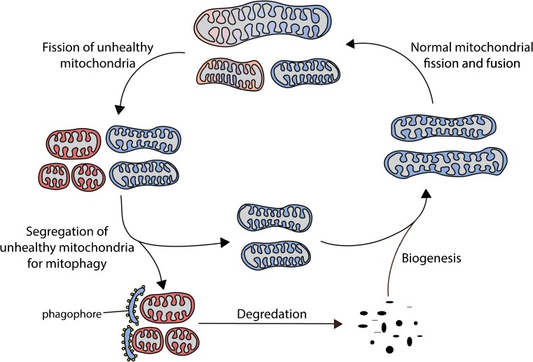
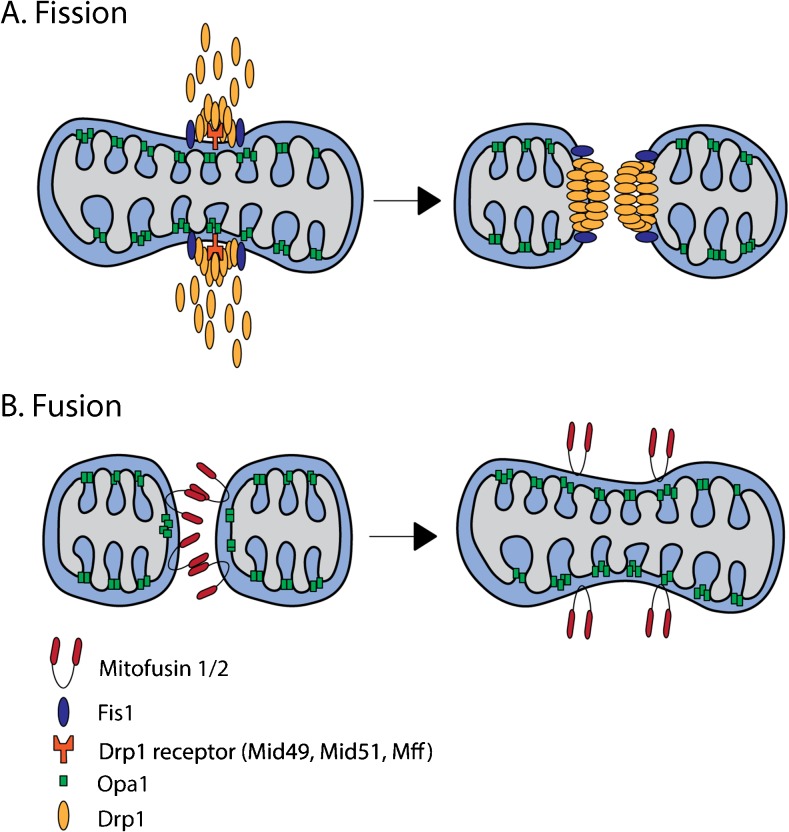
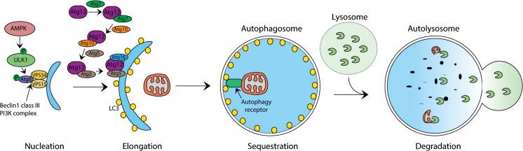
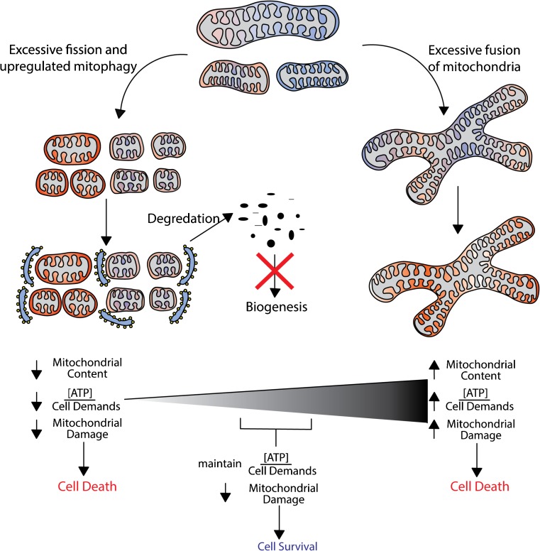
Mitochondria are key regulators of cell fate during disease. They control cell survival via the production of ATP that fuels cellular processes and, conversely, cell death via the induction of apoptosis through release of pro-apoptotic factors such as cytochrome C. Therefore, it is essential to have stringent quality control mechanisms to ensure a healthy mitochondrial network. Quality control mechanisms are largely regulated by mitochondrial dynamics and mitophagy. The processes of mitochondrial fission (division) and fusion allow for damaged mitochondria to be segregated and facilitate the equilibration of mitochondrial components such as DNA, proteins, and metabolites. The process of mitophagy are responsible for the degradation and recycling of damaged mitochondria. These mitochondrial quality control mechanisms have been well studied in chronic and acute pathologies such as Parkinson's disease, Alzheimer's disease, stroke, and acute myocardial infarction, but less is known about how these two processes interact and contribute to specific pathophysiologic states. To date, evidence for the role of mitochondrial quality control in acute and chronic disease is divergent and suggests that mitochondrial quality control processes can serve both survival and death functions depending on the disease state. This review aims to provide a synopsis of the molecular mechanisms involved in mitochondrial quality control, to summarize our current understanding of the complex role that mitochondrial quality control plays in the progression of acute vs chronic diseases and, finally, to speculate on the possibility that targeted manipulation of mitochondrial quality control mechanisms may be exploited for the rationale design of novel therapeutic interventions.

Keywords: Brain; Ischemia; Mitochondria; Mitochondrial dynamics; Mitophagy; Reperfusion.


Figures

Fig. 1
Fig. 1
The mitochondrial quality control cycle. The mitochondrial quality control cycle involves a dynamic process of fission, fusion, mitophagy, and biogenesis. When mitochondria become depolarized or dysfunctional, they are marked for degradation. Once marked, the unhealthy component of the mitochondria will undergo fission from the healthy mitochondrial network. Certain damaged mitochondria can fuse with other healthy mitochondria in an attempt to salvage that mitochondrion, but typically, dysfunctional mitochondria will undergo mitophagy. When the dysfunctional mitochondria are segregated from the healthy mitochondrial network, mitochondria will accumulate mitophagy markers that will recruit the phagophore. The phagophore will attach to the dysfunctional mitochondria, mature into an autophagosome, fuse with the lysosome to form the autolysosome, and degrade the mitochondria. Once degraded, the cell will recycle the amino acids and fatty acids to enable the remaining healthy mitochondrial network to grow and divide through biogenesis
Fig. 2
Fig. 2
Mitochondrial dynamics. a Fission is mediated by a family of dynamin-related proteins (Drp). Activated Drp1 translocates from the cytosol to the mitochondrial membrane where it interacts with Drp1 receptors (Mid 49, Mid51, Mff) and Fis1 to create the fission complex. Drp1 oligomers constrict and divide the mitochondria. b Fusion is mediated through the mitofusins (Mfn1/2) and optic atrophy 1 (Opa1). The mitofusins mediate the fusion of the outer mitochondrial membrane, while Opa1 is thought to mediate fusion of the inner mitochondrial membrane. Mitofusins are anchored to the outer mitochondrial membrane and interact with each other and form a hemifusion stalk. The stalk then grows into a lipidic hole and finally reestablishes membrane continuity. Opa1 forms a fusion pore for the inner mitochondrial membrane via its cardiolipin binding domain
Fig. 3
Fig. 3
Phases of Autophagy. Autophagy is carried out in four different phases: nucleation, elongation, sequestration, and degradation. Nucleation of the isolation membrane is initiated by the phosphorylation of the ULk1 complex by AMPK. ULK1 will then recruit several autophagosome-related proteins for nucleation of the phagophore via phosphorylation of beclin 1 in the PI3K complex. Elongation or the extension of the autophagosome membrane is mediated by two ubiquitin-like systems involving the transferring of Atg12 from Atg7 to Atg10 and finally Atg5 where it forms a dimeric complex with Atg16 on the phagophore membrane. LC3 is also thought to help mediate the extension of the phagophore. Sequestration occurs when damaged organelles are detected via LC3/autophagy receptor interactions. Autophagy receptors localized on damaged organelles will bind LC3 inducing elongation of the phagophore until the cargo is completely engulfed and matures into an autophagosome. In the degradation phase, the lysosome will fuse with the autophagosome (autolysosome), releasing acid hydrolase enzymes that degrade the contents
Fig. 4
Fig. 4
Mitophagic pathways. a In dysfunctional mitochondria, PINK1 accumulates in the outer mitochondrial membrane. Accumulations of PINK1 induce a concerted signaling cascade involving the simultaneous recruitment and phosphorylation of the E3 ubiquitin ligase Parkin, ubiquitin, and TBK1. Phosphorylation by PINK1 as well as phospho-Ser65-ubiquitin activates Parkin and leads to ubiquitination of outer mitochondrial membrane proteins in a feed forward process. Phagophore recruitment and binding are then mediated by OPTN and NDP52 with its ubiquitin and LC3 binding domains. b BNIP3/Nix are localized on the outer mitochondrial membrane and serve as mitophagy receptors and bind directly to the phagosome via LC3. c FUNDC1 localizes on the outer mitochondrial membrane and acts as a receptor for mitophagy under hypoxic condition. During hypoxia, PGAM5 dephosphorylates FUNDC1 and activates mitophagy via LC3 binding on the phagophore. d Cardiolipin localizes mainly in the inner leaflet of the inner mitochondrial membrane, specifically around the folds of the cristae. When cardiolipin is oxidized, it is externalized to the outer mitochondrial membrane where it is recognized by LC3 of the phagophore
Fig. 5
Fig. 5
Finding a balance of mitochondrial quality control. During I/R injury, there is excessive mitochondrial fragmentation, favoring an increase in mitophagy. Degradation of dysfunctional ROS-producing mitochondria is critical for survival; however, mitochondrial content decrease would compromise ATP production. Insufficient ATP production paired with inhibited biogenesis will ultimately lead to cell death. In contrast, if fission was completely inhibited, mitochondrial content would be maintained, but damaged mitochondria would not be segregated and could lead to accumulated mitochondrial dysfunction and exacerbate damage to the entire mitochondrial network. The increase in mitochondrial damage would augment pro-apoptotic stimuli and, ultimately, cause cell death. Therefore, a balance in mitochondrial quality control (i.e., an equilibrium between retaining adequate mitochondrial content for sufficient ATP production versus disposal of dysfunctional mitochondria) is optimal for cell survival after I/R injury

References

    1. Mozaffarian D, et al. Heart disease and stroke statistics—2015 update: a report from the American Heart Association. Circulation. 2015;131(4):e29–322. doi: 10.1161/CIR.0000000000000152. - DOI - PubMed
    1. Pulsinelli WA, Duffy TE. Regional energy balance in rat brain after transient forebrain ischemia. J Neurochem. 1983;40(5):1500–1503. doi: 10.1111/j.1471-4159.1983.tb13599.x. - DOI - PubMed
    1. Crack PJ, Taylor JM. Reactive oxygen species and the modulation of stroke. Free Radic Biol Med. 2005;38(11):1433–1444. doi: 10.1016/j.freeradbiomed.2005.01.019. - DOI - PubMed
    1. Aronowski J, Strong R, Grotta JC. Reperfusion injury: demonstration of brain damage produced by reperfusion after transient focal ischemia in rats. J Cereb Blood Flow Metab. 1997;17(10):1048–1056. doi: 10.1097/00004647-199710000-00006. - DOI - PubMed
    1. Sanderson TH, et al. Molecular mechanisms of ischemia-reperfusion injury in brain: pivotal role of the mitochondrial membrane potential in reactive oxygen species generation. Mol Neurobiol. 2013;47(1):9–23. doi: 10.1007/s12035-012-8344-z. - DOI - PMC - PubMed

MeSH terms

Substances

LinkOut - more resources