Review
. 2017 Jun;10(3):261-274.
doi: 10.1007/s12265-017-9736-2. Epub 2017 Apr 11.

Deep Phenotyping of Systemic Arterial Hemodynamics in HFpEF (Part 2): Clinical and Therapeutic Considerations

Affiliations
Review

Deep Phenotyping of Systemic Arterial Hemodynamics in HFpEF (Part 2): Clinical and Therapeutic Considerations

Julio A Chirinos. J Cardiovasc Transl Res. 2017 Jun.

Abstract

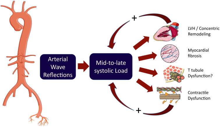
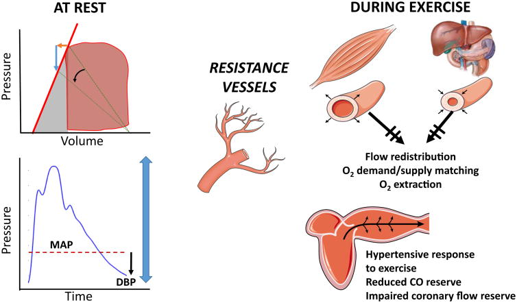
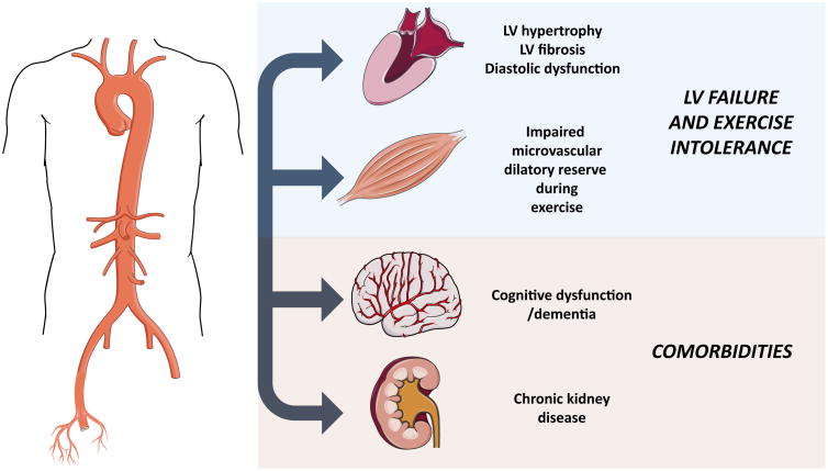
Multiple phase III trials over the last few decades have failed to demonstrate a clear benefit of various pharmacologic interventions in heart failure with a preserved left ventricular (LV) ejection fraction (HFpEF). Therefore, a better understanding of its pathophysiology is important. An accompanying review describes key technical and physiologic aspects regarding the deep phenotyping of arterial hemodynamics in HFpEF. This review deals with the potential of this approach to enhance our clinical, translational, and therapeutic approach to HFpEF. Specifically, the role of arterial hemodynamics is discussed in relation to (1) the pathophysiology of left ventricular diastolic dysfunction, remodeling, and fibrosis, (2) impaired oxygen delivery to peripheral skeletal muscle, which affects peripheral oxygen extraction, (3) the frequent presence of comorbidities, such as renal failure and dementia in this population, and (4) the potential to enhance precision medicine approaches. A therapeutic approach to target arterial hemodynamic abnormalities that are prevalent in this population (particularly, with inorganic nitrate/nitrite) is also discussed.

Keywords: Afterload; Arterial hemodynamics; Comorbidities; Dementia; Exercise intolerance; Heart failure with preserved ejection fraction; Pulsatile load; Renal disease; Wave reflections.


Figures

Fig. 1
Fig. 1
Role of arterial wave reflections and late systolic load on LV remodeling and diastolic dysfunction. Arterial wave reflections selectively increase mid-to-late systolic LV load. Late systolic LV load from wave reflections promotes LV hypertrophy, fibrosis, and myocardial dysfunction (longitudinal systolic dysfunction and slow relaxation). The effect of wave reflections on the myocardium is strongly influenced by the LV contraction pattern. Normally, systolic ejection determines a dynamic reconfiguration of LV geometry that reduces myocardial wall stress relative to LV pressure in mid-systole, thus protecting the cardiomyocytes against excessive load in late systole. Concentric LV hypertrophy/remodeling and impaired contractility (even in the presence of a normal EF) are associated with a blunted mid-systolic shift in the pressure-MWS relation, which is less effective to protect cardiomyocytes to load during a period of increased vulnerability. This may represent a “vicious circle” that facilitates the development and progression of HFpEF
Fig. 2
Fig. 2
Role of microvascular hemodynamic function in hemodynamics at rest and during exercise. At rest (left panels), the ventricular-arterial system of patients with HFpEF is characterized by increased LV systolic stiffness (i.e., increased EES, upper left panel, red line). Therefore, any therapeutic intervention that reduces vascular resistance at rest (black curved arrow) (which is the key arterial determinant of “effective arterial elastance,” represented in green lines) will induce a large pressure decrease (blue arrow) relative to the increase in stroke volume (orange arrow). Furthermore, given the presence of stiff conduit arteries and premature wave reflections that augment systolic, rather than diastolic, aortic pressure, a reduction in mean arterial pressure may decrease diastolic arterial pressure excessively (left lower panel), leading to a reduction of perfusion pressure in key organs. During exercise (right panels), however, a reduction is vascular resistance is essential. A combination of vasoconstriction in most vascular beds (splanchnic vasculature, non-exercising muscle, etc.) along with vasodilation in exercising muscle (local functional sympatholysis) is key for the optimization the cardiac output distribution and oxygen supply-demand matching in the periphery. In addition, the lack of reduction in SVR during exercise increases LV load and contributes to the limitation in cardiac output reserve. Microvascular function is also essential for enhancing coronary perfusion during exercise. However, pulsatile hemodynamics also affect myocardial oxygen demand (workload) and supply (diastolic perfusion pressure) during exercise (see text). MAP mean arterial pressure; DBP diastolic blood pressure
Fig. 3
Fig. 3
Effect of wave reflection on pulsatile pressure, flow, and power pulsatility into the carotid territory. The left-sided panel represents the effect of the forward traveling wave from the ascending aorta (blue arrows) into the aortic arch and carotid territory. The left-sided panel represents the effect of the reflected wave from the lower body traveling up the descending aorta (red arrows). Although a small reflection occurs at the aortic-carotid interface, the bulk of wave reflections arising from the lower body penetrate the carotid as a forward wave, increasing pressure and flow pulsatility in the carotid territory. Paradoxically, impedance matching at the aortic-carotid interface reduces local reflection but also promotes the penetration of distal reflected waves into the carotid territory
Fig. 4
Fig. 4
Role of arterial hemodynamic dysfunction in HFpEF. Abnormal macrovascular and microvascular hemodynamic dysfunction promotes LV diastolic dysfunction and LV maladaptive remodeling (hypertrophy, fibrosis) and causesa reduced vasodilatory reserve during exercise, which in turn impairs coronary flow, peripheral flow redistribution, oxygen delivery, and extraction in exercising muscle. Abnormal pulsatile arterial hemodynamics also contribute to microvascular disease in the brain and the kidney (dementia, chronic kidney disease, which are frequently encountered comorbidities in HFpEF)

Similar articles

Cited by

References

    1. Lam CS, Donal E, Kraigher-Krainer E, Vasan RS. Epidemiology and clinical course of heart failure with preserved ejection fraction. European Journal of Heart Failure. 2011;13:18–28. - PMC - PubMed
    1. Lloyd-Jones DM, Hong Y, Labarthe D, Mozaffarian D, Appel LJ, Van Horn L, Greenlund K, Daniels S, Nichol G, Tomaselli GF, Arnett DK, Fonarow GC, Ho PM, Lauer MS, Masoudi FA, Robertson RM, Roger V, Schwamm LH, Sorlie P, Yancy CW, Rosamond WD. Defining and setting national goals for cardiovascular health promotion and disease reduction: the American Heart Association's strategic Impact Goal through 2020 and beyond. Circulation. 2010;121:586–613. - PubMed
    1. Townsend RR, Wilkinson IB, Schiffrin EL, Avolio AP, Chirinos JA, Cockcroft JR, Heffernan KS, Lakatta EG, McEniery CM, Mitchell GF, Najjar SS, Nichols WW, Urbina EM, Weber T American Heart Association Council on H. Recommendations for improving and standardizing vascular research on arterial stiffness: a scientific statement from the American Heart Association. Hypertension. 2015;66:698–722. - PMC - PubMed
    1. Hoekstra T, Lesman-Leegte I, van Veldhuisen DJ, Sanderman R, Jaarsma T. Quality of life is impaired similarly in heart failure patients with preserved and reduced ejection fraction. European Journal of Heart Failure. 2011;13:1013–1018. - PubMed
    1. Lewis EF, Lamas GA, O'Meara E, Granger CB, Dunlap ME, McKelvie RS, Probstfield JL, Young JB, Michelson EL, Halling K, Carlsson J, Olofsson B, McMurray JJ, Yusuf S, Swedberg K, Pfeffer MA. Characterization of health-related quality of life in heart failure patients with preserved versus low ejection fraction in CHARM. European Journal of Heart Failure. 2007;9:83–91. - PubMed