. 2017 Aug;16(4):693-703.
doi: 10.1111/acel.12597. Epub 2017 Apr 12.

DNA damage and senescence in osteoprogenitors expressing Osx1 may cause their decrease with age

Affiliations

DNA damage and senescence in osteoprogenitors expressing Osx1 may cause their decrease with age

Ha-Neui Kim et al. Aging Cell. 2017 Aug.

Abstract

Age-related bone loss in mice results from a decrease in bone formation and an increase in cortical bone resorption. The former is accounted by a decrease in the number of postmitotic osteoblasts which synthesize the bone matrix and is thought to be the consequence of age-dependent changes in mesenchymal osteoblast progenitors. However, there are no specific markers for these progenitors, and conclusions rely on results from in vitro cultures of mixed cell populations. Moreover, the culprits of such changes remain unknown. Here, we have used Osx1-Cre;TdRFP mice in which osteoprogenitors express the TdRFP fluorescent protein. We report that the number of TdRFP-Osx1 cells, freshly isolated from the bone marrow, declines by more than 50% between 6 and 24 months of age in both female and male mice. Moreover, TdRFP-Osx1 cells from old mice exhibited markers of DNA damage and senescence, such as γH2AX foci, G1 cell cycle arrest, phosphorylation of p53, increased p21CIP1 levels, as well as increased levels of GATA4 and activation of NF-κB - two major stimulators of the senescence-associated secretory phenotype (SASP). Bone marrow stromal cells from old mice also exhibited elevated expression of SASP genes, including several pro-osteoclastogenic cytokines, and increased capacity to support osteoclast formation. These changes were greatly attenuated by the senolytic drug ABT263. Together, these findings suggest that the decline in bone mass with age is the result of intrinsic defects in osteoprogenitor cells, leading to decreased osteoblast numbers and increased support of osteoclast formation.

Keywords: ABT263; GATA4; NF-κB; osteoblasts; osteoporosis; p21; p53.


Figures

Figure 1
Figure 1
Cells targeted by Osx1‐Cre transgene in the bone marrow. (A) Representative femur sections from 6‐ and 24‐month‐old Osx1‐Cre;TdRFP mice. CB, cortical bone; BM, bone marrow; scale bar 20 μm. (B and C) Sorted cells from Osx1‐Cre;TdRFP mice (n = 6 mice) cultured with ascorbate and β‐glycerophosphate. (B) mRNA levels by qRTPCR in cells cultured for 7 days and (C) Alizarin Red S staining in cells cultured for 21 days, (top) 2.5× magnification (bottom) 20× magnification. *P < 0.05 by Student's t‐test. Bars represent mean and SD (error bars).
Figure 2
Figure 2
The number of Osx1‐Cre‐expressing cells decline with age. (A‐C) Osx1‐TdRFP + bone marrow cells were isolated from 4‐ and 23‐month‐old Osx1‐Cre;TdRFP mice (n = 4–6 mice/group). (A) Representative flow cytometric analysis of Osx1‐TdRFP + bone marrow cells. (B) Percentage of Osx1‐TdRFP + cells in the bone marrow. (C) Number of Osx1‐TdRFP + cells in the hindlimbs from each mouse. (D and E) Prx1‐TdRFP + bone marrow cells were isolated from 3‐ and 24‐month‐old Prx1‐TdRFP mice (n = 4–5 mice/group). (D) Representative flow cytometric analysis of Prx1‐TdRFP + bone marrow cells. (E) Percentage of Prx1‐TdRFP + cells in the bone marrow. (F and G) Bone marrow stromal cells from 6‐ and 26‐month‐old Osx1‐Cre;TdRFP mice cultured with ascorbate and β‐glycerophosphate. (F) mRNA levels by qRTPCR in cells cultured for 7 days and (G) Alizarin Red S staining in cells cultured for 21 days (triplicates), (top) 2.5× magnification (bottom) 20× magnification. *P < 0.05 by Student's t‐test. Bars represent mean and SD (error bars).
Figure 3
Figure 3
Osx1‐TdRFP + cells from old mice exhibit DNA damage and G1 arrest. (A) Representative FACS analysis of the cell cycle distribution of Osx1‐TdRFP + cells from 3‐ to 6‐month‐old and 23‐ to 26‐month‐old Osx1‐Cre;TdRFP mice (n = 5 mice/group). (B) Percentage of Osx1‐TdRFP + cells at G1 in mice described in A. (C and D) Analysis of DNA double‐strand breaks in TdRFP‐Osx1+ cells from 6‐ and 25‐month‐old Osx1‐Cre;TdRFP male mice (n = 5 mice/group). (C) Representative photomicrographs of γH2AX immunofluorescence staining (green) and nucleic counterstaining with Hoechst‐33342 (blue). (D) Percentage of γH2AX‐positive cells (left) and number of γH2AX foci per cell (right). *P < 0.05 by Student's t‐test. Bars represent mean and SD (error bars).
Figure 4
Figure 4
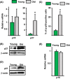
Osx1‐TdRFP + cells from old mice have increased p53/p21 levels. (A and B) Osx1‐TdRFP + cells from 6‐ and 24‐month‐old Osx1‐Cre;TdRFP female mice (n = 5 mice/group) (A) mRNA levels by qRTPCR and (B) protein levels by Western blot. (C) Percentage of p‐p53‐positive cells in Osx1‐TdRFP + cells from 3‐ and 24‐month‐old Osx1‐Cre;TdRFP male mice (n = 5 mice/group). (D and E) Bone marrow stromal cells from 6‐ and 26‐month‐old Osx1‐Cre;TdRFP female mice (n = 5 mice/group) cultured with ascorbate and β‐glycerophosphate. (D) Protein levels by Western blot in cells cultured for 10 days and (E) mRNA levels by qRTPCR in cells cultured for 7 days. *P < 0.05 by Student's t‐test. Bars represent mean and SD (error bars).
Figure 5
Figure 5
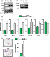
Osteoprogenitors in old mice exhibit elevated GATA4, active NF‐κB, SASP, and osteoclastogenic properties. Protein by Western blot in (A) Osx1‐TdRFP + cells from 6‐ and 24‐month‐old Osx1‐Cre;TdRFP female mice (n = 5 mice/group) and (B) bone marrow stromal cells from 6‐ and 26‐month‐old Osx1‐Cre;TdRFP female mice cultured with ascorbate and β‐glycerophosphate for 7 days (triplicates). (C) mRNA levels by qRTPCR in cells described in B. (D) Mouse bone marrow macrophages from 6‐month‐old wild‐type C57BL/6J male mice were co‐cultured with stromal cells from 6‐ and 26‐month‐old wild‐type C57BL/6J male mice in the presence of 1α,25(OH)2D3 for 7 days, and the cells were fixed and stained for TRAP. TRAP‐positive multinucleated cells containing three or more nuclei were counted as osteoclasts. *P < 0.05 by Student's t‐test. Bars represent mean and SD (error bars).
Figure 6
Figure 6
Senescent cell clearance by ABT263 attenuates GATA4 and SASP expression in stromal cell cultures from old mice. (A) Protein by Western blot in bone marrow stromal cells from 24‐month‐old wild‐type C57BL/6J female mice cultured with ascorbate and β‐glycerophosphate for 7 days, following pretreatment with ABT263 (triplicates). (B) mRNA levels by qRTPCR in cells described in A. (C) Bone marrow macrophages from 3‐month‐old wild‐type C57BL/6J female mice were co‐cultured for 7 days with stromal cells from 24‐month‐old wild‐type C57BL/6 female mice pretreated with vehicle or ABT263. TRAP‐positive multinucleated cells containing three or more nuclei were counted as osteoclasts. *P < 0.05 by Student's t‐test. Bars represent mean and SD (error bars).
Figure 7
Figure 7
IR induces senescence in osteoprogenitors. (A) SA‐β‐gal staining of IR‐induced senescent cells. (IR, 7 days after exposure to 10 Gy). (B) Gene expression in IR‐induced senescent cells. (C) Western blot analysis was performed in IR‐induced senescent cells. *P < 0.05 by Student's t‐test. Bars represent mean and SD (error bars).

Similar articles

Cited by

References

    1. Almeida M, O'Brien CA (2013) Basic biology of skeletal aging: role of stress response pathways. J. Gerontol. A Biol. Sci. Med. Sci. 68, 1197–1208. - PMC - PubMed
    1. Almeida M, Han L, Martin‐Millan M, Plotkin LI, Stewart SA, Roberson PK, Kousteni S, O'Brien CA, Bellido T, Parfitt AM, Weinstein RS, Jilka RL, Manolagas SC (2007) Skeletal involution by age‐associated oxidative stress and its acceleration by loss of sex steroids. J. Biol. Chem. 282, 27285–27297. - PMC - PubMed
    1. Baker DJ, Wijshake T, Tchkonia T, LeBrasseur NK, Childs BG, van de Sluis B, Kirkland JL, Van Deursen JM (2011) Clearance of p16Ink4a‐positive senescent cells delays ageing‐associated disorders. Nature 479, 232–236. - PMC - PubMed
    1. Baker DJ, Childs BG, Durik M, Wijers ME, Sieben CJ, Zhong J, Saltness RA, Jeganathan KB, Verzosa GC, Pezeshki A, Khazaie K, Miller JD, Van Deursen JM (2016) Naturally occurring p16(Ink4a)‐positive cells shorten healthy lifespan. Nature 530, 184–189. - PMC - PubMed
    1. Bellosta P, Masramon L, Mansukhani A, Basilico C (2003) p21(WAF1/CIP1) acts as a brake in osteoblast differentiation. J. Bone Miner. Res. 18, 818–826. - PubMed

MeSH terms