. 2017 Apr 12:7:45741.
doi: 10.1038/srep45741.

Acrolein acts as a neurotoxin in the nigrostriatal dopaminergic system of rat: involvement of α-synuclein aggregation and programmed cell death

Affiliations

Acrolein acts as a neurotoxin in the nigrostriatal dopaminergic system of rat: involvement of α-synuclein aggregation and programmed cell death

Yi-Ting Wang et al. Sci Rep. .

Abstract

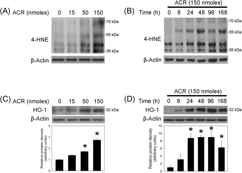
Clinical studies report significant increases in acrolein (an α,β-unsaturated aldehyde) in the substantia nigra (SN) of patients with Parkinson's disease (PD). In the present study, acrolein-induced neurotoxicity in the nigrostriatal dopaminergic system was investigated by local infusion of acrolein (15, 50, 150 nmoles/0.5 μl) in the SN of Sprague-Dawley rats. Acrolein-induced neurodegeneration of nigrostriatal dopaminergic system was delineated by reductions in tyrosine hydroxylase (TH) levels, dopamine transporter levels and TH-positive neurons in the infused SN as well as in striatal dopamine content. At the same time, apomorphine-induced turning behavior was evident in rats subjected to a unilateral infusion of acrolein in SN. Acrolein was pro-oxidative by increasing 4-hydroxy-2-nonenal and heme oxygenase-1 levels. Furthermore, acrolein conjugated with proteins at lysine residue and induced α-synuclein aggregation in the infused SN. Acrolein was pro-inflammatory by activating astrocytes and microglia. In addition, acrolein activated caspase 1 in the infused SN, suggesting acrolein-induced inflammasome formation. The neurotoxic mechanisms underlying acrolein-induced neurotoxicity involved programmed cell death, including apoptosis and necroptosis. Compared with well-known Parkinsonian neurotoxins, including 1-methyl-4-phenyl-1,2,3,6-tetrahydropyridine and rotenone which do not exist in the SN of PD patients, our in vivo study shows that acrolein acts as a Parkinsonian neurotoxin in the nigrostriatal dopaminergic system of rat brain.


Conflict of interest statement

The authors declare no competing financial interests.

Figures

Figure 1
Figure 1. Intranigral infusion of acrolein induced neurodegeneration of the nigrostriatal dopaminergic system of rat brain.
Acrolein (ARC, 15–150 nmoles) was locally infused in the substantia nigra (SN) and rats were sacrificed 7 days after intranigral infusion of acrolein. (A) Striatal dopamine content was measured using HPLC-ECD. Values are the mean ± S.E.M. (n = 5–6/group) *p < 0.05 in the striatum ipsilateral to acrolein-infused SN compared with that to the intact SN by one-way analysis of variance (one-way ANOVA) and followed by the LSD test as a post-hoc analysis. (B) Tyrosine hydroxylase (TH) in the SN was measured using Western blot. Each lane contained 3 μg proteins for all experiments. Graphs show statistic results from relative optical density of bands on the blots. Values are the mean ± S.E.M. (n = 3/group). *p < 0.05 in the acrolein-infused SN compared with the intact SN by one-way analysis of variance (one-way ANOVA) and followed by the LSD test as a post-hoc analysis. (C) Dopamine transporter in the SN was measured using Western blot. Each lane contained 25 μg proteins for all experiments. Values are the mean ± S.E.M. (n = 7/group). *p < 0.05 in the acrolein-infused SN compared with the intact SN by t-test. (D) Representative confocal microscopic data showed TH-positive neurons in the SN of rat subjected to intranigral infusion of acrolein (150 nmoles). TH-positive cells in acrolein-infused SN were counted and expressed as % of that in the contralateral intact SN. Values are the mean ± S.E.M. (n = 5). *p < 0.05 in the acrolein-infused SN compared with the control SN by t-test. (E) A behavioral study of apomorphine-induced rotations in rats subjected to a unilateral infusion of acrolein (n = 6) or vehicle (n = 4) in the SN. Values are the mean ± S.E.M. *p < 0.05 in the contralateral rotations compared with the ipsilateral rotations in rats with acrolein-infused SN by Mann-Whitney Rank Sum Test. n.s.: not significant.
Figure 2
Figure 2. Intranigral infusion of acrolein induced oxidative stress in the nigrostriatal dopaminergic system of rat brain.
(A) and (C): For a dose-dependent effect, ARC (15–150 nmoles) was locally infused in the substantia nigra (SN) and rats were sacrificed 7 days after intranigral infusion of acrolein. (B) and (D): For a time-dependent effect, acrolein (150 nmoles) was locally infused in the SN and rats were sacrificed at the time indicated. Western blot assay was employed to detect 4-HNE (A,B) and HO-1 (C,D) in the SN. Each lane contained 25 μg proteins for all experiments. Graphs show statistical results from relative optical density of bands on the blots estimated by Image J. Values are the mean ± S.E.M. (n = 3/group). *p < 0.05 in the acrolein-infused SN compared with the intact SN by one-way analysis of variance (one-way ANOVA) and followed by the LSD test as a post-hoc analysis.
Figure 3
Figure 3. Intranigral infusion of acrolein induced protein conjugation and α-synuclein aggregation in the nigrostriatal dopaminergic system of rat brain.
(A and C): For a dose-dependent effect, ARC (15–150 nmoles) was locally infused in the substantia nigra (SN) and rats were sacrificed 7 days after intranigral infusion of acrolein. (B and D): For a time-dependent effect, acrolein (150 nmoles) was locally infused in the SN and rats were sacrificed at the time indicated. Western blot assay was employed to detect FDP-lysine (A,B) and α-synuclein (C,D) in the SN. Each lane contained 25 μg proteins for all experiments. Graphs show statistical results from relative optical density of bands on the blots estimated by Image J. Values are the mean ± S.E.M. (n = 3/group). *p < 0.05 in the acrolein-infused SN compared with the intact SN by one-way analysis of variance (one-way ANOVA) and followed by the LSD test as a post-hoc analysis. (E) Representative confocal microscopic data showed co-localization of FDP-lysine and α-synuclein immunoreactivities in the infused SN of rat brain. Acrolein (150 nmoles) was locally infused in the SN and rats were sacrificed 48 hrs after intranigral infusion of acrolein. Scale bars: 10 μm. Results were repeated in independent experiments.
Figure 4
Figure 4. Intranigral infusion of acrolein induced neuroinflammation in the nigrostriatal dopaminergic system of rat brain.
(A and C), For a dose-dependent effect, ARC (15–150 nmoles) was locally infused in the substantia nigra (SN) and rats were sacrificed 7 days after intranigral infusion of acrolein. (B and D): For a time-dependent effect, acrolein (150 nmoles) was locally infused in the SN and rats were sacrificed at the time indicated. Western blot assay was employed to detect GFAP and ED-1 (A,B) as well as caspase 1 activation (C,D) in the SN. Each lane contained 25 μg protein for all experiments. Graphs show statistic results from relative optical density of bands on the blots estimated by Image J. Values are the mean ± S.E.M. (n = 3/group). *p < 0.05 in the acrolein-infused SN compared with the intact SN by one-way analysis of variance (one-way ANOVA) and followed by the LSD test as a post-hoc analysis.
Figure 5
Figure 5. Intranigral infusion of acrolein induced programmed cell death in the nigrostriatal dopaminergic system of rat brain.
(A and C), For a dose-dependent effect, ARC (15–150 nmoles) was locally infused in the substantia nigra (SN) and rats were sacrificed 7 days after intranigral infusion of acrolein. (B and D): For a time-dependent effect, acrolein (150 nmoles) was locally infused in the SN and rats were sacrificed at the time indicated. Western blot assay was employed to detect caspase 3 activation (A,B) as well as RIP-1 and RIP-3 (C,D) in the SN. Each lane contained 25 μg protein for all experiments. Graphs show statistic results from relative optical density of bands on the blots estimated by Image J. Values are the mean ± S.E.M. (n = 5/group for A, n = 3/group for BD). *p < 0.05 in the acrolein-infused SN compared with the intact SN by one-way analysis of variance (one-way ANOVA) and followed by the LSD test as a post-hoc analysis.

Similar articles

Cited by

References

    1. Feng Z., Hu W., Hu Y. & Tang M. S. Acrolein is a major cigarette-related lung cancer agent: Preferential binding at p53 mutational hotspots and inhibition of DNA repair. Proceedings of the National Academy of Sciences of the United States of America 103, 15404–15409, doi: 10.1073/pnas.0607031103 (2006). - DOI - PMC - PubMed
    1. Magnusson R., Nilsson C. & Andersson B. Emissions of aldehydes and ketones from a two-stroke engine using ethanol and ethanol-blended gasoline as fuel. Environmental science & technology 36, 1656–1664 (2002). - PubMed
    1. Ho S. S., Yu J. Z., Chu K. W. & Yeung L. L. Carbonyl emissions from commercial cooking sources in Hong Kong. Journal of the Air & Waste Management Association (1995) 56, 1091–1098 (2006). - PubMed
    1. Esterbauer H., Schaur R. J. & Zollner H. Chemistry and biochemistry of 4-hydroxynonenal, malonaldehyde and related aldehydes. Free radical biology & medicine 11, 81–128 (1991). - PubMed
    1. Uchida K. et al.. Acrolein is a product of lipid peroxidation reaction. Formation of free acrolein and its conjugate with lysine residues in oxidized low density lipoproteins. The Journal of biological chemistry 273, 16058–16066 (1998). - PubMed

Publication types

MeSH terms

LinkOut - more resources