Review
. 2017 Aug;22(4):10.1111/hel.12386.
doi: 10.1111/hel.12386. Epub 2017 Apr 12.

Survival of Helicobacter pylori in gastric acidic territory

Affiliations
Review

Survival of Helicobacter pylori in gastric acidic territory

Shamshul Ansari et al. Helicobacter. 2017 Aug.

Abstract

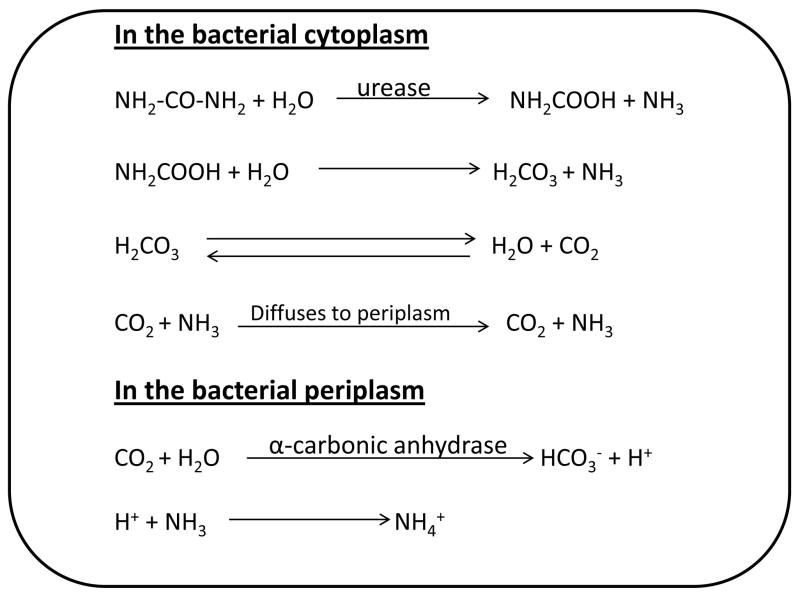
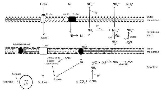
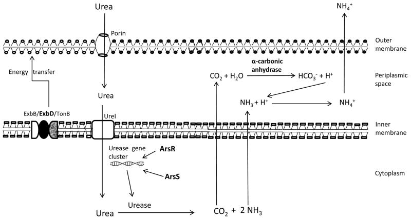
Background: Helicobacter pylori is well adapted to colonize the epithelial surface of the human gastric mucosa and can cause persistent infections. In order to infect the gastric mucosa, it has to survive in the gastric acidic pH. This organism has well developed mechanisms to neutralize the effects of acidic pH.

Objective: This review article was designed to summarize the various functional and molecular aspects by which the bacterium can combat and survive the gastric acidic pH in order to establish the persistent infections.

Methods: We used the keywords (acid acclimation, gastric acidic environment, H. pylori and survival) in combination or alone for pubmed search of recent scientific literatures. One hundred and forty one papers published between 1989 and 2016 were sorted out. The articles published with only abstracts, other than in English language, case reports and reviews were excluded.

Results: Many literatures describing the role of several factors in acid survival were found. Recently, the role of several other factors has been claimed to participate in acid survival.

Conclusion: In conclusion, this organism has well characterized mechanisms for acid survival.

Keywords: Helicobacter pylori; acid acclimation; gastric acidic environment; survival.


Conflict of interest statement

disclosure: The authors declare that they have no any conflict of interests.

Figures

Figure 1
Figure 1
Hydrolysis of urea by urease. Urease breaks down the urea to form carbamate and NH3. Carbamate is further decomposed to produce carbonic acid and NH3. Carbonic acid is broken down spontaneously aqueous to produce H2O and CO2. Therefore, one molecule of urea after hydrolysis produces two molecules of ammonia (NH3) and one molecule of carbon dioxide (CO2). CO2 and NH3 are defused to periplasm where α-carbonic anhydrase catalyzes the breakdown of CO2 to produce HCO3 and H+. The H+ produced combines with NH3 to produce NH4+.
Figure 2
Figure 2
The metallo-enzyme, urease requires nickel (Ni) for its activity. Ni slowly enters the outer membrane through porin (outer membrane protein) and actively enters through FecA3/FrpB4 proteins anchored in outer membrane and crosses inner membrane through NixA (inner membrane protein). However, in acidic pH more Ni is required for increased activity of urease and for the active transport of essential molecules (Ni), ExbD provides energy to the outer membrane. Urea enters the outer membrane through the porin and inner membrane through UreI (found in inner membrane) which opens in acid exposure of bacteria. Endogenously produced by arginase in urea cycle (during in vitro survival) and exogenously entered urea molecules are hydrolyzed by urease to ammonia (NH3) and carbon dioxide (CO2). ArsR regulates urease gene cluster expression. Ammonia (NH3) produced is diffused out and binds with proton (H+) leading to the neutralization of acidic pH. However, NH3 is also converted to ammonium (NH4+) inside the cell (cytoplasm and periplasm) which is toxic to the bacteria. The cytoplasmic NH4+ is assimilated to glutamate (GLT) by GDH and to glutamine (GLN) by GS. GLN can diffuse to periplasmic space or can be converted to aspergine (ASN) by GatCAB aminoacyl-tRNA amidotransferase (GatCAB) and then can diffuse to periplasmic space. GLN and ASN bind with water molecule and led to the formation of NH4+ and catalyzed by Ggt and AsnB respectively. Finally the NH4+ exits to the bacterial cell and increases the pH that leads to the survival of bacteria at acidic pH.
Figure 3
Figure 3. Acid acclimation
Exposure of bacterial cells to acidic pH opens the urea channel, UreI and urea enters the cytoplasm to be hydrolyzed by urease to NH3 and CO2. However, the outer membrane proteins (porins) need energy for active transport of essential molecules (urea) which is provided by ExbD located in inner membrane in the form of complex of three proteins (ExbB/ExbD/TonB). ArsR and ArsS regulate the expression of urease gene cluster and increase the urease activity at strong acidic pH. Ammonia (NH3) and carbon dioxide (CO2) diffuse to the periplasmic space. In the periplasmic space, CO2 binds with water molecule to synthesize HCO3 and H+ catalyzed by periplasmic α-carbonic anhydrase. The synthesized HCO3 acts as a strong buffer and can maintain the periplasmic pH to 6.1. Protons (H+), entering to the periplasm and synthesized by α-carbonic anhydrase, are neutralized by NH3 converting to NH4+ and diffuses out of the bacterial cell membrane.

References

    1. Blaser MJ. Who are we? Indigenous microbes and the ecology of human diseases. EMBO Reports. 2006;7(10):956–60. - PMC - PubMed
    1. Ruggiero P. Helicobacter pylori and inflammation. Curr Pharm Des. 2010;16:4225–4236. - PubMed
    1. IARC Working-Group; World Health Organization, International Agency for Research on Cancer, editor. Schistosomes, Liver Flukes and Helicobacter pylori. Lyon, France: 1994.
    1. IARC Working Group. Helicobacter pylori Eradication as a Strategy for Preventing Gastric Cancer. Lyon, France: International Agency for Research on Cancer; 2014. (IARC Working Group Reports, No. 8)
    1. Hardbower DM, de Sablet T, Chaturvedi R, Wilson KT. Chronic inflammation and oxidative stress: the smoking gun for Helicobacter pylori-induced gastric cancer? Gut Microbes. 2013;4:475, e481. - PMC - PubMed

LinkOut - more resources