. 2017 Apr 27;376(17):1647-1658.
doi: 10.1056/NEJMoa1611930. Epub 2017 Apr 5.

Autoantibodies against GPIHBP1 as a Cause of Hypertriglyceridemia

Affiliations

Autoantibodies against GPIHBP1 as a Cause of Hypertriglyceridemia

Anne P Beigneux et al. N Engl J Med. .

Abstract

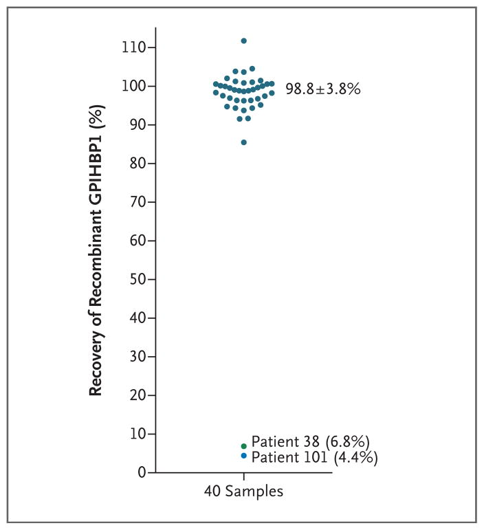
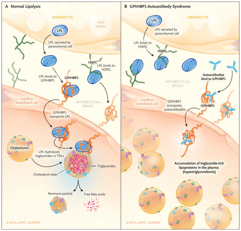
Background: A protein that is expressed on capillary endothelial cells, called GPIHBP1 (glycosylphosphatidylinositol-anchored high-density lipoprotein binding protein 1), binds lipoprotein lipase and shuttles it to its site of action in the capillary lumen. A deficiency in GPIHBP1 prevents lipoprotein lipase from reaching the capillary lumen. Patients with GPIHBP1 deficiency have low plasma levels of lipoprotein lipase, impaired intravascular hydrolysis of triglycerides, and severe hypertriglyceridemia (chylomicronemia). During the characterization of a monoclonal antibody-based immunoassay for GPIHBP1, we encountered two plasma samples (both from patients with chylomicronemia) that contained an interfering substance that made it impossible to measure GPIHBP1. That finding raised the possibility that those samples might contain GPIHBP1 autoantibodies.

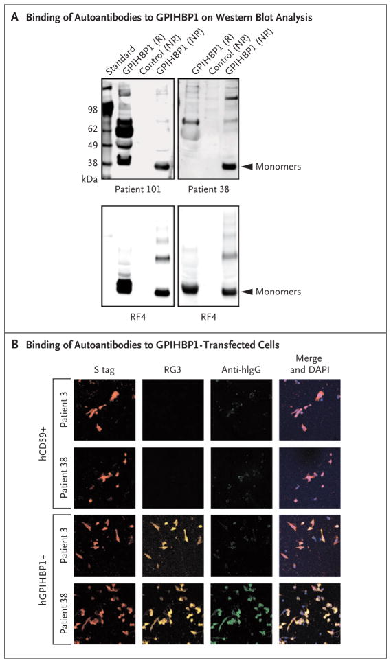
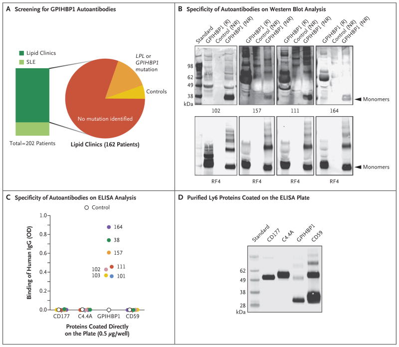
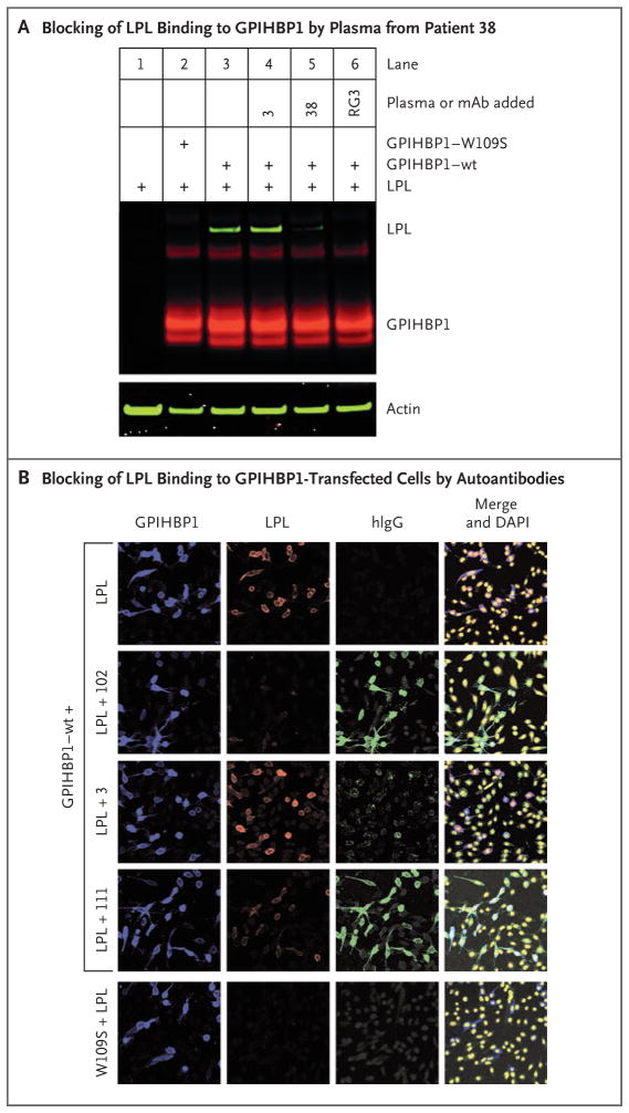
Methods: Using a combination of immunoassays, Western blot analyses, and immunocytochemical studies, we tested the two plasma samples (as well as samples from other patients with chylomicronemia) for the presence of GPIHBP1 autoantibodies. We also tested the ability of GPIHBP1 autoantibodies to block the binding of lipoprotein lipase to GPIHBP1.

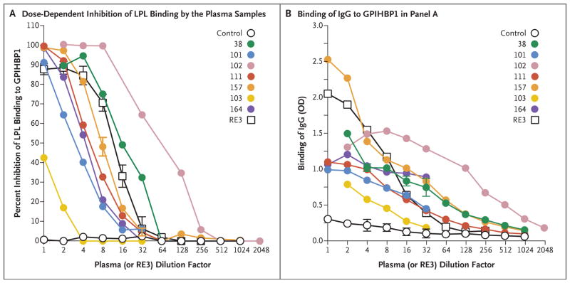
Results: We identified GPIHBP1 autoantibodies in six patients with chylomicronemia and found that these autoantibodies blocked the binding of lipoprotein lipase to GPIHBP1. As in patients with GPIHBP1 deficiency, those with GPIHBP1 autoantibodies had low plasma levels of lipoprotein lipase. Three of the six patients had systemic lupus erythematosus. One of these patients who had GPIHBP1 autoantibodies delivered a baby with plasma containing maternal GPIHBP1 autoantibodies; the infant had severe but transient chylomicronemia. Two of the patients with chylomicronemia and GPIHBP1 autoantibodies had a response to treatment with immunosuppressive agents.

Conclusions: In six patients with chylomicronemia, GPIHBP1 autoantibodies blocked the ability of GPIHBP1 to bind and transport lipoprotein lipase, thereby interfering with lipoprotein lipase-mediated processing of triglyceride-rich lipoproteins and causing severe hypertriglyceridemia. (Funded by the National Heart, Lung, and Blood Institute and the Leducq Foundation.).


Figures

Figure 1
Figure 1. GPIHBP1 Immunoassay Interference
To validate the enzyme-linked immunosorbent assay (ELISA) analysis, recombinant GPIHBP1 was spiked into 40 plasma samples; the results of that analysis are shown here. (Details are provided in Methods Section S2 in the Supplementary Appendix.) Briefly, GPIHBP1 levels in 1:20 dilutions of plasma were measured before and after spiking of the sample with 62.5 pg of recombinant GPIHBP1. In 38 of 40 plasma samples, the mean (±SD) recovery of spiked recombinant GPIHBP1 was nearly complete (98.8±3.8%). In these samples, the mean GPIHBP1 values were 21.0±3.0 pg per milliliter before spiking with recombinant GPIHBP1 and 82.5±3.0 pg per milliliter after spiking. The recovery of recombinant GPIHBP1 in samples obtained from Patients 38 and 101 (two patients with chylomicronemia and low plasma GPIHBP1 levels) was very low (6.8% and 4.4%, respectively). The GPIHBP1 level in the plasma sample obtained from Patient 38 was 3.0 pg per milliliter in the 1:20 dilution and 4.5 pg per milliliter after the sample was spiked; in the sample obtained from Patient 101, the GPIHBP1 levels before and after spiking were 1.0 pg per milliliter and 3.0 pg per milliliter, respectively. Because of the low coefficient of variation in the ELISA analysis, a few of the samples had an apparent recovery rate that was slightly more than 100%.
Figure 2
Figure 2. GPIHBP1 Autoantibodies in Plasma Samples Obtained from Patients 38 and 101
Panel A shows the results of Western blot analysis of GPIHBP1 autoantibodies in plasma samples obtained from Patients 38 and 101. Proteins in the medium of GPIHBP1-transfected drosophila S2 cells were fractionated according to size by means of sodium dodecyl sulfate–polyacrylamide-gel electrophoresis (SDS-PAGE) under reducing (R) and nonreducing (NR) conditions. (Details are provided in Methods Section S9 in the Supplementary Appendix.) GPIHBP1 contains a tightly folded cysteine-rich domain (Ly6 domain) that is essential for the binding of lipoprotein lipase. The hypothesis was that some of the autoantibodies might bind to the Ly6 domain and that disrupting the disulfide bonds with reducing reagents might disrupt the epitope of some of the autoantibodies. The autoantibodies in plasma obtained from Patient 101 (containing 20 arbitrary units [AU] of GPIHBP1 autoantibodies per milliliter) bound to both reduced and nonreduced GPIHBP1; additional nonspecific binding was seen under reducing conditions. The autoantibodies in plasma obtained from Patient 38 (containing 20 AU of GPIHBP1 autoantibodies per milliliter) bound avidly only to nonreduced GPIHBP1. In the lower row, the panels show the same blots incubated with the GPIHBP1-specific monoclonal antibody RF4 (4 μg per milliliter) (see Methods Section S10 in the Supplementary Appendix). The monoclonal antibody RF4 binds to the acidic domain of GPIHBP1 and therefore binds both reduced and nonreduced human GPIHBP1; it also binds to GPIHBP1 dimers and multimers; the latter forms are artifacts of insect-cell overexpression and are due to inappropriate intermolecular disulfide bonds. The control culture medium was obtained from S2 cells that do not express human GPIHBP1. Panel B shows the results of an immunocytochemical experiment documenting that GPIHBP1 autoantibodies in plasma obtained from Patient 38 (1:20 dilution; green) bound to cells that had been transfected with an S-protein–tagged version of human GPIHBP1 (hGPIHBP1) but did not bind to nontransfected cells or to cells that were transfected with S-protein–tagged human CD59 (hCD59) (see Methods Section S7 in the Supplementary Appendix). CD59-transfected and GPIHBP1-transfected cells were detected with an antibody against the S-protein tag (red); GPIHBP1-expressing cells were also detected with a GPIHBP1-specific monoclonal antibody RG3 (orange). DNA was stained with 4,6-diamidino-2-phenylindole (DAPI, blue) to reveal all cells (both transfected and nontransfected) on the coverslip. The immunoglobulins in plasma obtained from Patient 3 (who was homozygous for a GPIHBP1 deletion) did not bind to GPIHBP1-transfected cells.
Figure 3
Figure 3. Identification of Four Additional Plasma Samples with GPIHBP1 Autoantibodies
Panel A summarizes the plasma samples screened for GPIHBP1 autoantibodies in 202 deidentified archived plasma samples from lipid clinics or a rheumatology clinic in which patients with systemic lupus erythematosus (SLE) were being treated. Forty patients with SLE, half of whom were receiving immunosuppressive therapy, were included; 10 of those patients had plasma triglyceride levels ranging from 350 to 750 mg per deciliter (4.0 to 8.5 mmol per liter). The 162 patients from various lipid clinics included 130 patients who had hypertriglyceridemia with no known genetic cause, 23 patients who had hypertriglyceridemia and a mutation in GPIHBP1 or LPL, and 9 controls. (Details are provided in Table S1 in the Supplementary Appendix.) Panel B shows Western blot analysis demonstrating that the immunoglobulins in plasma samples obtained from Patients 102, 157, 111, and 164 (containing 20 AU of GPIHBP1 autoantibody per milliliter) bind preferentially to nonreduced human GPIHBP1. The lower panels show the same blots incubated with the human GPIHBP1–specific monoclonal antibody RF4 (4 μg per milliliter). The monoclonal antibody RF4, which binds to reduced and nonreduced forms of GPIHBP1 and to GPIHBP1 multimers, was used as a loading control. The control culture medium was obtained from S2 cells that do not express human GPIHBP1. Panel C shows the results from ELISA analyses revealing that the immunoglobulin (IgG) in plasma samples obtained from Patients 38, 101, 102, 103, 111, 157, and 164 bind to wells of an ELISA plate coated with purified human GPIHBP1 but not to wells coated with other human Ly6 proteins (CD177, C4.4A, and CD59) (see Methods Section S4 in the Supplementary Appendix). The dilution was 1:12,500 for the sample obtained from Patient 102 and 1:500 for the other samples. A control plasma sample did not bind to any of the Ly6 proteins. The optical density (OD) was measured at a wavelength of 450 nm. Panel D shows Western blot analysis (performed with the use of monoclonal antibody R24) of purified Ly6 proteins (CD177, C4.4A, GPIHBP1, and CD59) that had been fractionated according to size by means of SDS-PAGE. All four proteins had an amino-terminal urokinase-type plasminogen activator receptor (uPAR) tag that could be detected with monoclonal antibody R24. This Western blot analysis documents that similar amounts of all four Ly6 proteins were used to coat 96-well plates for the ELISA analysis shown in Panel C.
Figure 4
Figure 4. Blocking of Binding of Lipoprotein Lipase to GPIHBP1 by Autoantibodies
Panel A shows a Western blot analysis of cell extracts from Chinese hamster ovary (CHO) cells transfected with S-protein–tagged wild-type (wt) GPIHBP1 or a mutant GPIHBP1 (W109S), which lacks the ability to bind lipoprotein lipase (LPL) (see Methods Section S6 in the Supplementary Appendix). The lipoprotein lipase that was used in these experiments was V5-tagged. Western blot analyses were performed with a goat antibody against the S-protein tag (Abcam, 5 μg per milliliter), followed by infrared dye (IRDye) 680–donkey antigoat IgG at 1:2000 dilution (red) and an IRDye 800-V5 antibody at 1:500 dilution (green). Actin was used as a loading control and was detected with a rabbit antibody against actin (Abcam, 5 μg per milliliter) followed by an IRDye 800–donkey antirabbit IgG at 1:2000 dilution (green). In lane 3, binding of lipoprotein lipase to wild-type GPIHBP1 is shown. In lane 4, a control plasma sample from Patient 3 did not block binding of lipoprotein lipase to wild-type GPIHBP1. In lane 5, preincubation of cells with plasma obtained from Patient 38 (1:20 dilution) blocked lipoprotein lipase binding. In lane 6, the GPIHBP1-specific monoclonal antibody (mAb) RG3 (20 μg per milliliter) also blocked the binding of lipoprotein lipase to wild-type GPIHBP1. In lane 2, mutant W109S was included as a control. Panel B shows an immunocytochemical study indicating that the binding of immunoglobulins (green) in plasma samples obtained from Patients 102 and 111 blocked the binding of lipoprotein lipase (red) to GPIHBP1-expressing cells (blue). Under the same conditions, the immunoglobulins in a control plasma sample from Patient 3 did not block binding of lipoprotein lipase to GPIHBP1-transfected cells. Cells expressing the W109S mutant did not bind lipoprotein lipase. DNA was stained with DAPI (yellow).
Figure 5
Figure 5. Blocking of Binding of Lipoprotein Lipase to GPIHBP1 by Autoantibodies in a Dose-Dependent Manner
Panel A shows the results of a solid-phase assay of binding of lipoprotein lipase to GPIHBP1 (see Methods Section S5 in the Supplementary Appendix). Briefly, ELISA plates were coated with the monoclonal antibody R24 and incubated with uPAR-tagged human GPIHBP1, followed by an overnight incubation with serial dilutions of human plasma samples or the GPIHBP1-specific monoclonal antibody RE3. The next day, after incubating the plates with V5-tagged human lipoprotein lipase, the amount of GPIHBP1-bound lipoprotein lipase was detected with a horseradish peroxidase (HRP)–labeled V5 antibody and compared with the amount of bound lipoprotein lipase in the absence of human plasma or monoclonal antibody RE3 (set at 100% binding). These ELISA studies showed that GPIHBP1 autoantibodies in the plasma of six adult patients with chylomicronemia (Patients 38, 101, 102, 111, 157, and 164) and the plasma of an infant of Patient 102 (Patient 103) blocked binding of lipoprotein lipase to GPIHBP1, as did the monoclonal antibody RE3. (The 1:1 dilution for monoclonal antibody RE3 corresponds to 20 μg per milliliter.) A control plasma sample did not block binding of lipoprotein lipase to GPIHBP1. Panel B shows the amount of IgG binding to GPIHBP1 for each of the dilutions tested in Panel A. Plasma samples obtained from Patients 38, 102, and 157 had the highest titers of GPIHBP1 autoantibody.
Figure 6
Figure 6. Normal Lipolysis and Defective Triglyceride Processing in the GPIHBP1-Autoantibody Syndrome
Panel A shows normal intravascular processing of triglycerides in a healthy person, and Panel B shows defective triglyceride processing in a patient with the GPIHBP1-autoantibody syndrome. Normally, the lipoprotein lipase that is secreted by parenchymal cells (e.g., adipocytes and myocytes) is captured by GPIHBP1 on the basolateral surface of endothelial cells. GPIHBP1 then transports lipoprotein lipase across endothelial cells to the capillary lumen, where the lipoprotein lipase hydrolyzes triglycerides in triglyceride-rich lipoproteins (e.g., very-low-density lipoproteins and chylomicrons). GPIHBP1 autoantibodies block the binding of lipoprotein lipase to GPIHBP1 and therefore block the transport of lipoprotein lipase to the capillary lumen, resulting in an accumulation of triglyceride-rich lipoproteins in the plasma (hypertriglyceridemia). HSPG denotes heparan sulfate proteoglycan, and TRL triglyceride-rich lipoprotein.

Comment in

References

    1. Beigneux AP, Davies BS, Gin P, et al. Glycosylphosphatidylinositol-anchored high-density lipoprotein-binding protein 1 plays a critical role in the lipolytic processing of chylomicrons. Cell Metab. 2007;5:279–91. - PMC - PubMed
    1. Davies BS, Beigneux AP, Barnes RH, II, et al. GPIHBP1 is responsible for the entry of lipoprotein lipase into capillaries. Cell Metab. 2010;12:42–52. - PMC - PubMed
    1. Franssen R, Young SG, Peelman F, et al. Chylomicronemia with low postheparin lipoprotein lipase levels in the setting of GPIHBP1 defects. Circ Cardiovasc Genet. 2010;3:169–78. - PMC - PubMed
    1. Olivecrona G, Ehrenborg E, Semb H, et al. Mutation of conserved cysteines in the Ly6 domain of GPIHBP1 in familial chylomicronemia. J Lipid Res. 2010;51:1535–45. - PMC - PubMed
    1. Charrière S, Peretti N, Bernard S, et al. GPIHBP1 C89F neomutation and hydrophobic C-terminal domain G175R mutation in two pedigrees with severe hyperchylomicronemia. J Clin Endocrinol Metab. 2011;96:E1675–E1679. - PubMed