. 2017 Apr 12:6:e23785.
doi: 10.7554/eLife.23785.

Constitutive activation of kappa opioid receptors at ventral tegmental area inhibitory synapses following acute stress

Affiliations

Constitutive activation of kappa opioid receptors at ventral tegmental area inhibitory synapses following acute stress

Abigail M Polter et al. Elife. .

Abstract

Stressful experiences potently activate kappa opioid receptors (κORs). κORs in the ventral tegmental area regulate multiple aspects of dopaminergic and non-dopaminergic cell function. Here we show that at GABAergic synapses on rat VTA dopamine neurons, a single exposure to a brief cold-water swim stress induces prolonged activation of κORs. This is mediated by activation of the receptor during the stressor followed by a persistent, ligand-independent constitutive activation of the κOR itself. This lasting change in function is not seen at κORs at neighboring excitatory synapses, suggesting distinct time courses and mechanisms of regulation of different subsets of κORs. We also provide evidence that constitutive activity of κORs governs the prolonged reinstatement to cocaine-seeking observed after cold water swim stress. Together, our studies indicate that stress-induced constitutive activation is a novel mechanism of κOR regulation that plays a critical role in reinstatement of drug seeking.

Keywords: GABA; Kappa Opioid Receptor; Stress; Synaptic Plasticity; VTA; dopamine; neuroscience; rat.


Conflict of interest statement

The authors declare that no competing interests exist.

Figures
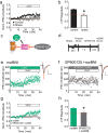
Figure 1.
Figure 1.. norBNI rescues LTPGABA through activation of JNK.
(A) Summary data showing the blockade of LTPGABA after stress. (B) Comparison of the magnitude of LTPGABA10–15 min after SNAP application. (IPSC amplitudes, control: 140 ± 5% of baseline values, n = 13; stress: 94 ± 11% of baseline values, n = 6; unpaired t-test, *p=0.0005. (C) Schematic of norBNI’s competitive and non-competitive inhibition of κOR signaling. (D) Experimental design. (E) Representative single experiment showing that bath application of norBNI (100 nM) rescues LTPGABA in a slice prepared 24 hr after stress. (F) Representative single experiment from a slice prepared 24 hr after stress showing that norBNI does not rescue LTPGABA in the presence of the JNK inhibitor SP600125 (20 µM). (G) Summary data from both groups. (H) Comparison of the magnitude of LTPGABA10–15 min after SNAP application. (IPSC amplitudes, norBNI only: 139 ± 7% of baseline values, n = 6; norBNI+SP600125: 106 ± 9% of baseline values, n = 11; unpaired t-test, *p=0.029.) Insets for this and all figures: IPSCs before (black trace, control) and 15 min after drug application (red trace, SNAP, 400 µM). Scale bars: 20 ms, 100 pA. Insets are averages of 12 IPSCs. DOI: http://dx.doi.org/10.7554/eLife.23785.003
Figure 1—figure supplement 1.
Figure 1—figure supplement 1.. Inhibition of JNK does not affect LTPGABA or its block by stress in the absence of norBNI.
(A) Summary data showing that LTPGABA is expressed in slices from naïve animals in the presence of the JNK inhibitor SP600125 (20 µM). Average magnitude of LTPGABA10–15 min after SNAP = 144 ± 13% of baseline values, n = 5; one-sample t-test, p=0.0257. (B) Summary data showing that LTPGABA remains blocked in slices from stressed animals in the presence of the JNK inhibitor SP600125 (20 µM). Average magnitude of LTPGABA10–15 min after SNAP = 111 ± 11% of baseline values, n = 6; one-sample t-test, p=0.38. DOI: http://dx.doi.org/10.7554/eLife.23785.004
Figure 2.
Figure 2.. The neutral antagonist 6β-naltrexol fails to rescue LTPGABA in slices from stressed animals.
(A) Schematic of norBNI and 6β-naltrexol inhibition of κOR signaling. (B) Experimental design. (C) Representative experiment showing that bath application of norBNI (100 nM) rescues LTPGABA in a slice prepared 24 hr after stress. (D) Representative experiment from a cell 24 hr after stress showing that 6β-naltrexol (10 µM) fails to rescue LTPGABA. (E) Summary data from both groups. (F) Comparison of the magnitude of LTPGABA10–15 min after SNAP application. (IPSC amplitudes, norBNI: 141 ± 20% of baseline values, n = 10; 6β-naltrexol: 100 ± 8% of baseline values, n = 10; unpaired t-test, *p=0.048). DOI: http://dx.doi.org/10.7554/eLife.23785.005
Figure 2—figure supplement 1.
Figure 2—figure supplement 1.. 6β-naltrexol does not affect basal inhibitory synaptic transmission but does block κORs.
(A) Summary data showing that 6β-naltrexol (10 µM) does not affect basal inhibitory transmission in cells from control or stressed rats. Normalized IPSC amplitude 5–10 min after 6β-naltrexol: control = 97 ± 6% of baseline values, n = 6 (4 animals); stress = 92 ± 17% of baseline values, n = 4 (2 animals); unpaired t-test p=0.79. (B) Summary data showing that U50488 depresses EPSC amplitudes from VTA dopamine neurons and that 6β-naltrexol (10 µM) prevents this depression. Normalized EPSC amplitude 5–10 min after U50488: U50488 alone = 75 ± 2% of baseline values, n = 3; U50488 + 6β-naltrexol = 95 ± 1% of baseline values, n = 4; unpaired t-test *p=0.0003. DOI: http://dx.doi.org/10.7554/eLife.23785.006
Figure 3.
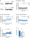
Figure 3.. 6β-naltrexol rescues LTPGABA when administered pre-stress, but not post-stress.
(A) Experimental design. (B) Representative experiment showing that a cell from a vehicle-treated stressed animal does not exhibit LTPGABA. (C) Representative experiment showing that a cell from an animal treated with 6β-naltrexol (10 mg/kg) 30 min pre-stress exhibits LTPGABA. (D) Representative experiment showing that a cell from an animal treated with 6β-naltrexol 24 hr post-stress does not exhibit LTPGABA. (E) Summary data showing compiled data from all groups. (F) Comparison of the magnitude of LTPGABA 10–15 min after SNAP application. (1-way ANOVA followed by Dunnett’s multiple comparison test. F2, 30=4.231,p=0.024. IPSC amplitudes, 6β-naltrexol pre-stress: 136 ± 12% of baseline values, n = 12, p<0.05 from vehicle; 6β-naltrexol post-stress: 100 ± 9% of baseline values, n = 11, n.s. from vehicle; vehicle+stress: 102 ± 8% of baseline values, n = 10). DOI: http://dx.doi.org/10.7554/eLife.23785.007
Figure 4.
Figure 4.. Single treatment with a κOR agonist leads to prolonged blockade of LTPGABA.
(A) Experimental design. (B) Representative experiment showing that a cell from a saline-treated animal exhibits LTPGABA. (C) Representative single experiment showing a cell prepared 24 hr after a single treatment with U50488 (5 mg/kg) does not exhibit LTPGABA. (D) Representative experiment showing that a cell prepared five days after a single treatment with U50488 does not exhibit LTPGABA. (E) Summary data from all groups. (F) Comparison of the magnitude of LTPGABA10–15 min after SNAP application. (1-way ANOVA followed by Dunnett’s multiple comparison test. F2, 27=12.21, p=0.0002. IPSC amplitudes, Saline: 137 ± 6% of baseline values, n = 11; U50488 1 day: 108 ± 6% of baseline values, n = 9, p<0.05 vs. saline; U50488 5 days: 100 ± 5% of baseline values, n = 10, p<0.05 vs. saline). DOI: http://dx.doi.org/10.7554/eLife.23785.008
Figure 5.
Figure 5.. κORs at VTA excitatory synapses are not constitutively activated by stress.
(A) Representative experiment showing that norBNI (100 nM) does not potentiate excitatory synapses on Ih+ VTA neurons in a slice prepared from a control animal. (B) Representative experiment showing that norBNI does not potentiate excitatory synapses on Ih+ VTA neurons in a slice prepared from a stressed animal. (C) Summary data from Ih+ neurons. No significant difference in IPSC amplitude 10–15 min after norBNI application (t-test p=0.81 IPSC amplitudes, control: 94 ± 2% of baseline values, n = 5; stressed: 92 ± 6% of baseline values, n = 6). (D) Representative experiment showing that norBNI does not potentiate excitatory synapses on Ih− VTA neurons in a slice prepared from a control animal. (E) Representative single experiment showing that norBNI does not potentiate excitatory synapses on Ih− VTA neurons in a slice prepared from a stressed animals. (F) Summary data from Ih− neurons. No significant difference in IPSC amplitude 10–15 min after norBNI application (t-test p=0.49 IPSC amplitudes, control: 112 ± 2% of baseline values, n = 5; stressed: 110 ± 4% of baseline values, n = 5). DOI: http://dx.doi.org/10.7554/eLife.23785.009
Figure 6.
Figure 6.. Post-stress rescue of reinstatement by norBNI but not 6β-naltrexol.
(A) Experimental design. (B) Lever pressing during the final extinction session (white bar) and reinstatement session (colored bar). Saline (black): last extinction session: 6.4 ± 1.7 lever presses; reinstatement session: 13.8 ± 2.4 lever presses; n = 8, *p=0.011, paired t-test. norBNI (green): last extinction session: 4.8 ± 1.4 lever presses; reinstatement session: 5.2 ± 1.1 lever presses; n = 6, p=0.76, paired t-test. 6β-naltrexol (blue): last extinction session: 5.7 ± 1.8 lever presses; reinstatement session: 11.2 ± 3.4 lever presses; n = 11, *p=0.033, paired t-test. DOI: http://dx.doi.org/10.7554/eLife.23785.010
Figure 7.
Figure 7.. Constitutive activation of κORs by stress.
(A) During stress, dynorphin binding to the κOR triggers a shift to a constitutively active state. By blocking dynorphin binding, both norBNI and 6β-naltrexol prevent the loss of LTPGABA during this time. (B) After stress, the block of LTPGABA is maintained by constitutive activity of κORs and is no longer dependent on dynorphin binding. (C) norBNI reverses the stress-induced block of LTPGABA by activating the JNK signaling pathway which non-competitively reduces κOR activity. DOI: http://dx.doi.org/10.7554/eLife.23785.011

References

    1. Baimel C, Bartlett SE, Chiou LC, Lawrence AJ, Muschamp JW, Patkar O, Tung LW, Borgland SL. Orexin/hypocretin role in reward: implications for opioid and other addictions. British Journal of Pharmacology. 2015;172:334–348. doi: 10.1111/bph.12639. - DOI - PMC - PubMed
    1. Baimel C, Lau BK, Qiao M, Borgland SL. Projection-Target-Defined effects of orexin and dynorphin on VTA dopamine neurons. Cell Reports. 2017;18:1346–1355. doi: 10.1016/j.celrep.2017.01.030. - DOI - PubMed
    1. Becker JA, Wallace A, Garzon A, Ingallinella P, Bianchi E, Cortese R, Simonin F, Kieffer BL, Pessi A. Ligands for kappa-opioid and ORL1 receptors identified from a conformationally constrained peptide combinatorial library. Journal of Biological Chemistry. 1999;274:27513–27522. doi: 10.1074/jbc.274.39.27513. - DOI - PubMed
    1. Briand LA, Vassoler FM, Pierce RC, Valentino RJ, Blendy JA. Ventral tegmental afferents in stress-induced reinstatement: the role of cAMP response element-binding protein. Journal of Neuroscience. 2010;30:16149–16159. doi: 10.1523/JNEUROSCI.2827-10.2010. - DOI - PMC - PubMed
    1. Bruchas MR, Chavkin C. Kinase cascades and ligand-directed signaling at the kappa opioid receptor. Psychopharmacology. 2010;210:137–147. doi: 10.1007/s00213-010-1806-y. - DOI - PMC - PubMed

Publication types

Substances

LinkOut - more resources