. 2017 Jun 1;157(2):399-409.
doi: 10.1093/toxsci/kfx057.

From the Cover: ImpairedProliferation and Differentiation of the Conducting Airway Epithelium Associated With Bronchiolitis Obliterans After Sulfur Mustard Inhalation Injury in Rats

Affiliations

From the Cover: ImpairedProliferation and Differentiation of the Conducting Airway Epithelium Associated With Bronchiolitis Obliterans After Sulfur Mustard Inhalation Injury in Rats

Matthew D McGraw et al. Toxicol Sci. .

Abstract

Sulfur mustard (SM) is a chemical warfare agent that causes chronic airway remodeling. This study's objective was to assess for changes to the bronchiolar epithelium after SM exposure to explain its contribution to chronic airway remodeling.

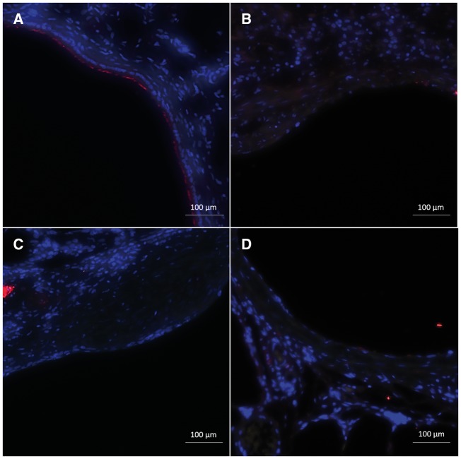
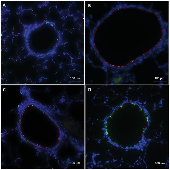
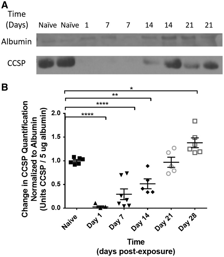
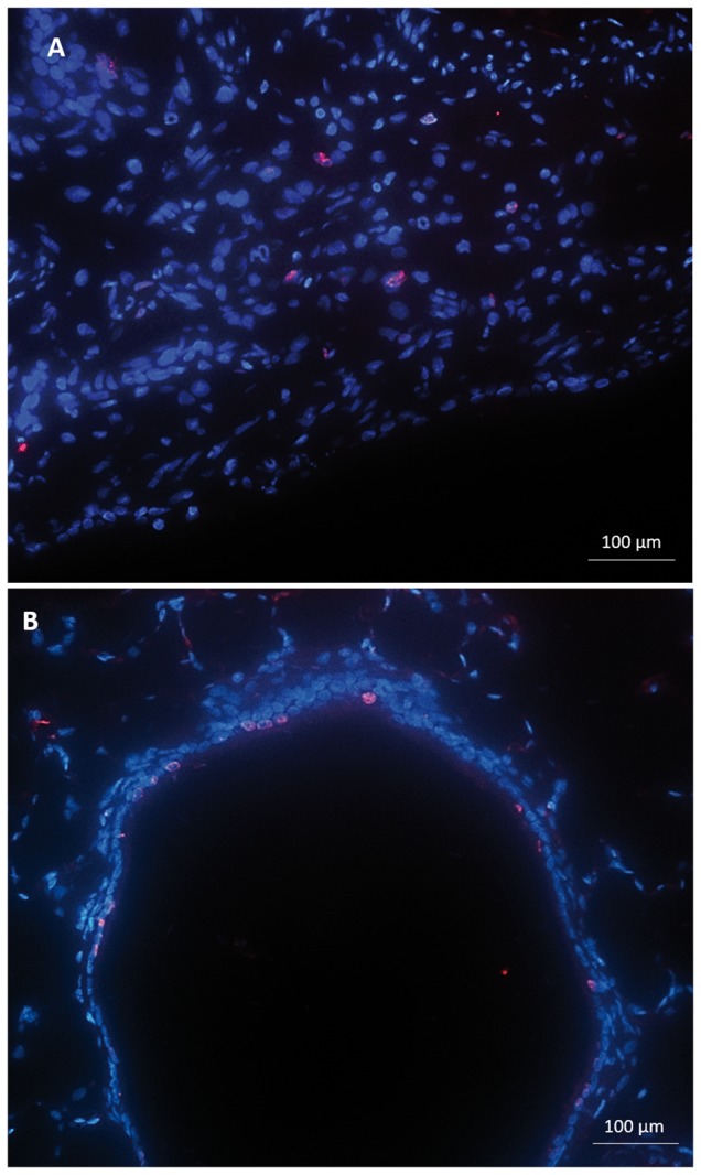
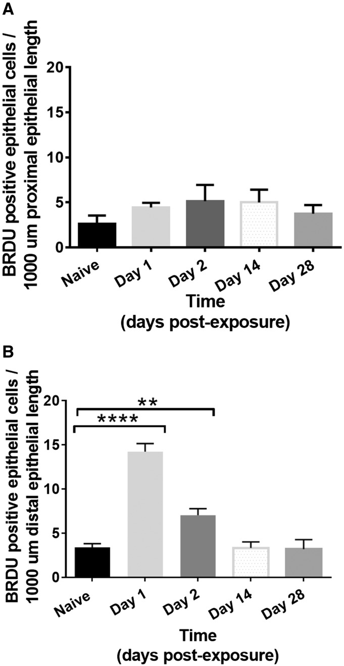
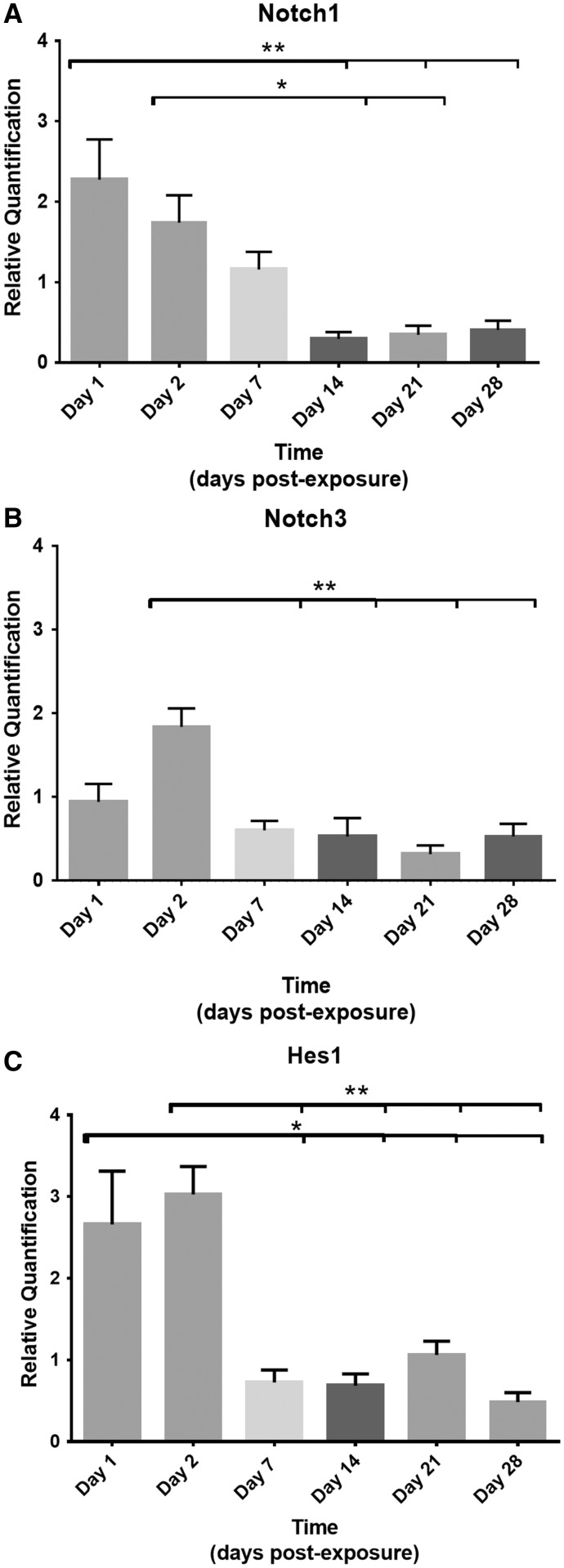
Materials and methods: Adult male rats were exposed to a sublethal dose of SM inhalation (1.0-1.2 mg/kg) for 50 min. Histological sections of the bronchiolar epithelium were analyzed for changes using hematoxylin and eosin, trichrome, and immunofluorescent staining for acetylated tubulin (AT) and club cell secretory protein (CCSP). CCSP in bronchoalveolar lavage fluid was assessed using western blot. A bromodeoxyuridine (BRDU) assay was used to assess for epithelial proliferation, and real-time PCR measured changes in Notch mRNA expression.

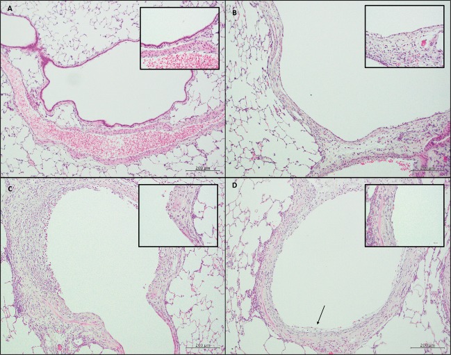
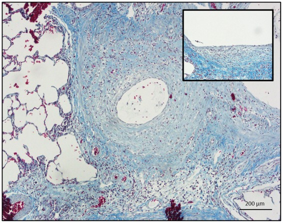
Results: SM caused significant proximal bronchiolar epithelial injury with epithelial denudation, loss of acetylated tubulin and CCSP staining, and reduced bronchoalveolar lavage fluid CCSP levels. bromodeoxyuridine (BRDU) + staining of proximal bronchiolar epithelial cells was not increased, but staining was increased in the distal bronchiolar epithelium. One month after injury, the proximal bronchiolar epithelium was not fully repaired. Significant collagen deposition surrounded proximal bronchioles with luminal obstruction, consistent with bronchiolitis obliterans. These changes corresponded with a downregulation of Notch1, Notch3, and Hes1 mRNA expressions.

Conclusions: This study demonstrates that SM exposure resulted in severe proximal airway epithelial injury, persistent morphological changes, impaired epithelial proliferation and, ultimately, bronchiolitis obliterans. These changes occurred at the same time that the Notch signaling genes were downregulated. Thus, the lung epithelium and the Notch signaling pathway may be worthy targets for the prevention of chronic airway remodeling after SM inhalation injury.

Keywords: Notch; bronchiolitis obliterans; epithelium; proliferation; regeneration; sulfur mustard.


Figures

FIG. 1
FIG. 1
Histological sections of proximal and distal conducting airways. (size bar: 200 µm.) Inset: Higher magnification of epithelial layer. (size bar: 50 µm.) A, Naïve rat section with the proximal bronchiolar epithelium with a single layer of epithelium with a uniform underlying basement membrane. B, Two days after SM exposure, the epithelium was patchy with areas of complete epithelial denudation and flattened, squamous-appearing epithelium without cilia. C, Two weeks after exposure, areas of proximal epithelium appeared with persistent areas of denudation and metaplastic, squamous epithelium. The surrounding mesenchyme was disorganized with significant inflammatory infiltrates. D, One month after SM injury, the epithelial layer of the proximal bronchioles was still not restored with epithelial sloughing (black arrow).
FIG. 2
FIG. 2
Trichrome staining of a proximal rat bronchiole 21 days after SM exposure with significant collagen deposition surrounding an injured and denuded proximal epithelium (size bar: 200 µm.). Inset: Higher magnification demonstrating a patchy and squamous-appearing epithelium overlying dense, subepithelial collagen (blue) staining (size bar: 50 µm).
FIG. 3
FIG. 3
Immunofluorescent staining for common epithelial markers, including AT (red) and CCSP (green) of proximal bronchiolar epithelium (size bar: 100 µm). A, Naïve rat proximal bronchiolar epithelium with the majority of cells staining positive for AT but with a few, sporadic cells positive for CCSP. B, Two days after SM exposure, there was near complete loss of staining for both AT and CCSP. C, Fourteen days after SM injury, staining for ciliated and club cells remained absent with areas of complete denudation. D, Twenty-eight days after SM injury, staining of the proximal epithelium remained absent for AT and CCSP.
FIG. 4
FIG. 4
Immunofluorescent staining for common epithelial markers, including AT (red) and CCSP (green), of distal bronchiolar epithelium (size bar: 100 µm). A, In naïve control rats, the distal bronchiolar epithelium showed well organized staining with both ciliated and CCSP. B, Two days after SM, the distal epithelium showed normal architectural integrity and persistent AT staining, but with complete loss of CCSP staining. C, Fourteen days after injury, CCSP staining returned, but was less uniform than naïve control. D, Twenty-eight days postexposure, the epithelium’s structure appeared similar to naïve bronchioles with uniform AT and CCSP staining.
FIG. 5
FIG. 5
A, Western blot of BALF for CCSP quantification (bottom line). Albumin was used as the positive standard control (top line). B: Quantification of change in BALF CCSP normalized to albumin on postexposure day to amount of BALF CCSP normalized to naïve control. CCSP was significantly reduced on day 1 (0.03 ± 0.13 vs. 1.00 ± 0.03; P < .0001) and remained reduced on day 7 (0.29 ± 0.12; P < .0001) and day 14 (0.51 ± 0.13; P < .01) after SM exposure. By day 21 postexposure, CCSP returned to levels equivalent to that of naïve controls (0.97 ± 0.13; P > .05). By day 28 postexposure, BALF CCSP was increased in exposed animals compared with controls (1.38 ± 0.13 vs 1.00 ± 0.03; P < .05).
FIG. 6
FIG. 6
Immunofluorescent staining for BRDU (pink) and DAPI nuclear (blue) staining at day 1 after SM exposure in both the proximal (A) and distal conducting airway (B). (Size bar: 100 µm). (A) In the proximal bronchioles, there was no significant increase in the number of proliferative epithelial cells per 1000 µm of epithelial length. BRDU staining was present in the adjacent, subepithelial mesenchyme. (B) In the distal bronchioles, there was a significant increase in number of proliferative epithelial cells per 1000 µm of epithelial length.
FIG. 7
FIG. 7
Number of BRDU positive epithelial cells per 1000 µm of epithelial length in proximal (A) and distal (B) bronchiolar epithelium. 5 proximal and distal bronchioles were assessed for each animal with 3–4 animals at each time point (naïve, days 1, 2, 14, and 28). (A) There was no significant increase in the number of BRDU positive epithelial cells per 1000 µm of the proximal bronchiolar epithelial length on days 1 or 2 after exposure compared with naïve controls (Naïve: 2.6 ± 0.5; day 1: 4.4 ± 0.3; day 2: 5.1 ± 1.0; day 14: 5.0 ± 0.5; day 28: 3.8 ± 0.5; P > .05). (B) The number of BRDU+ epithelial cells was significantly increased in the distal bronchiolar epithelium on day 1 after injury compared with naïve controls (14.2 ± 0.9 vs 3.4 ± 0.4; P = .0001) and remained significantly elevated on day 2 after injury (7.0 ± 0.7 vs 3.4 ± 0.4; P = .01), but was equivalent to naïve controls on day 14 and 28 (day 14: 3.3 ± 0.4; day 28: 3.8 ± 0.4; P > .05).
FIG. 8
FIG. 8
Relative quantification of RNA expression of 2 Notch receptor genes, Notch1 (A), Notch3 (B), and 1 downstream effector gene, Hes1 (C). The Ct value of each sample was normalized to HPRT1 as the endogenous control gene and relative quantification was calculated by the 2−ΔΔCt method. (A) On days 1 and 2 postexposure, Notch 1 levels were upregulated (2.27 ± 0.40 and 1.74 ± 0.43, respectively), but downregulated on days 14, differing significantly from day 1 and 2 (0.30 ± 0.38, P < .001 and P < .05). RNA expression levels remained significantly downregulated at days 21 and 28 compared with day 1 expression levels (0.35 ± 0.38, P < .001; 0.41 ± 0.40, P < .05). (B) Receptor Notch3 RNA levels were significantly upregulated on day 2 postexposure (1.84 ± 0.24, P < .05) but downregulated on day 7 (0.60 ± 0.22, P < .001), day 14 (0.53 ± 0.24, P < .001), day 21 (0.32 ± 0.22, P < .0001), and day 28 (0.53 ± 0.24, P < .01) compared with day 2. (C) Hes1 levels were significantly upregulated on days 1 and 2 (2.66 ± 0.45, P < .05 and 3.03 ± 0.45, P < .01. By day 7, Hes1 RNA levels were downregulated (0.73 ± 0.51) and remained significantly downregulated at days 14, 21, and 28 (P < .05) compared with days 1 and 2.

Similar articles

Cited by

References

    1. Allon N., Amir A., Manisterski E., Rabinovitz I., Dachir S., Kadar T. (2009). Inhalation exposure to sulfur mustard in the guinea pig model: Clinical, biochemical and histopathological characterization of respiratory injuries. Toxicol. Appl. Pharmacol. 241, 154–162. - PubMed
    1. Anderson DR, Byers SL, Vesely KR. 2000. Treatment of sulfur mustard (HD)-induced lung injury. J Appl Toxicol. 20 Suppl 1, S129–S132. - PubMed
    1. Beheshti J., Mark E. J., Akbaei H. M., Aslani J., Ghanei M. (2006). Mustard lung secrets: Long term clinicopathological study following mustard gas exposure. Pathol. Res. Pract. 202, 739–744. - PubMed
    1. Bracher A., Doran S. F., Squadrito G. L., Postlethwait E. M., Bowen L., Matalon S. (2012). Targeted aerosolized delivery of ascorbate in the lungs of chlorine-exposed rats. J. Aerosol. Med. Pulm. Drug Deliv. 25, 333–341. doi:10.1089/jamp.2011.0963 - PMC - PubMed
    1. Cabiati M., Raucci S., Caselli C., Guzzardi M. A., D’Amico A., Prescimone T., Del Ry S. (2012). Tissue-specific selection of stable reference genes for real-time PCR normalization in an obese rat model. J. Mol. Endocrinol. 48, 251–260. - PubMed

MeSH terms