. 2017 Jun;65(6):335-346.
doi: 10.1369/0022155417702849. Epub 2017 Apr 12.

The MicroRNA miR-210 Is Expressed by Cancer Cells but Also by the Tumor Microenvironment in Triple-Negative Breast Cancer

Affiliations

The MicroRNA miR-210 Is Expressed by Cancer Cells but Also by the Tumor Microenvironment in Triple-Negative Breast Cancer

Isabelle Bar et al. J Histochem Cytochem. 2017 Jun.

Abstract

The triple-negative breast cancer (TNBC) subtype occurs in about 15% of breast cancer and is an aggressive subtype of breast cancer with poor outcome. Furthermore, treatment of patients with TNBC is more challenging due to the heterogeneity of the disease and the absence of well-defined molecular targets. Microribonucleic acid (RNA) represents a new class of biomarkers that are frequently dysregulated in cancer. It has been described that the microRNA miR-210 is highly expressed in TNBC, and its overexpression had been linked to poor prognosis. TNBC are often infiltrated by immune cells that play a key role in cancer progression. The techniques traditionally used to analyze miR-210 expression such as next generation sequencing or quantitative real-time polymerase chain reaction (PCR) do not allow the precise identification of the cellular subtype expressing the microRNA. In this study, we have analyzed miR-210 expression by in situ hybridization in TNBC. The miR-210 signal was detected in tumor cells, but also in the tumor microenvironment, in a region positive for the pan-leucocyte marker CD45-LCA. Taken together, our results demonstrate that miR-210 is expressed in tumor cells but also in the tumor microenvironment. Our results also highlight the utility of using complementary approaches to take into account the cellular context of microRNA expression.

Keywords: TNBC; in situ hybridization; miR-210; tumor microenvironment.


Conflict of interest statement

Competing Interests: The author(s) declared no potential conflicts of interest with respect to the research, authorship, and/or publication of this article.

Figures

Figure 1.
Figure 1.
Analysis of miR-210 expression level by qRT-PCR in TNBC. A: distribution of the normalized expression level of miR-210 in 26 TNBC and 13 normal breast samples. MiR-210 is highly expressed in TNBC compared with normal samples (**, p=0.0016, bars represent means ± SEM). B: distribution of the normalized expression level of miR-210 in 13 TNBC with mutated BRCA1 and 10 TNBC with wild type BRCA1 gene: the miR-210 expression is similar between mutated and non-mutated samples (NS, p=0.5399, bars represent means ± SEM). C: Western blot analysis of BRCA1 protein expression in SUM-159PT cells transfected with scramble or BRCA1 siRNA for 72 hr. β-actin is used as loading control. D: analysis of miR-210 expression by RT-qPCR in SUM-159PT cells transfected with scramble or BRCA1 siRNA for 72 hr (normalized to RNU44): The depletion of BRCA1 by siRNA does not influence miR-210 expression (NS, p=0.3391, bars represent means ± SEM of three independent experiments). Abbreviations: qRT-PCR, quantitative real-time polymerase chain reaction; TNBC, triple-negative breast cancer; RNU44, small nucleolar RNA 44; SEM, standard error of the mean.
Figure 2.
Figure 2.
Validation of miR-210 in situ hybridization. A and B: The miR-210 signal is detected in epithelial cancer (A), and no signal is detected with the negative control probe (scramble probe) (B). C and D: Normal glandular cells do not express miR-210 (C) but express the positive control probe U6 in the nucleus (D). (Scale bar: 50 µm.) Abbreviations: TME, tumor microenvironment; Tu, epithelial tumor cells; Nl, normal glandular cells.
Figure 3.
Figure 3.
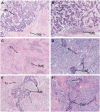
Detection of miRNA-210 expression in triple-negative breast cancer (TNBC) by in situ hybridization (ISH). A–D: miR-210 expression is detected in tumor cells. C–F. Strong miR-210 expression is detected in the tumor microenvironment. (Scale bar: 50 µm.) Abbreviations: Tu, tumor; TME, tumor microenvironment.
Figure 4.
Figure 4.
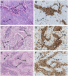
miR-210 positive cells in the tumor microenvironment co-localize with CD45-LCA expressing cells. A-C-E: detection of miR-210 by ISH; B-D-F: detection of CD45-LCA by IHC in adjacent sections of TNBC. (Scale bar: 100 µm.) Abbreviations: CD45-LCA, CD45-leukocyte common antigen; ISH, in situ hybridization; TNBC, triple-negative breast cancer; Tu, tumor; TME, tumor microenvironment.
Figure 5.
Figure 5.
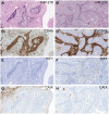
Detection of miR-210, CD45-LCA, HIF1-alpha and CAIX expression in adjacent sections of two representative TNBC samples. On the left (Fig. 5A-C-E-G), miR-210 is expressed in CD45-LCA positive cells, in the absence of HIF1-alpha expression. CAIX is expressed in the tumor cells. On the right (Fig. 5B-D-F-H), HIF1-alpha is detected in the tumor cells but not in the immune cells expressing miR-210. CAIX is not detected in the tumor cells. (Scale bar: 250 µm.) Abbreviation: CD45-LCA, CD45-leukocyte common antigen; HIF1-alpha, hypoxia inducible factor 1 alpha; TNBC, triple-negative breast cancer; CAIX, carbonic anhydrase IX.

References

    1. Lehmann BD, Bauer JA, Chen X, Sanders ME, Chakravarthy AB, Shyr Y, Pietenpol JA. Identification of human triple-negative breast cancer subtypes and preclinical models for selection of targeted therapies. J Clin Invest. 2011;121:2750–67. - PMC - PubMed
    1. Burstein MD, Tsimelzon A, Poage GM, Covington KR, Contreras A, Fuqua SAW, Savage MI, Osborne CK, Hilsenbeck SG, Chang JC, Mills GB, Lau CC, Brown PH. Comprehensive genomic analysis identifies novel subtypes and targets of triple-negative breast cancer. Clin Cancer Res. 2015;21:1688–98. - PMC - PubMed
    1. García-Teijido P, Cabal ML, Fernández IP, Pérez YF. Tumor-infiltrating lymphocytes in triple negative breast cancer: the future of immune targeting. Clin Med Insights Oncol. 2016;10:31–9. - PMC - PubMed
    1. Pruneri G, Vingiani A, Bagnardi V, Rotmensz N, De Rose A, Palazzo A, Colleoni AM, Goldhirsch A, Viale G. Clinical validity of tumor-infiltrating lymphocytes analysis in patients with triple-negative breast cancer. Ann Oncol. 2016;27:249–56. - PubMed
    1. Yuan Z-Y, Luo R-Z, Peng R-J, Wang S-S, Xue C. High infiltration of tumor-associated macrophages in triple-negative breast cancer is associated with a higher risk of distant metastasis. Onco Targets Ther. 2014;7:1475–80. - PMC - PubMed

Publication types