. 2017 Apr 11;19(2):235-245.
doi: 10.1016/j.celrep.2017.03.054.

The 150-Loop Restricts the Host Specificity of Human H10N8 Influenza Virus

Affiliations

The 150-Loop Restricts the Host Specificity of Human H10N8 Influenza Virus

Netanel Tzarum et al. Cell Rep. .

Abstract

Adaptation of influenza A viruses to new hosts are rare events but are the basis for emergence of new influenza pandemics in the human population. Thus, understanding the processes involved in such events is critical for anticipating potential pandemic threats. In 2013, the first case of human infection by an avian H10N8 virus was reported, yet the H10 hemagglutinin (HA) maintains avian receptor specificity. However, the 150-loop of H10 HA, as well as related H7 and H15 subtypes, contains a two-residue insert that can potentially block human receptor binding. Mutation of the 150-loop on the background of Q226L and G228S mutations, which arose in the receptor-binding site of human pandemic H2 and H3 viruses, resulted in acquisition of human-type receptor specificity. Crystal structures of H10 HA mutants with human and avian receptor analogs, receptor-binding studies, and tissue staining experiments illustrate the important role of the 150-loop in H10 receptor specificity.

Keywords: 150-loop; H10N8; HA; crystal structure; glycan array; hemagglutinin; host specificity; influenza A virus.


Figures

Figure 1
Figure 1. The Role of the 150-loop in Receptor Specificity of H10 HA
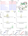
(A) Sequence alignment of the HA 150-loop sequences of representative IAV from all HA subtypes. The alignment shows the elongation of the 150-loop by two amino acids (designated 158a and 158b) in H7, H10 and H15 HAs (insert colored in black). (B) Superposition of the RBS subdomains of H10 HA (red), pandemic H1 (yellow, PDB 3AL4), pandemic H2 (green, PDB 3KU5), pandemic H3 (cyan, PDB 4FNK), VN1203 H5 (pink, PDB 2FK0), A/Taiwan/2/2013 H6 (magenta, PDB 4XKD) and Sh2H7 (gray, PDB 4N5J) HAs. The conserved secondary structural elements of the HA RBS (130-loop, 150-loop, 190-helix and 220-loop) and H10 Lys158a are labeled. (C) Glycan microarray analysis of recombinant wild-type and H10 HA RBS mutants. The H10K158aA (C2) and H10K158aG (C3) mutants bind specifically to α2–3 linked sialosides, similar to H10 wt HA (C1). (C4) The H10LS double mutant binds poorly to α2–3 and α2–6 linked sialosides. (C5) Mutating Lys158a to Ala on the background of H10LS now results in binding to α2–6 linked sialosides with only weak binding to α2–3 linked sialosides. (C6) Mutating Asp193 to Thr on the background of the H10LS-K158aA mutant reveals a complete switch in binding from α2–3 to α2–6 linked sialosides. The mean signal and standard error were calculated from six independent replicates on the array. α2–3 linked sialosides are shown in yellow bars (# 11 to 79 on the x axis) and α2–6 linked sialosides in green (# 80 to 135). Glycans 1 to 10 are non-sialylated controls (gray). (D) Binding of H10N8 wt and H10LS-K158aA-D193T mutant HAs to α2–3 and α2–6 linked receptors, as determined by glycan ELISA. H10 wt (left panels) binds selectively to glycans terminating in α2–3-linked sialic acids with no significant binding to α2–6 receptors. H10LS-K158aA-D193T (right panels) shows only slightly reduced avidity for avian α2–3 receptors but gain of strong binding to human α2–6 sialoglycans. All tested glycans are biantennary, N-linked receptor analogs with one to four Gal-GlcNAc repeats (LN1-4) after the terminal sialic acid. An asialo, mono-LN (LN-L) receptor was used as a negative binding control for each HA. The numbers of the glycans in the list of glycans imprinted on the microarray (Table S1) are indicated in brackets. See also Figure S1.
Figure 2
Figure 2. Crystal Structure of the H10LS-K158aA-D193T Mutant in Complex with Human and Avian Receptor Analogs
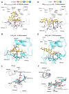
(A–C) The interactions between human receptor analog 6′-SLNLN and H10 HA RBS. The glycan structure of the 6′-SLNLN analog is represented in the upper part of the figure. The RBS conserved secondary structural elements are labeled and shown as ribbons. Selected RBS residues and the receptor analog are labeled and shown in sticks. Hydrogen bonds are indicated by black dashes between the human analog and the RBS of the H10LS-K158aA-D193T mutant (A) and H10 wt (B). (C) Superposition of the human receptor analog from the H10 wt HA complex (cyan) onto that of the H10LS-K158aA-D193T mutant (white) indicating slight displacement of the human analog in the RBS and conformational changes arising from a slight rotation around the linkage between Sia-1 and Gal-2. The superposition was done on the RBS subdomain. For clarity, only the RBS of the H10LS-K158aA-D193T mutant is presented. (D–F) Interactions between avian analog 3′-SLN and the H10 HA RBS. Hydrogen bonds are indicated between 3′-SLN and the RBS of H10LS-K158aA-D193T mutant (D) and H10 wt (E). (F) Superposition of the 3′-SLN analog from the H10 wt HA complex (cyan) onto that of the H10LS-K158aA-D193T mutant (white) indicates displacement of the avian analog in the RBS and isomerization of the Sia-Gal bond (from trans to cis in the mutant complex) as a consequence of the Q226L and G228S mutations. See also Figure S2.
Figure 3
Figure 3. The Role of the 150-loop in Receptor Specificity of H10, H7 and H3 HAs
Glycan microarray analysis was used to determine the role of the 150-loop in the binding specificity of group-2 HAs. (A) Deletion of K158a on the background of H10LS and H10LS-D193T mutants results in binding to α2–6 linked sialosides with only weak binding to α2–3 linked sialosides. (B) Sh2H7 HA binds to avian receptor analogs. Mutation of Asp158a to Ala on the background of the G228S mutation results in gain of binding to human receptors analogs, but no loss in binding to avian analogs. (C) Schematic representation of insertion of Ser and Lys (between positions 157 and 158) into the 150-loop of pandemic HK68 and A/Wyoming/3/2003 H3 HAs. (D) The insertion reveals no change in binding specificity compared to wt. See also Figure S3.
Figure 4
Figure 4. Binding of H10 Mutants to Human Trachea Tissue
Binding of recombinant H10 HAs to sections of human trachea tissue. The VN1203 H5N1 (A1) and A/Cal/04 H1N1 (A2) HAs were used as controls for avian and human HAs. Whereas the H10K158aA mutant (B2) shows no binding to human trachea tissue and the H10 wt (B1 and C1) and the H10LS-K158aA (B3) mutants show weak binding, the H10LS-K158aA-D193T mutant (B4 and C2) exhibits increased binding to human trachea tissue. HA binding of H10 wt and the H10LS-K158aA-D193T mutant to tissue sections was destroyed by treatment of the tissue with sialidase (C3 and C4). Each panel (A, B and C) represents results from an independent experiment. See also Figure S4.

References

    1. Air GM. Sequence relationships among the hemagglutinin genes of 12 subtypes of influenza A virus. Proc Natl Acad Sci USA. 1981;78:7639–7643. - PMC - PubMed
    1. Bateman AC, Karamanska R, Busch MG, Dell A, Olsen CW, Haslam SM. Glycan analysis and influenza A virus infection of primary swine respiratory epithelial cells: the importance of NeuAcα2–6 glycans. J Biol Chem. 2010;285:34016–34026. - PMC - PubMed
    1. Belser JA, Katz JM, Tumpey TM. The ferret as a model organism to study influenza A virus infection. Dis Model Mech. 2011;4:575–579. - PMC - PubMed
    1. Chandrasekaran A, Srinivasan A, Raman R, Viswanathan K, Raguram S, Tumpey TM, Sasisekharan V, Sasisekharan R. Glycan topology determines human adaptation of avian H5N1 virus hemagglutinin. Nat Biotechnol. 2008;26:107–113. - PubMed
    1. Chen H, Yuan H, Gao R, Zhang J, Wang D, Xiong Y, Fan G, Yang F, Li X, Zhou J, et al. Clinical and epidemiological characteristics of a fatal case of avian influenza A H10N8 virus infection: a descriptive study. Lancet. 2014;383:714–721. - PubMed

Publication types