. 2017 Apr 11;112(7):1366-1373.
doi: 10.1016/j.bpj.2017.02.025.

Fragment-Based NMR Study of the Conformational Dynamics in the bHLH Transcription Factor Ascl1

Affiliations

Fragment-Based NMR Study of the Conformational Dynamics in the bHLH Transcription Factor Ascl1

Lorenzo Baronti et al. Biophys J. .

Abstract

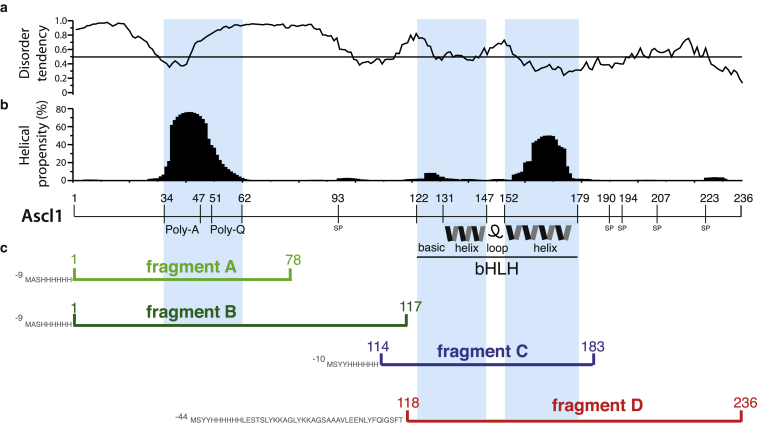
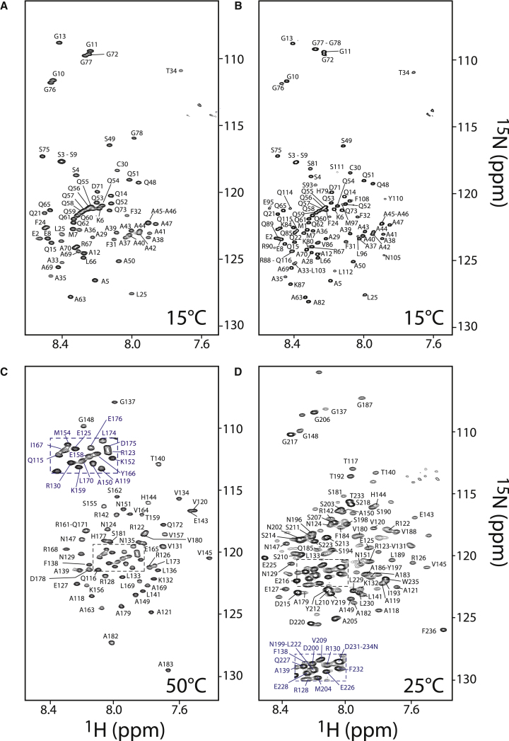
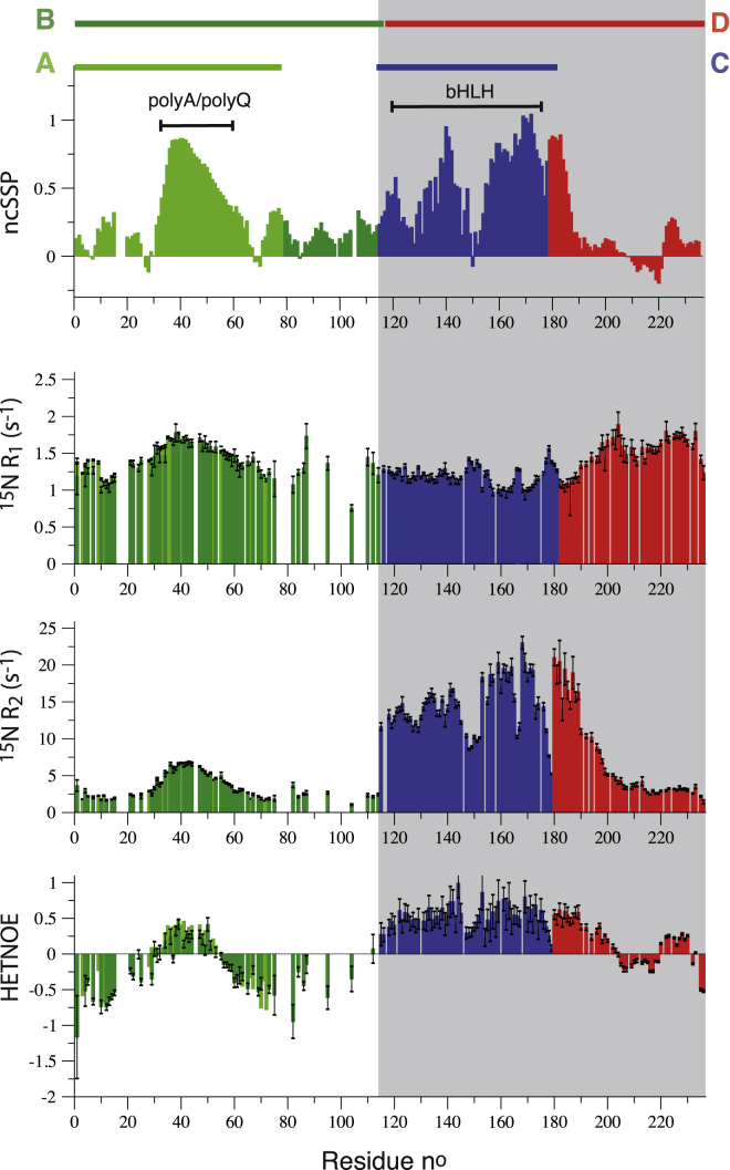
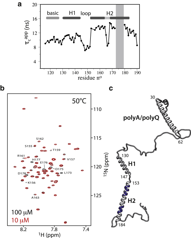
The Achaete-scute homolog 1 (Ascl1) protein regulates a large subset of genes that leads neuronal progenitor cells to distinctive differentiation pathways during human brain development. Although it is well known that Ascl1 binds DNA as a homo- or heterodimer via its basic helix-loop-helix (bHLH) motif, little is known about the conformational sampling properties of the DNA-free full-length protein, and in particular about the bHLH domain-flanking N- and C-terminal segments, which are predicted to be highly disordered in solution. The structural heterogeneity, low solubility, and high aggregation propensity of Ascl1 in aqueous buffer solutions make high-resolution studies of this protein a challenging task. Here, we have adopted a fragment-based strategy that allowed us to obtain high-quality NMR data providing, to our knowledge, the first comprehensive high-resolution information on the structural propensities and conformational dynamics of Ascl1. The emerging picture is that of an overall extended and highly dynamic polypeptide chain comprising three helical segments and lacking persistent long-range interactions. We also show that the C-terminal helix of the bHLH domain is involved in intermolecular interactions, even in the absence of DNA. Our results contribute to a better understanding of the mechanisms of action that govern the regulation of proneural transcription factors.


Figures

Figure 1
Figure 1
Ascl1 domain organization, structure prediction, and fragment design. (a) The disorder tendency was computed with the IUPred software (18, 29), whereas (b) the helical propensity prediction was obtained from Agadir (19). Peculiar sequence features of Ascl1, such as the polyA, polyQ, and Ser-Pro regions, as well as the bHLH domain, are highlighted. (c) The different fragments used in this study are shown at the bottom using a color code that is maintained throughout the manuscript. To see this figure in color, go online.
Figure 2
Figure 2
1H-15N BEST-TROSY correlation spectra of the different Ascl1 fragments designed and used for this study. (A) Residues 1–78. (B) Residues 1–117. (C) Residues 114–183 (the bHLH domain). (D) Residues 118–236. The spectra shown were recorded at an optimized sample temperature at which the NMR assignment was performed. Correlation peaks in the different NMR spectra are annotated by the corresponding residue number and type. To see this figure in color, go online.
Figure 3
Figure 3
NMR data collected for the four fragments of Ascl1, reporting on conformational features (structure and dynamics) along the polypeptide chain. From top to bottom are shown neighbor-corrected secondary structural propensity (ncSSP) values, calculated from the measured 13C chemical shifts, 15N R1 and R2 relaxation rate constants, and HETNOE intensity ratios. 15N relaxation data were recorded at 25°C, and the magnetic field strength was 600 MHz 1H frequency (700 MHz for fragment D). Data measured for the highly flexible terminal residues at the nonnative fragment ends are not shown. All graphs are color-coded to make clear that the underlying data originate from different NMR samples. White (grey) backgrounds indicate that the data have been measured on sampels without (with) detergent. To see this figure in color, go online.
Figure 4
Figure 4
NMR characterization of the bHLH domain in the absence of DNA. (a) Residue-specific apparent rotational correlation times computed from the 15N relaxation rate constants R2 and R1 as τc=(6R2/R1)7/(4πνN). (b) Overlaid 1H-15N correlation spectra (50°C) of Ascl1 fragment C recorded on samples of 100 μM and 10 μM concentration. Correlation peaks that show concentration-dependent peak shifts or line broadening are annotated by the corresponding residue number and amino acid type. (c) Graphical sketch of full-length Ascl1 representing an example of a transiently occurring α-helix-rich conformer. In addition, residues involved in dimerization, as identified in (b), are highlighted in blue. To see this figure in color, go online.

References

    1. Imayoshi I., Kageyama R. Review and fate choice of neural progenitor cells. Neuron. 2014;82:9–23. - PubMed
    1. Wilkinson G., Dennis D., Schuurmans C. Proneural genes in neocortical development. Neuroscience. 2013;253:256–273. - PubMed
    1. Castro D.S., Guillemot F. Old and new functions of proneural factors revealed by the genome-wide characterization of their transcriptional targets. Cell Cycle. 2011;10:4026–4031. - PMC - PubMed
    1. Huang Y., Cai S., Chen Z. Intermolecular single-quantum coherence sequences for high-resolution NMR spectra in inhomogeneous fields. J. Magn. Reson. 2010;203:100–107. - PubMed
    1. Vasconcelos F.F., Castro D.S. Transcriptional control of vertebrate neurogenesis by the proneural factor Ascl1. Front. Cell. Neurosci. 2014;8:412. - PMC - PubMed

MeSH terms

LinkOut - more resources