. 2017 Apr 12;15(4):e2000420.
doi: 10.1371/journal.pbio.2000420. eCollection 2017 Apr.

Bordetella bronchiseptica exploits the complex life cycle of Dictyostelium discoideum as an amplifying transmission vector

Affiliations

Bordetella bronchiseptica exploits the complex life cycle of Dictyostelium discoideum as an amplifying transmission vector

Dawn L Taylor-Mulneix et al. PLoS Biol. .

Abstract

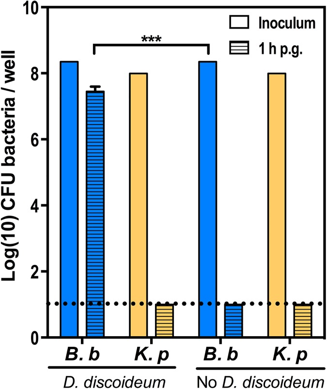
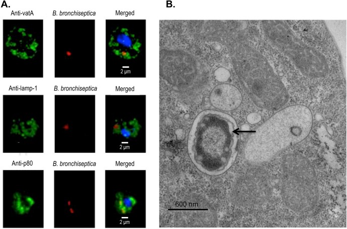
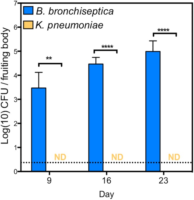
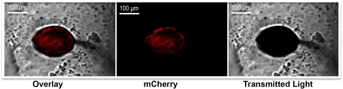
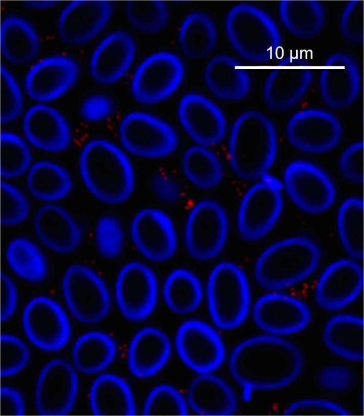
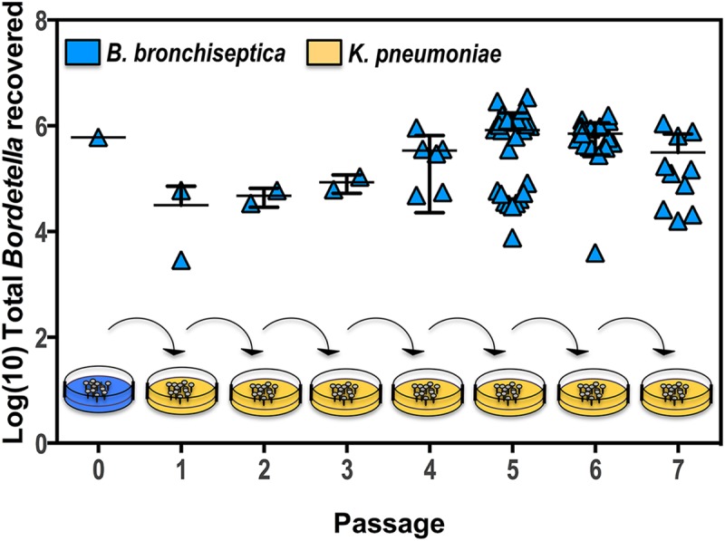
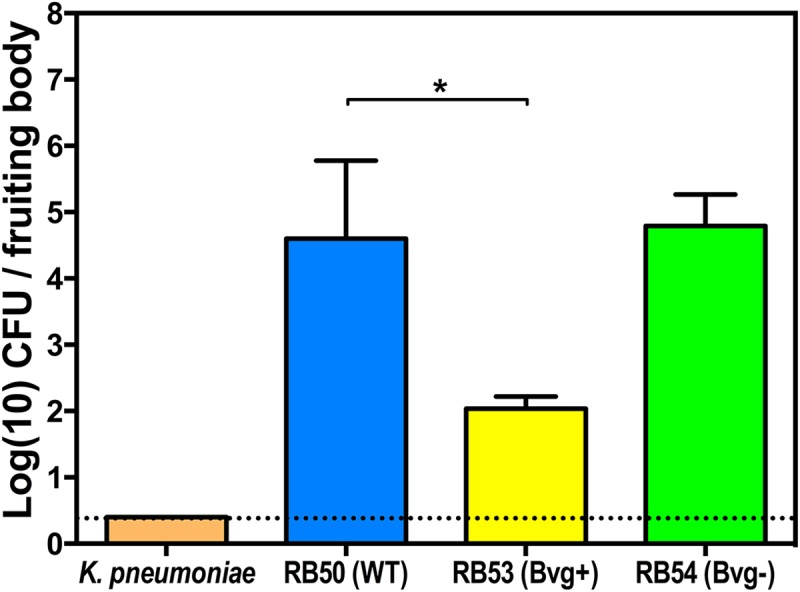
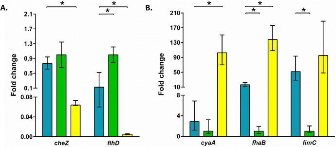
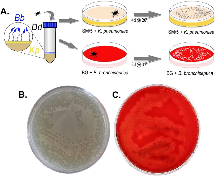
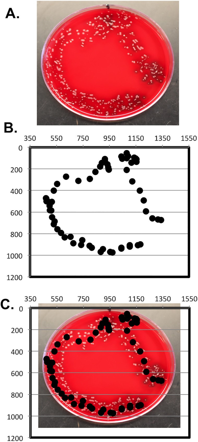
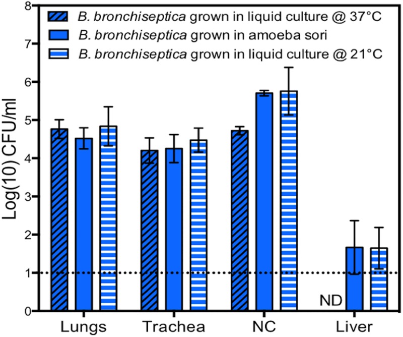
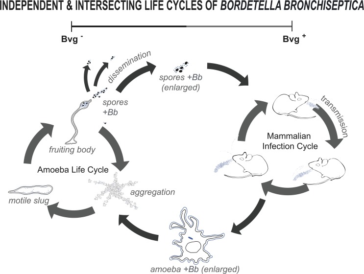
Multiple lines of evidence suggest that Bordetella species have a significant life stage outside of the mammalian respiratory tract that has yet to be defined. The Bordetella virulence gene (BvgAS) two-component system, a paradigm for a global virulence regulon, controls the expression of many "virulence factors" expressed in the Bvg positive (Bvg+) phase that are necessary for successful respiratory tract infection. A similarly large set of highly conserved genes are expressed under Bvg negative (Bvg-) phase growth conditions; however, these appear to be primarily expressed outside of the host and are thus hypothesized to be important in an undefined extrahost reservoir. Here, we show that Bvg- phase genes are involved in the ability of Bordetella bronchiseptica to grow and disseminate via the complex life cycle of the amoeba Dictyostelium discoideum. Unlike bacteria that serve as an amoeba food source, B. bronchiseptica evades amoeba predation, survives within the amoeba for extended periods of time, incorporates itself into the amoeba sori, and disseminates along with the amoeba. Remarkably, B. bronchiseptica continues to be transferred with the amoeba for months, through multiple life cycles of amoebae grown on the lawns of other bacteria, thus demonstrating a stable relationship that allows B. bronchiseptica to expand and disperse geographically via the D. discoideum life cycle. Furthermore, B. bronchiseptica within the sori can efficiently infect mice, indicating that amoebae may represent an environmental vector within which pathogenic bordetellae expand and disseminate to encounter new mammalian hosts. These data identify amoebae as potential environmental reservoirs as well as amplifying and disseminating vectors for B. bronchiseptica and reveal an important role for the Bvg- phase in these interactions.


Conflict of interest statement

The authors have declared that no competing interests exist.

Figures

Fig 1
Fig 1. Intracellular survival in amoeba cells.
B. bronchiseptica (B. b, blue) or K. pneumoniae (K. p, orange) were incubated for 1 h in HL/5 media containing D. discoideum at a multiplicity of infection (MOI) of 100 or in HL/5 media alone. Bars represent the initial inocula (open bars) and bacteria recovered 1 h post gentamicin (p.g.) application (hatched bars). *** denotes p < 0.00005. The dotted line indicates the limit of detection. CFU, colony forming unit. For further details, please see S1 Data.
Fig 2
Fig 2. Visualization of B. bronchiseptica within D. discoideum.
(A) Fluorescent confocal imaging of B. bronchiseptica within D. discoideum. D. discoideum were exposed to B. bronchiseptica RB50 pLC018 (mCherry, red) for 1 h at a multiplicity of infection (MOI) of 100 prior to 1 h gentamicin treatment. D. discoideum were then fixed in 4% paraformaldehyde and permeabilized with 0.1% saponin prior to incubation with the following primary antibodies: mouse monoclonal anti-vatA, rat monoclonal anti-lamp-1, and mouse monoclonal anti-p80. Secondary antibodies, specifically goat anti-mouse and goat anti-rat, were conjugated to either fluorescein or a green fluorescent dye (green). D. discoideum were also stained with DAPI for visualization of nuclei (blue). Z-stacks were captured at 60× magnification and zoomed to 300%. Individual panels and merged fluorescent images are displayed. (B) Electron microscopy images of intracellular B. bronchiseptica within D. discoideum. D. discoideum were exposed to B. bronchiseptica RB50 for 1 h at a MOI of 100 prior to 1 h gentamicin treatment. Following exposure and gentamicin treatment, D. discoideum harboring intracellular bacteria were fixed for 1 h in 2% glutaraldehyde and then processed for transmission electron microscopy. The image shows a single intracellular bacterium (arrow) within D. discoideum.
Fig 3
Fig 3. B. bronchiseptica survives and replicates in amoeba sori.
Recovery of B. bronchiseptica (blue) and K. pneumoniae (orange) from D. discoideum sori on days 9, 16, and 23 post addition of amoebae to lawns of B. bronchiseptica or K. pneumoniae, respectively. ** denotes a p-value < 0.005; **** denotes a p-value < 0.00005. A grey dotted line indicates the limit of detection. ND signifies "not detected." CFU, colony forming unit. For further details, please see S1 Data.
Fig 4
Fig 4. B. bronchiseptica localizes to the amoeba sorus.
Fluorescence confocal microscopy imaging of D. discoideum fruiting body grown 16 d on a lawn of B. bronchiseptica RB50 pLC003 (mCherry, red) at 10× magnification.
Fig 5
Fig 5. B. bronchiseptica localizes to the amoeba sorus, distributed between the D. discoideum spores.
Confocal microscopy image of D. discoideum sori grown on a lawn of B. bronchiseptica RB50 pLC003 (mCherry, red) at 60× magnification and zoomed to 200%. Amoeba spores were stained with calcofluor (blue).
Fig 6
Fig 6. B. bronchiseptica maintains a stable association with D. discoideum.
D. discoideum sori grown on a lawn of B. bronchiseptica strain RB50 (blue) were collected in 1 mL PBS. One aliquot was enumerated (Passage 0), while another aliquot was used to inoculate a lawn of K. pneumoniae (Passage 1). Amoeba sori that developed on the K. pneumoniae lawn (orange) were then collected, enumerated, and used to inoculate a new lawn of K. pneumoniae (Passage 2). Collection, enumeration, and passaging were repeated as indicated. For further details, please see S1 Data.
Fig 7
Fig 7. The Bvg- phase is advantageous for B. bronchiseptica recovery from sori.
Recovery of wild-type (RB50, blue), Bvg- (RB54, green) phase-locked, and Bvg+ (RB53, yellow) phase-locked B. bronchiseptica and K. pneumoniae (orange) from D. discoideum sori on day 10 post addition of amoebae to the respective bacterial lawns. * denotes a p-value < 0.05. CFU, colony-forming unit. For further details, please see S1 Data.
Fig 8
Fig 8. Gene expression of B. bronchiseptica in sori.
Genes associated with Bvg- (A) and Bvg+ (B) were selected, and the expression of those gene transcripts was compared with B. bronchiseptica isolated from sori (blue) against Bvg- (green) and Bvg+ (yellow) phase-locked mutants grown in liquid culture. Data represent the mean fold change in gene expression levels (with standard deviations) relative to the Bvg- phase-locked mutant RB54 from three independent experiments. An asterisk (*) indicates significantly different expression levels (p-value ≤ 0.05) in pair-wise comparisons. For further details, please see S2 Data.
Fig 9
Fig 9. B. bronchiseptica in amoeba sori can be transmitted via an intermediary to a new geographical location.
(A) Schematic showing the experimental setup in which flies were added to conical tubes containing B. bronchiseptica–filled fruiting bodies. Flies were then transferred to new plates containing either a lawn of K. pneumoniae (yellow plates, top) to assess amoeba transmission or Bordet-Gengou (BG) agar with streptomycin (red plates, bottom) to assess Bordetella transmission. (B) Representative image showing amoeba plaque growth along the fly’s footprints. (C) Representative image showing B. bronchiseptica colony growth along the fly’s footprints. Bb, B. bronchiseptica; Dd, D. discoideum; Kp, K. pneumoniae.
Fig 10
Fig 10. Ant transmitted B. bronchiseptica after exposure to amoeba.
(A) B. bronchiseptica growth on a Bordet-Gengou (BG) agar plate with streptomycin inoculated by an ant carrying amoebae. (B) Scatter plot graph showing the location of the ant’s thorax as it walked on the BG agar plate, recorded at quarter-second intervals using a custom software package. (C) BG agar plate overlaid with the scatter plot graph. For further details, please see S3 Data.
Fig 11
Fig 11. B. bronchiseptica recovered from sori efficiently infects mice.
Bacterial numbers recovered from respiratory tracts and livers of mice (n = 4) on day 3 post inoculation with B. bronchiseptica recovered from sori of D. discoideum (clear bars) or grown in culture at 37°C (black hatched bars) or 21°C (white hatched bars). A grey dotted line indicates the limit of detection. NC signifies “nasal cavity.” ND signifies “not detected.” CFU, colony-forming unit. For further details, please see S1 Data.
Fig 12
Fig 12. Model illustrating how BvgAS may regulate two independent but interconnected life cycles of B. bronchiseptica.
The model illustrates the survival and transmission of B. bronchiseptica (blue) both in the mammalian host (in Bvg+ phase) and along with the amoeba (in Bvg- phase) and the connections between these cycles. Infected mice shed B. bronchiseptica, which can both transmit to colonize other mammalian hosts and spread in the environment. The Bvg+ phase genes are known to be necessary for B. bronchiseptica colonization and transmission between mammalian hosts. Outside the mammalian host, in the Bvg- phase, B. bronchiseptica can form a stable association with the amoebae, like D. discoideum, such that it is incorporated into the fruiting body sori and transmitted from sorus to sorus. This association may constitute an alternative life cycle for bordetellae, involving the many Bvg- specific genes that are highly conserved yet not apparently expressed during the mammalian infection cycle. Importantly, B. bronchiseptica recovered from amoeba sori can efficiently infect mice, indicating that these two independent life cycles are interlinked. Bb, B. bronchiseptica.

Similar articles

Cited by

References

    1. Diavatopoulos DA, Cummings CA, Schouls LM, Brinig MM, Relman DA, Mooi FR. Bordetella pertussis, the causative agent of whooping cough, evolved from a distinct, human-associated lineage of B. bronchiseptica. PLoS Pathog. 2005;1: e45 doi: 10.1371/journal.ppat.0010045 - DOI - PMC - PubMed
    1. Dworkin MS, Sullivan PS, Buskin SE, Harrington RD, Olliffe J, MacArthur RD, et al. Bordetella bronchiseptica infection in human immunodeficiency virus-infected patients. Clin Infect Dis Off Publ Infect Dis Soc Am. 1999;28: 1095–1099. - PubMed
    1. Bendor L, Weyrich LS, Linz B, Rolin OY, Taylor DL, Goodfield LL, et al. Type Six Secretion System of Bordetella bronchiseptica and Adaptive Immune Components Limit Intracellular Survival During Infection. PLoS ONE. 2015;10: e0140743 doi: 10.1371/journal.pone.0140743 - DOI - PMC - PubMed
    1. Cummings CA, Bootsma HJ, Relman DA, Miller JF. Species- and strain-specific control of a complex, flexible regulon by Bordetella BvgAS. J Bacteriol. 2006;188: 1775–1785. doi: 10.1128/JB.188.5.1775-1785.2006 - DOI - PMC - PubMed
    1. Weiss AA, Falkow S. Genetic Analysis of Phase Change in Bordetella pertussis. Infect Immun. 1984;43: 263–69. - PMC - PubMed

MeSH terms

Substances