. 2017 Apr 12;12(4):e0175273.
doi: 10.1371/journal.pone.0175273. eCollection 2017.

Direct fluorescence detection of VirE2 secretion by Agrobacterium tumefaciens

Affiliations

Direct fluorescence detection of VirE2 secretion by Agrobacterium tumefaciens

Noga Yaakov et al. PLoS One. .

Abstract

VirE2 is a ssDNA binding protein essential for virulence in Agrobacterium tumefaciens. A tetracysteine mutant (VirE2-TC) was prepared for in vitro and in vivo fluorescence imaging based on the ReAsH reagent. VirE2-TC was found to be biochemically active as it binds both ssDNA and the acidic secretion chaperone VirE1. It was also biologically functional in complementing virE2 null strains transforming Arabidopsis thaliana roots and Nicotiana tabacum leaves. In vitro experiments demonstrated a two-color fluorescent complex using VirE2-TC/ReAsH and Alexa Fluor 488 labeled ssDNA. In vivo, fluorescent VirE2-TC/ReAsH was detected in bacteria and in plant cells at time frames relevant to transformation.


Conflict of interest statement

Competing Interests: The authors have declared that no competing interests exist.

Figures

Fig 1
Fig 1. Expression of TC-modified VirE2 mutant.
(A) wtVirE2 and VirE2-TC purified from inclusion bodies co-migrate in SDS-PAGE analysis. (B) Co-expression of 6xHis-tagged VirE1 with wtVirE2 or VirE2-TC led to a soluble product containing both proteins.
Fig 2
Fig 2. Electrophoretic Mobility Shift Analysis (EMSA) confirms binding of VirE2 to M13 ssDNA substrate.
M13 ssDNA concentration was held constant, while relative protein concentration increased. Lanes: (A) M13 alone, B-E) wtVirE2 at 1:1, 1:5, 1:10, 1:15 ratios wt:wt, (F-I) VirE2-TC at equal ratios. VirE2-TC binds M13 ssDNA less avidly than the wild-type, but leaves no evidence of completely unbound ssDNA.
Fig 3
Fig 3. Transmission electron microscopy confirms the formation of VirE2-DNA complexes with similar structure.
(A) wild-type and (B) TC-mutant VirE2. Scale bar 50 nm.
Fig 4
Fig 4. GUS expression in Arabidopsis roots.
(A) Infection by VirE2-null strain with both wild-type VirE2 expression and GUS gene transfer from binary plasmid pCAMBIA2301. (B) Infection by VirE2-null strain with both VirE2-TC expression and GUS gene transfer from binary plasmid pCAMBIA2301. (C) Positive control: GUS expression in roots infected by wild type strain transferring GUS gene from pCAMBIA2301 vector. (D) Negative control: roots infected by VirE2-null strain carrying the same GUS gene on pCAMBIA2301 vector.
Fig 5
Fig 5. Complementation assay in Nicotiana tabacum leaves.
(A) Positive control: GFP expression in leaf infiltrated with wild-type Agrobacterium carrying binary plasmid pCAMBIA2300-GFP. (B) Complementation by VirE2-TC: GFP expression in leaf tissue infiltrated with two VirE2-null Agrobacterium strains (AT12516), one carrying pCAMBIA2300-GFP and the other expressing VirE2-TC from a binary plasmid pCAMBIA2301:VirE2-TC. (C) Complementation by wild-type VirE2: GFP expression in leaf infiltrated with two VirE2-null Agrobacterium strains (AT12516), one carrying pCAMBIA2300-GFP and the other expressing wtVirE2 from binary plasmid pCAMBIA2301:VirE2. autofluorescence of chlorophyll in red. Scale bar 50 μm.
Fig 6
Fig 6. Confocal microscopy images of ssDNA-protein particles formed by M13 circular ssDNA labeled with Alexa 488.
(A) wild type VirE2, (B) VirE2-TC. Scale bar 50 μm.
Fig 7
Fig 7. In vivo labeling of VirE2-TC.
ReAsH labeling of virulence activated bacteria expressing VirE2-TC. Inset: zoom of a single fluorescent bacterium (white arrow) shows polar accumulation of the fluorescent label. Scale bar 10 μm.
Fig 8
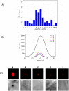
Fig 8. Analysis of ReAsH labeling in leaf tissue to distinguish bacteria from fluorescent VirE2 puncta.
(A) Integrated density values of VirE2-TC particles and bacteria labeled with ReAsH reagent. Bars 6 and 8 represent bacteria while the others are VirE2 particles. Bar 7 shows a number of clustered VirE2 particles. Note that the scale is logarithmic in order to display the small particles and bacteria together. (B) Line profiles across a bacterium and several VirE2 particles accentuate the differences in size and total brightness. Note that fluorescent spots of VirE2 are diffraction-limited by the microscope optics. (C) A subset of cropped images showing a bacterium (left) and several VirE2 particles at the same brightness scale. The bacterium image reaches saturation in the figure in order to provide visible contrast in the faint particles. The corresponding region in the differential interference contrast image (acquired simultaneously) is shown below each box. The bacterium appears as a large refractive object while the VirE2 particles are too small to produce a visible signal.
Fig 9
Fig 9. Overlay views of fluorescent puncta in leaf tissue.
(A) infiltrated Agrobacterium labeled with ReAsH, localized near the upper surface of the Nicotiana tabacum leaf. (B-E) VirE2 puncta labeled with ReAsH reagent in Nicotiana tabacum (B,E) and Nicotiana benthamiana (C,D). Brightness settings are adjusted individually for visibility in the overlays. Scale bar 10 μm.
Fig 10
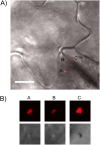
Fig 10. A virB mutant strain does not secrete fluorescent puncta.
(A) Large, motile fluorescent spots, visible also in the bright field images (B, lower panels), indicate bacteria. Smaller spots representing secreted VirE2 in the host cells were not observed. Scale bar 10 μm.

Similar articles

Cited by

References

    1. Chilton MD, Drummond MH, Merio DJ, Sciaky D, Montoya AL, Gordon MP, et al. Stable incorporation of plasmid DNA into higher plant cells: the molecular basis of crown gall tumorigenesis. Cell. 1977;11: 263–271. - PubMed
    1. Pitzschke A, Hirt H. New insights into an old story: Agrobacterium-induced tumour formation in plants by plant transformation. EMBO J. 2010;29: 1021–1032. 10.1038/emboj.2010.8 - DOI - PMC - PubMed
    1. Citovsky V, Wong ML, Zambryski P. Cooperative interaction of Agrobacterium VirE2 protein with single-stranded DNA: implications for the T-DNA transfer process. Proc Natl Acad Sci. 1989;86: 1193–1197. - PMC - PubMed
    1. Das A. Agrobacterium tumefaciens virE operon encodes a single-stranded DNA-binding protein. Proc Natl Acad Sci U S A. 1988;85: 2909–2913. - PMC - PubMed
    1. Gietl C, Koukolíková-Nicola Z, Hohn B. Mobilization of T-DNA from Agrobacterium to plant cells involves a protein that binds single-stranded DNA. Proc Natl Acad Sci U S A. 1987;84: 9006–9010. - PMC - PubMed

MeSH terms

LinkOut - more resources