. 2017 Apr 12;12(4):e0175585.
doi: 10.1371/journal.pone.0175585. eCollection 2017.

Amplification of a transgene within a long array of replication origins favors higher gene expression in animal cells

Affiliations

Amplification of a transgene within a long array of replication origins favors higher gene expression in animal cells

Kiwamu Ohsaki et al. PLoS One. .

Abstract

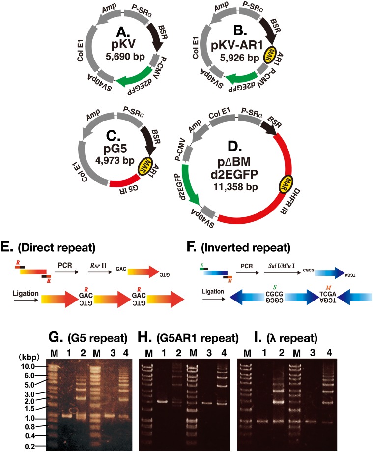
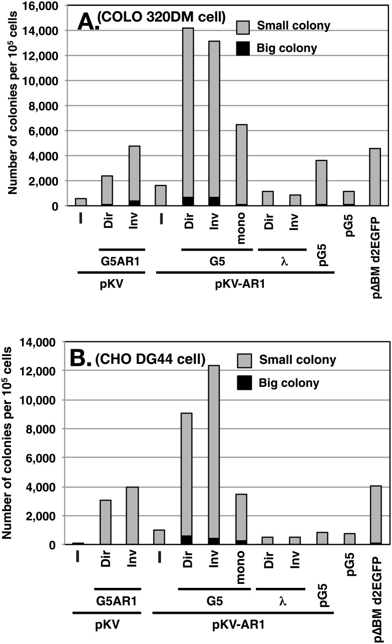
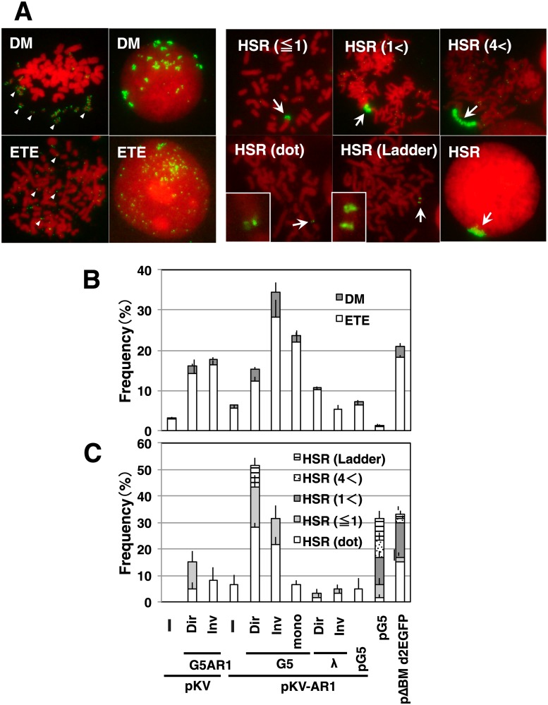
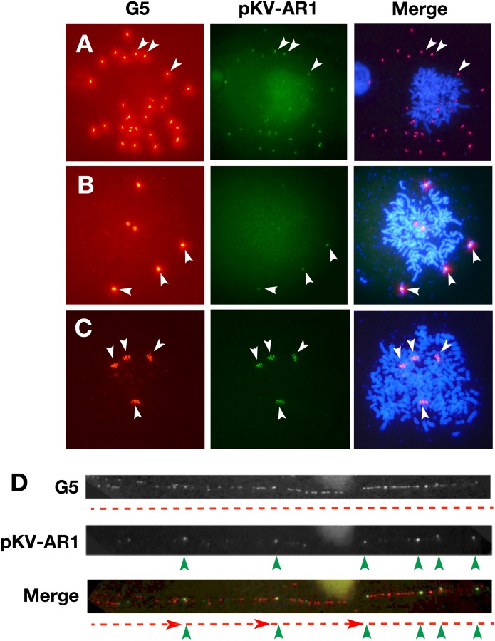
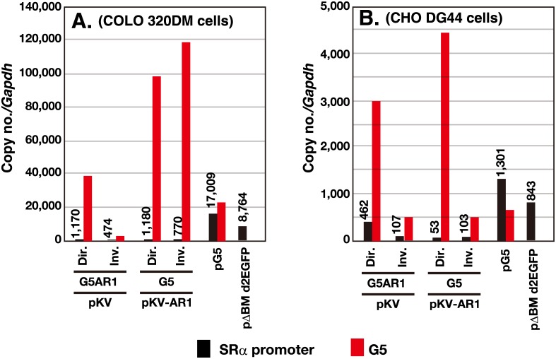
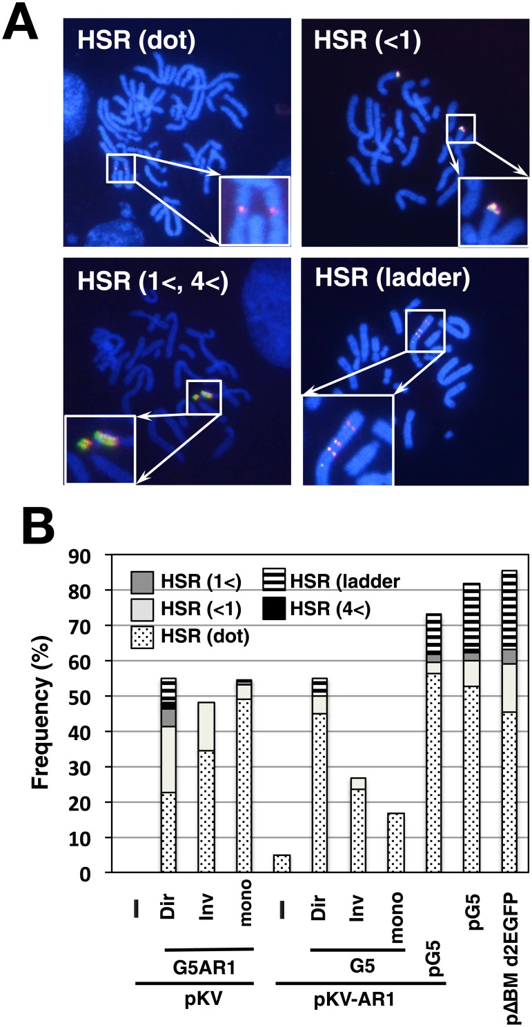
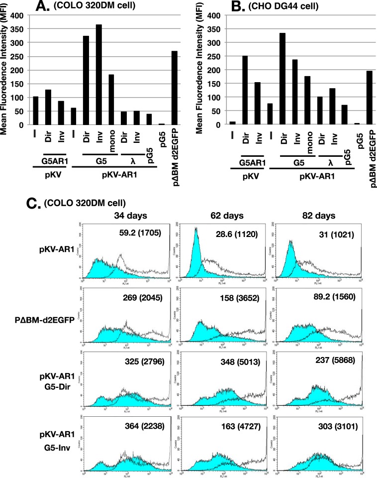
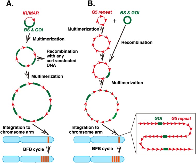
Plasmids with both a mammalian replication initiation region (IR) and a matrix attachment region (MAR) are spontaneously amplified in transfected cells, and generate extrachromosomal double minute (DM) or chromosomal homogeneously staining region (HSR). We previously isolated the shortest core IR (G5) required for gene amplification. In this study, we ligated the G5 DNA to create direct or inverted repeats, mixed the repeats with an expression plasmid, and transfected the mixture into human COLO 320DM or hamster CHO DG44 cells. Consequently, we found that the transfected sequence generated DMs or HSR where, surprisingly, the plasmid sequence was embedded within a long stretch of G5 sequences. The amplified structure from the direct G5 repeats was stable, whereas that from the inverted repeats was not. The amplification might be explained by the efficient replication/multimerization of the G5 repeat and recombination with the co-transfected plasmid in an extrachromosomal context. The product might then be integrated into a chromosome arm to generate a HSR. The expression from the plasmid within the long G5 array was much higher than that from a simple plasmid repeat. Because G5 is a core IR that favors gene expression, a long array of G5 provides an excellent environment for gene expression from the embedded plasmid.


Conflict of interest statement

Competing Interests: The authors have declared that no competing interests exist.

Figures

Fig 1
Fig 1. Preparation of repeat DNA and construction of plasmids.
(A to D) Structure of the plasmids used in this study. (E and F) Direct or inverted repeat DNA was prepared by PCR amplification, digestion with Rsr II (R) or double digestion with Sal I (S) and Mlu 1 (M), respectively, followed by ligation. (G to I) G5 (G), G5AR1 (H), or a portion of λ (I) DNA before (lanes 1 and 3) and after ligation to direct repeats (lane 2) or inverted repeats (lane 4). DNA was separated by agarose gel electrophoresis. M; molecular weight marker.
Fig 2
Fig 2. Effect of co-transfected repeat DNA on the transformed colony number.
COLO 320DM cells (A) or CHO DG44 cells (B) were transfected with the plasmid pKV or pKV-AR1 or a mixture of the plasmid and the direct (Dir) or the inverted (Inv) repeats of G5AR1, G5, or a part of λ-phage. As a control, pG5 or pΔBM d2EGFP plasmid was transfected. Two days after the transfection, 5 μg/ml blasticidin was added, and the cells were cultured for a further week. The number of large (more than ca. 150 cells) or small (ca. 50 to 150 cells) colonies per 105 transfected cells was counted and plotted. A representative result of three independent experiments that gave similar results is shown.
Fig 3
Fig 3. Effect of co-transfected repeat DNA on gene amplification in COLO 320DM cells.
After single transfection or co-transfection of the indicated DNA into COLO 320DM cells, the stable transformants were selected by blasticidin for 1 month. The metaphase chromosome spreads from the transformants were analyzed by FISH using a DIG-probe prepared from pG5 plasmid DNA. The hybridized probes were detected by green (FITC) fluorescence, and DNA was counterstained red with propidium iodide. Representative images for each type of amplification are shown in A. The frequency of cells having each type of extrachromosomal (B) or chromosomal (C) amplification was scored by examination of more than 30 metaphase cells in triplicate, and mean +/- standard deviations are plotted.
Fig 4
Fig 4. Simultaneous detection of co-transfected sequences in COLO 320DM cells.
A mixture of pKV-AR1 DNA and G5 inverted (A and B) or direct (C and D) repeat DNA was co-transfected into COLO 320DM cells, and the stable transformants were selected by blasticidin for a month. Metaphase chromosome spreads (A to C) or chromatin fibers (D) were prepared from these cells. The slides were simultaneously hybridized with biotin-labeled G5 probe and DIG-labeled pKV-AR1 plasmid probe, and the hybridized probes were detected by red (Alexa594) or green (FITC) fluorescence, respectively. DNA was counterstained blue with DAPI in panels A to C. Among metaphase chromosomes, strong G5 signals and weak pKV-AR1 signals were co-localized at multiple DMs (A) or HSRs (B and C). In panel D, small pKV-AR1 signals (green arrowheads) were periodically embedded in the long stretch of G5 signals (red broken lines).
Fig 5
Fig 5. Copy number of the transfected sequence in the stable transformants.
After transfection of the indicated DNA into COLO 320DM or CHO DG44 cells, the stable transformants were selected by blasticidin for 1 month. The genomic DNA was isolated and subjected to real-time PCR to determine the amount of SRα promoter, G5, λ-phage, and Gapdh sequence. The PCR reaction was done in triplicate, and the mean value was used to calculate the copy number of each sequence relative to Gapdh (see graph). The deviation between triplicate reactions was too small to be represented with error bars.
Fig 6
Fig 6. Simultaneous detection of co-transfected sequences in CHO DG44 cells.
After transfection of the indicated DNA into CHO DG44 cells, the stable transformants were selected by blasticidin for 1 month, and metaphase chromosome spreads were prepared. The slides were simultaneously hybridized with biotin-labeled G5 probe and DIG-labeled pKV-AR1 plasmid probe, and the hybridized probes were detected by red (Alexa594) and green (FITC) fluorescence, respectively. DNA was counterstained blue using DAPI. Representative images showing various HSRs are shown in A. The frequency of cells having each type of amplification was determined by examining more than 30 metaphase cells with each amplification and is plotted in B.
Fig 7
Fig 7. Plasmid-encoded d2EGFP expression in the stable transformants.
After transfection of the indicated DNA into COLO 320DM (A) or CHO DG44 (B) cells, the stable transformants were selected by blasticidin for 34 or 29 days, respectively. The d2EGFP expression was measured using flow cytometry, and the mean fluorescence intensity in arbitrary units was plotted. (C) The indicated COLO 320DM transformants were cultured for 34, 62, and 82 days after transfection, and analyzed by flow cytometry in the absence (blue filled line) or presence (unfilled line) of 2 mM sodium butyrate during the last 3 days. The mean fluorescence intensity for butyrate (-) and (+; parenthesized) culture was noted in each chart.
Fig 8
Fig 8. Models that explain amplification of the IR/MAR plasmid (A) or the co-transfected plasmid and G5 repeats (B).
(A) The IR/MAR plasmid is maintained and multimerized at an extrachromosomal site. It frequently recombines with co-transfected DNA. After integration of the large plasmid multimer into a chromosome arm, it initiates the BFB cycle, which further amplifies the plasmid sequence and generates a large HSR. (B) G5 is a core IR, and the circular G5 repeat is efficiently multimerized at an extrachromosomal site. It may frequently recombine with the co-transfected plasmid DNA that has the blasticidin-resistance gene (BS) and the gene of interest (GOI). The multimerized large circle may integrate into a chromosome arm, and generate a HSR after the BFB cycle.

Similar articles

Cited by

References

    1. Andersson DI, Hughes D. Gene amplification and adaptive evolution in bacteria. Annu Rev Genet. 2009;43:167–195. 10.1146/annurev-genet-102108-134805 - DOI - PubMed
    1. Albertson DG. Gene amplification in cancer. Trends Genet. 2006;22(8):447–455. Epub 2006 Jun 19. 10.1016/j.tig.2006.06.007 - DOI - PubMed
    1. Omasa T. Gene Amplification and Its Application in Cell and Tissue Engineering. J Biosci Bioeng. 2002;94(6):600–605. - PubMed
    1. Shimizu N, Miura Y, Sakamoto Y, Tsutsui K. Plasmids with a mammalian replication origin and a matrix attachment region initiate the event similar to gene amplification. Cancer Res. 2001;61(19):6987–6990. - PubMed
    1. Shimizu N, Hashizume T, Shingaki K, Kawamoto JK. Amplification of plasmids containing a mammalian replication initiation region is mediated by controllable conflict between replication and transcription. Cancer Res. 2003;63(17):5281–5290. - PubMed

Substances