. 2017 Apr 12;13(4):e1006301.
doi: 10.1371/journal.ppat.1006301. eCollection 2017 Apr.

A novel Meloidogyne graminicola effector, MgGPP, is secreted into host cells and undergoes glycosylation in concert with proteolysis to suppress plant defenses and promote parasitism

Affiliations

A novel Meloidogyne graminicola effector, MgGPP, is secreted into host cells and undergoes glycosylation in concert with proteolysis to suppress plant defenses and promote parasitism

Jiansong Chen et al. PLoS Pathog. .

Abstract

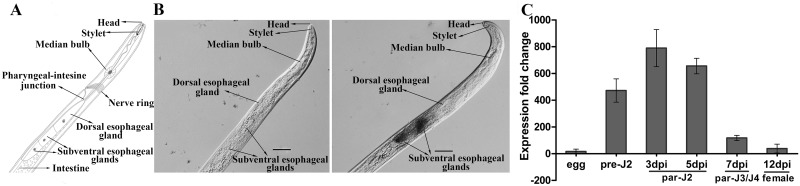
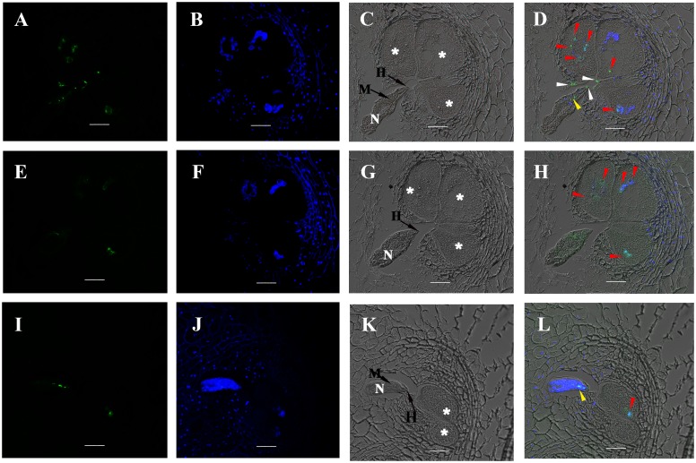
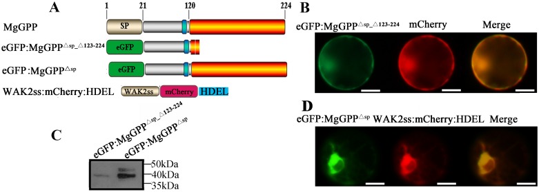
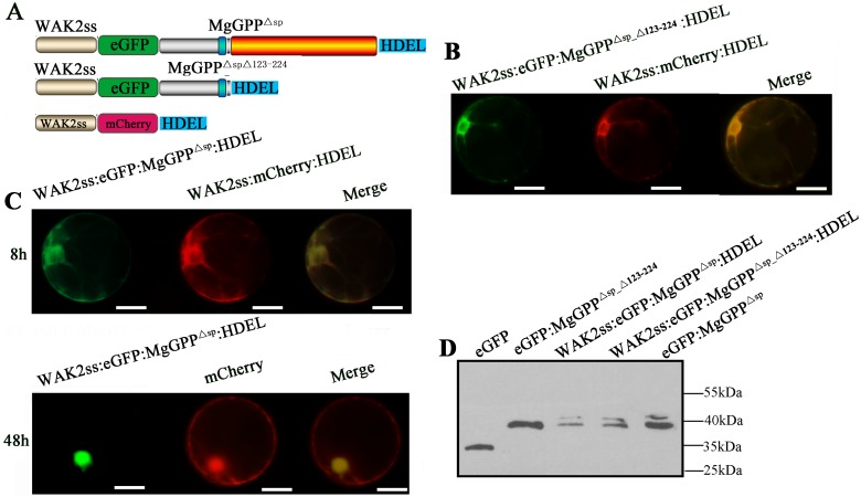
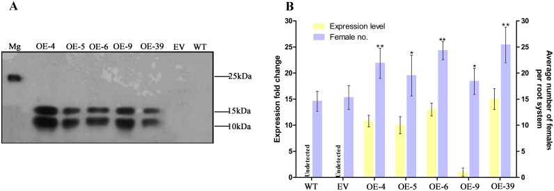
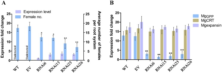
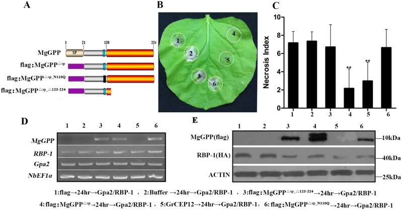
Plant pathogen effectors can recruit the host post-translational machinery to mediate their post-translational modification (PTM) and regulate their activity to facilitate parasitism, but few studies have focused on this phenomenon in the field of plant-parasitic nematodes. In this study, we show that the plant-parasitic nematode Meloidogyne graminicola has evolved a novel effector, MgGPP, that is exclusively expressed within the nematode subventral esophageal gland cells and up-regulated in the early parasitic stage of M. graminicola. The effector MgGPP plays a role in nematode parasitism. Transgenic rice lines expressing MgGPP become significantly more susceptible to M. graminicola infection than wild-type control plants, and conversely, in planta, the silencing of MgGPP through RNAi technology substantially increases the resistance of rice to M. graminicola. Significantly, we show that MgGPP is secreted into host plants and targeted to the ER, where the N-glycosylation and C-terminal proteolysis of MgGPP occur. C-terminal proteolysis promotes MgGPP to leave the ER, after which it is transported to the nucleus. In addition, N-glycosylation of MgGPP is required for suppressing the host response. The research data provide an intriguing example of in planta glycosylation in concert with proteolysis of a pathogen effector, which depict a novel mechanism by which parasitic nematodes could subjugate plant immunity and promote parasitism and may present a promising target for developing new strategies against nematode infections.


Conflict of interest statement

The authors have declared that no competing interests exist.

Figures

Fig 1
Fig 1. Expression patterns of MgGPP in Meloidogyne graminicola.
(A) Schematic representation of M. graminicola pre-J2. (B) Localization of MgGPP in the subventral esophageal glands of M. graminicola pre-J2s by in situ hybridization. Fixed nematodes were hybridized with (left) sense and (right) antisense cDNA probes from MgGPP. Scale bars, 10 μm. (C) The developmental expression pattern of MgGPP by RT-qPCR analysis in five different life stages of M. graminicola. The fold change values were calculated using the 2-ΔΔCT method and presented as the change in mRNA level at various nematode developmental stages relative to that of the egg stage. The data shown are the means of three repeats plus standard deviation (SD), and three independent experiments were performed with similar results. dpi, days post-infection; pre-J2, pre-parasitic second-stage juvenile; par-J2, par-J3 and par-J4, parasitic second-, third- and fourth-stage juveniles, respectively.
Fig 2
Fig 2. MgGPP localization in sectioned rice root galls at 5 dpi.
(A-D) Localization of the secreted MgGPP protein in the giant cell nuclei (red arrows), the cell wall of adjacent giant cells (white arrows) and the lumen of the anterior esophagus of the nematode (yellow arrows). (E-H) Localization of the secreted MgGPP protein in the giant cell nuclei (red arrows). (I-L) Localization of the secreted MgGPP protein in the giant cell nucleus (red arrow) and the lumen of the anterior esophagus of the nematode (yellow arrows). Micrographs A, E and I are observations of the Alexa Fluor 488-conjugated secondary antibody. Micrographs B, F and J are images of 4,6-diamidino-2-phenylindole (DAPI)-stained nuclei. Micrographs C, G and K are images of differential interference contrast. Micrographs D, H and L are superpositions of images of the Alexa Fluor 488-conjugated secondary antibody, DAPI-stained nuclei and differential interference contrast. N, nematode; asterisks, giant cells; M, metacorpus; H, the head of M. graminicola; Scale bars, 20 μm.
Fig 3
Fig 3. Subcellular localization of MgGPP in the rice root protoplast cells.
(A) Schematic diagram showing the fusion protein structures of MgGPP. (B) eGFP:MgGPPΔsp was transformed into rice root protoplasts. HDEL is a signal for retention in the ER, and WAK2ss-mCherry-HDEL was used as a marker to indicate the ER. The NH2-terminal signal sequence (WAK2ss) from Arabidopsis thaliana wall-associated kinase 2 was used to direct the fusion protein to secretory compartments. Signals that colocalized with the ER marker WAK2ss-mCherry-HDEL were observed in the ER at ~8 h after cotransformation, and signals that colocalized with mCherry were observed in the nuclei at ~48 h after cotransformation. (C) MgGPPΔsp:eGFP was transformed into rice root protoplasts. Signals that colocalized with mCherry were observed in the cytoplasm and nuclei at ~8 h and ~48 h after cotransformation. (D) Free eGFP was transformed into rice root protoplasts. Signals that colocalized with mCherry were observed in the whole transformed cells. (E) Using an anti-GFP antibody, western blot analysis of proteins from transformed cells showed the ~27 kDa size of MgGPPΔsp:eGFP, which was much smaller than the expected size of MgGPPΔsp:eGFP (~50 kDa) but identical to the size of eGFP, and two protein forms of ~43 and ~39 kDa of eGFP:MgGPPΔsp that were both smaller than the expected size of eGFP:MgGPPΔsp (~50 kDa). These indicated that MgGPP may be processed and cleaved. Scale bar, 50 μm.
Fig 4
Fig 4. Assays for glycosylation of MgGPP.
(A) Using an anti-MgGPP antibody, western blot analysis of proteins from pre-J2s, par-J3s/J4s and females of Meloidogyne graminicola treated with or without PNGase F all showed the ~25 kDa size band, indicating that MgGPP is not glycosylated in nematodes. (B) Using an anti-GFP antibody, western blot analysis of proteins from the transformed cells of rice and tobacco showed two protein forms of ~43 and ~39 kDa of eGFP:MgGPPΔsp, the ~39 kDa size of MgGPPΔsp:eGFP treated with PNGase A, and ~39 kDa size of the point mutation eGFP:MgGPPΔsp_N110Q, indicating that N-glycosylation of MgGPP occurred in host plants.
Fig 5
Fig 5. MgGPP123-224 is processed proteolytically in multiple loci.
(A) Schematic diagram showing the protein structures of MgGPP mutants and the bluish bar represents the glycosylation site (N110). (B) Using an anti-GFP antibody, western blot analysis of proteins from transformed cells showed the ~27 kDa size of MgGPPΔsp_Δ201–224:eGFP, MgGPPΔsp_Δ161–224:eGFP and MgGPPΔsp_Δ141–224:eGFP, which were identical to the size of eGFP, and the ~39 kDa size of MgGPPΔsp_Δ121–224:eGFP that was identical to the size of eGFP:MgGPPΔsp_Δ121–224. (C) Using an anti-GFP antibody, western blot analysis of proteins from transformed cells all showed the ~27 kDa size of MgGPPΔsp_Δ121–140:eGFP, MgGPPΔsp_Δ121–160:eGFP and MgGPPΔsp_Δ121–200:eGFP that were identical to the size of eGFP. (D) Using an anti-GFP antibody, western blot analysis of proteins from transformed cells showed the ~39 kDa size of MgGPPΔsp_Δ121–224:eGFP, MgGPPΔsp_Δ122–224:eGFP and MgGPPΔsp_Δ123–224:eGFP, and ~27 kDa size of MgGPPΔsp_Δ124–224:eGFP, MgGPPΔsp_Δ125–224:eGFP and MgGPPΔsp_Δ126–224:eGFP that were identical to the size of eGFP. These indicated that MgGPP was cleaved in multiple loci from 123 to 224 aa.
Fig 6
Fig 6. Subcellular localization of MgGPPΔsp_Δ123–224 in the rice root protoplast cells.
(A) Schematic diagram showing the protein structures of MgGPP mutants. (B) eGFP:MgGPPΔsp_Δ123–224 was transformed into rice root protoplasts. Signals that colocalized with mCherry were observed in the cytoplasm and nuclei at ~8 h after cotransformation. (C) Using an anti-GFP antibody, western blot analysis of proteins from transformed cells showed a ~39-kDa band in cells transformed with eGFP:MgGPPΔsp_Δ123–224 and two protein forms of ~43 and ~39 kDa in the cells transformed with eGFP:MgGPPΔsp. (D) As a control, eGFP:MgGPPΔsp was transformed into rice root protoplasts. Signals were observed in the ER at ~8 h after cotransformation. These indicated MgGPP lacking amino acids 123–224 could not be imported into the ER and glycosylated. Scale bar, 50 μm.
Fig 7
Fig 7. Subcellular localization of eGFP:MgGPPΔsp_Δ123–224:HDEL and eGFP:MgGPPΔsp:HDEL in the rice root protoplast cells.
(A) Schematic diagram showing the protein structures of WAK2ss:eGFP:MgGPPΔsp_Δ123–224:HDEL and WAK2ss:eGFP:MgGPPΔsp:HDEL. (B) WAK2ss:eGFP:MgGPPΔsp_Δ123–224:HDEL was transformed into rice root protoplasts. HDEL is a signal for retention in the ER, and WAK2ss-mCherry-HDEL was used as a marker to indicate the ER. The NH2-terminal signal sequence (WAK2ss) from Arabidopsis thaliana wall-associated kinase 2 was used to direct fusion proteins to secretory compartments. Signals that colocalized with the ER marker WAK2ss-mCherry-HDEL were consistently observed in the ER after cotransformation. (C) WAK2ss:eGFP:MgGPPΔsp:HDEL was transformed into rice root protoplasts. Signals that colocalized with the ER marker WAK2ss-mCherry-HDEL were observed in the ER at ~8 h after cotransformation, and signals that colocalized with mCherry were observed in the nuclei at ~48 h after cotransformation. (D) Using an anti-GFP antibody, western blot analysis of proteins from transformed cells showed two protein forms of ~43 and ~39 kDa of both eGFP:MgGPPΔsp_Δ123–224:HDEL and eGFP:MgGPPΔsp:HDEL. These indicated that the glycosylation of MgGPP occurred in the ER, and proteolysis of MgGPP123-224 led MgGPPΔsp_Δ123–224 to leave the ER. Scale bar, 50 μm.
Fig 8
Fig 8. Transgenic lines expressing MgGPP in rice exhibit enhanced susceptibility to Meloidogyne graminicola.
(A) Western blot confirmation of the MgGPP product with an anti-MgGPP antibody. Two protein forms of ~12 and ~ 16 kDa were detected because of the glycosylation and proteolysis of MgGPP. As a control, one protein of ~25 kDa was detected in M. graminicola. (B) qRT-PCR analysis was used to confirm the MgGPP mRNA expression level in transgenic-MgGPP lines. Transgenic rice expressing MgGPP showed an increased number of females in roots compared with the controls. The data are presented as the means ± standard deviation (SD) from fifteen plants. *P < 0.05; **P < 0.01, Student’s t test. OE-4, 5, 6, 9 and 39, five transgenic rice lines; Mg, M. graminicola; EV, empty vector; WT, wild type.
Fig 9
Fig 9. In planta RNAi of MgGPP attenuates Meloidogyne graminicola parasitism.
(A) qRT-PCR analysis to detect the GUS intron fragment was used to confirm dsRNA expression levels in roots of RNAi lines. Transgenic RNAi rice lines showed a decreased number of females in roots compared with the controls. The data are presented as the means ± standard deviation (SD) from fifteen plants. (B) qRT-PCR assays of the expression levels of MgGPP in M. graminicola collected from RNAi lines, transgenic empty vector (EV) plants and wild type (WT) plants. The expression levels of Mg-CRT and Mg-expansin from M. graminicola were used to determine the specificity of the MgGPP-targeting RNAi. *P < 0.05; **P < 0.01, Student’s t test. RNAi6, 15, 25 and 26, different transgenic RNAi rice lines.
Fig 10
Fig 10. Suppression of Gpa2/RBP-1-triggered cell death by MgGPP.
(A) Schematic diagram showing the protein structures of MgGPP mutants. (B) Assay of the suppression of Gpa2/RBP-1-triggered cell death in Nicotiana benthamiana by MgGPP. N. benthamiana leaves were infiltrated with buffer or Agrobacterium tumefaciens cells carrying flag:MgGPPΔsp_Δ123–224, flag:MgGPPΔsp, GrCEP12, flag:MgGPPΔsp_N110Q and the flag control gene, followed 24 h later with A. tumefaciens cells carrying the Gpa2/RBP-1 genes. The cell death phenotype was scored and photographs were taken 5 days after the last infiltration. (C) The average areas of cell death of in leaves infected with MgGPP and other proteins followed by Gpa2/RBP-1. The columns with asterisks indicates a highly statistically significant reduction of the necrosis index of MgGPP and GrCEP12 compared with that of the negative control flag. Each column represents the mean with the standard deviation (n = 55). **P<0.01, Student’s t test. (D) RT-PCR confirmation of the expression of MgGPP, RBP-1 and Gpa2. (E) Western blot analysis was used to confirm the expression of RBP-1, MgGPP and MgGPP mutants with an anti-GFP antibody.

Similar articles

Cited by

References

    1. Blok VC, Jones JT, Phillips MS, Trudgill DL. Parasitism genes and host range disparities in biotrophic nematodes: the conundrum of polyphagy versus specialisation. Bioessays. 2008;30(3):249–59. 10.1002/bies.20717 - DOI - PubMed
    1. Trudgill DL, Blok VC. Apomictic, polyphagous root-knot nematodes: exceptionally successful and damaging biotrophic root pathogens. Annu Rev Phytopathol. 2001;39:53–77. 10.1146/annurev.phyto.39.1.53 - DOI - PubMed
    1. Caboni P, Ntalli NG, Aissani N, Cavoski I, Angioni A. Nematicidal activity of (E,E)-2,4-decadienal and (E)-2-decenal from Ailanthus altissima against Meloidogyne javanica. J Agric Food Chem. 2012;60(4):1146–51. 10.1021/jf2044586 - DOI - PubMed
    1. Davis EL, Hussey RS, Baum TJ. Getting to the roots of parasitism by nematodes. Trends Parasitol. 2004;20(3):134–41. 10.1016/j.pt.2004.01.005 - DOI - PubMed
    1. Gheysen G, Mitchum MG. How nematodes manipulate plant development pathways for infection. Curr Opin Plant Biol. 2011;14(4):415–21. 10.1016/j.pbi.2011.03.012 - DOI - PubMed

MeSH terms

Substances

LinkOut - more resources