. 2017 Apr 12;12(4):e0173873.
doi: 10.1371/journal.pone.0173873. eCollection 2017.

Spontaneous diseases in captive ratites (Struthioniformes) in northwestern Germany: A retrospective study

Affiliations

Spontaneous diseases in captive ratites (Struthioniformes) in northwestern Germany: A retrospective study

Aimara Bello et al. PLoS One. .

Abstract

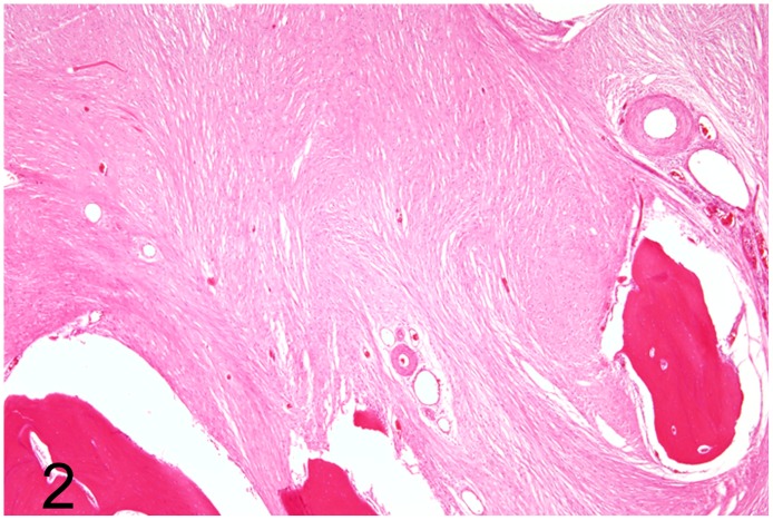
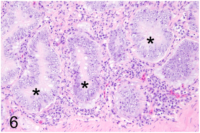
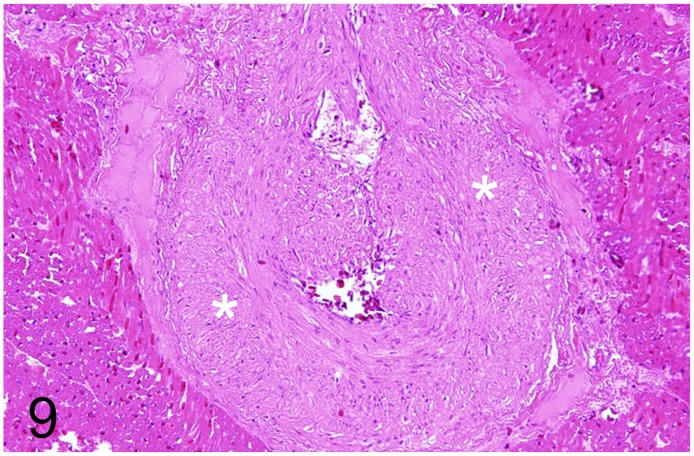
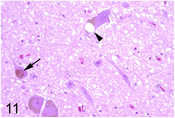
A retrospective study was carried out to define the spectrum of spontaneous diseases in ostriches and few other captive ratites, order Struthioniformes, in northwestern Germany. The investigation included 71 ratites necropsied between 1968 and 2014. They consisted of 54 ostriches, 5 emus, and 12 rheas with 37 adults, 23 juveniles and 11 neonates and embryonated eggs. Necropsy reports were reviewed, histologic preparations were re-examined and additional histochemical and immunohistochemical stains were carried out in selected cases. In many animals more than one morphologic diagnosis attributable to different disease processes was found. In adult animals (n = 37), the most commonly altered organ systems were the musculoskeletal system (49%), the digestive system (46%), and the cardiovascular system (46%) affected by traumatic lesions, inflammatory and degenerative changes, respectively. A spongy degeneration was found in the brain (35%); however, immunohistochemistry and western blotting failed to detect pathological prion protein. In juvenile animals (n = 23), the musculoskeletal (44%) and the digestive system (43%) were mainly affected by traumatic and inflammatory lesions, respectively. In embryonated eggs and neonates (n = 11) the major cause of death was circulatory failure associated with generalized subcutaneous edema as described for improper incubation conditions (64%). Summarized, most of the findings observed in adult and juvenile ratites in northwestern Germany are related to trauma, inflammatory and degenerative disorders, whereas death in embryonated eggs and neonates was most likely related to breeding conditions. A spongy encephalopathy awaits further studies to elucidate cause and pathogenesis.


Conflict of interest statement

Competing Interests: The authors have declared that no competing interests exist.

Figures

Fig 1
Fig 1. Intraosseous fibrosarcoma, left tarso-metatarsal bone, emu, case No. 53.
Adjacent to the gray-white neoplastic tissue that occupies partially the marrow cavity (asterisk) the oblique fracture line of the bone is visible (arrow).
Fig 2
Fig 2. Intraosseous fibrosarcoma, left tarso-metatarsal bone, emu, case No. 53.
The fibrosarcoma is composed of spindle-shaped neoplastic cells embedded in abundant amounts of collagen fibers. Hematoxylin and eosin (HE).
Fig 3
Fig 3. Proliferative enteritis caused by Lawsonia intracellularis, intestine, rhea, case No. 41.
Red-brown discoloration of the intestinal mucosa with fleshy appearance of the duodenal wall. P = pancreas.
Fig 4
Fig 4. Proliferative enteritis caused by Lawsonia intracellularis, intestine, rhea, case No. 41.
Marked thickening of the intestinal mucosa (asterisks).
Fig 5
Fig 5. Proliferative enteritis caused by Lawsonia intracellularis, intestine, rhea, case No. 41.
Subgross magnification reveals severe elongation of the intestinal crypts. HE.
Fig 6
Fig 6. Proliferative enteritis caused by Lawsonia intracellularis, intestine, rhea, case No. 41.
Severe crypt hyperplasia (asterisks) and moderate inflammatory infiltration of the adjacent lamina propria. HE.
Fig 7
Fig 7. Fibrino-necrotizing typhlocolitis caused by Brachyspira sp., intestine, rhea, case No. 31.
Brown-greenish tinged fibrin attached to the cecal mucosa (arrows).
Fig 8
Fig 8. Fibrino-necrotizing typhlocolitis caused by Brachyspira sp., intestine, rhea, case No. 31.
Multifocal fibrino-necrotic mucosal lesions (asterisks). HE. Inset: Immunolabelling of spirochetes using a polyclonal antiserum against B. hyodysenteriae. Immunofluorescence.
Fig 9
Fig 9. Hypertrophy and hyperplasia of the tunica media, cardiac artery, rhea, case No. 22.
Marked increase in thickness of the tunica media (asterisks). HE.
Fig 10
Fig 10. Spongy degeneration, brain, ostrich, case No. 19.
Oval to spherical vacuoles of variable diameter (arrows) are present in the perikaryon of several neurons of the red nucleus. HE.
Fig 11
Fig 11. Spongy degeneration and lipofuscin deposition, brain, ostrich, case No. 34.
Oval vacuole (arrowhead) and deposition of a coarse granular golden-brown pigment (arrow) in the perikaryon of some brain stem neurons. HE.
Fig 12
Fig 12. Spongy degeneration and lipofuscin deposition, brain, ostrich, case No. 34.
PAS-positive coarse granular pigment deposition suggestive of lipofuscin (arrow) in a brain stem neuron. PAS-reaction.
Fig 13
Fig 13. Avian influenza, larynx and trachea, rhea, case No. 62.
Severe necro-suppurative laryngo-tracheitis with diffuse hyperemia and numerous white, necrotic foci.
Fig 14
Fig 14. Avian influenza, trachea, rhea, case No. 62.
Severe, diffuse, necro-suppurative tracheitis. HE.
Fig 15
Fig 15. Avian influenza, trachea, rhea, case No. 62.
Intralesional immunolabelling of influenza A virus nucleoprotein (arrows). Immunohistochemistry.
Fig 16
Fig 16. Avian mycobacteriosis and aspergillosis, trachea, emu, case No. 63.
Multifocal yellow granulomas (arrows) in the trachea caused by M. avium subsp. avium and focal white granuloma caused by Aspergillus sp. (arrowhead).
Fig 17
Fig 17. Avian mycobacteriosis and aspergillosis, lung, emu, case No. 63.
Necro-granulomatous pneumonia. HE.
Fig 18
Fig 18. Neonatal death, subcutis, ostrich, case No. 35.
Generalized subcutaneous edema (anasarca) and purulent omphalitis (arrow).

Similar articles

Cited by

References

    1. IUCN. The IUCN Red List of Threatened Species: International Union for Conservation of Nature and Natural Resources 2016 [cited 09/25/2016]. http://www.iucnredlist.org.
    1. Reiner G. Straußenhaltung in Deutschland—eine Übersicht (Teil I) [In German]: Lohmann Information; 2000. www.lohmann-information.com/archive_year_2000.html.
    1. Wöhr AC, Schulz A, Erhard MH. Tierschutzaspekte bei der Haltung von Zuchtstraußen in Deutschland [in German]. DTW Deutsche tierarztliche Wochenschrift. 2005;112(3):87–91. - PubMed
    1. Korbel R, Schubert M, Erhard M, Wöhr C, Bergmann S, Rückschloss S, et al. Betrachtungen und Empfehlungen zur artgemäßen und tierschutzgerechten Haltung von Straußenvögeln in Deutschland [in German]. Tierärztliche Praxis (G). 2015;43(4):232–44. - PubMed
    1. Anonymous. Vom Geschäft mit der afrikanischen "Primaballerina": Welt; 2013. https://www.welt.de/newsticker/news3/article113478763/Vom-Geschaeft-mit-....

LinkOut - more resources