

Mobile Genome Express (MGE): A comprehensive automatic genetic analyses pipeline with a mobile device
- PMID: 28403238
- PMCID: PMC5389793
- DOI: 10.1371/journal.pone.0174696
Mobile Genome Express (MGE): A comprehensive automatic genetic analyses pipeline with a mobile device
Abstract
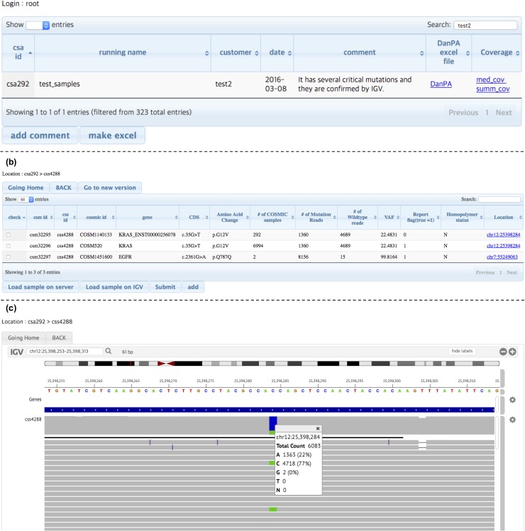
The development of next-generation sequencing (NGS) technology allows to sequence whole exomes or genome. However, data analysis is still the biggest bottleneck for its wide implementation. Most laboratories still depend on manual procedures for data handling and analyses, which translates into a delay and decreased efficiency in the delivery of NGS results to doctors and patients. Thus, there is high demand for developing an automatic and an easy-to-use NGS data analyses system. We developed comprehensive, automatic genetic analyses controller named Mobile Genome Express (MGE) that works in smartphones or other mobile devices. MGE can handle all the steps for genetic analyses, such as: sample information submission, sequencing run quality check from the sequencer, secured data transfer and results review. We sequenced an Actrometrix control DNA containing multiple proven human mutations using a targeted sequencing panel, and the whole analysis was managed by MGE, and its data reviewing program called ELECTRO. All steps were processed automatically except for the final sequencing review procedure with ELECTRO to confirm mutations. The data analysis process was completed within several hours. We confirmed the mutations that we have identified were consistent with our previous results obtained by using multi-step, manual pipelines.
Conflict of interest statement
Figures


Similar articles
-
ClinQC: a tool for quality control and cleaning of Sanger and NGS data in clinical research.BMC Bioinformatics. 2016 Feb 2;17:56. doi: 10.1186/s12859-016-0915-y. BMC Bioinformatics. 2016. PMID: 26830926 Free PMC article.
-
TOGGLE: toolbox for generic NGS analyses.BMC Bioinformatics. 2015 Nov 9;16:374. doi: 10.1186/s12859-015-0795-6. BMC Bioinformatics. 2015. PMID: 26552596 Free PMC article.
-
Clinical evaluation of panel testing by next-generation sequencing (NGS) for gene mutations in myeloid neoplasms.Diagn Pathol. 2016 Jan 22;11:11. doi: 10.1186/s13000-016-0456-8. Diagn Pathol. 2016. PMID: 26796102 Free PMC article.
-
Introduction to the analysis of next generation sequencing data and its application to venous thromboembolism.Thromb Haemost. 2015 Nov;114(5):920-32. doi: 10.1160/TH15-05-0411. Epub 2015 Oct 8. Thromb Haemost. 2015. PMID: 26446408 Review.
-
A review of bioinformatic methods for forensic DNA analyses.Forensic Sci Int Genet. 2018 Mar;33:117-128. doi: 10.1016/j.fsigen.2017.12.005. Epub 2017 Dec 12. Forensic Sci Int Genet. 2018. PMID: 29247928 Review.
Cited by
-
Identification of the Rice Wines with Different Marked Ages by Electronic Nose Coupled with Smartphone and Cloud Storage Platform.Sensors (Basel). 2017 Oct 31;17(11):2500. doi: 10.3390/s17112500. Sensors (Basel). 2017. PMID: 29088076 Free PMC article.
References
-
- Watson JD, Crick FHC. Molecular structure of nucleic acids. Nature. 1953. pp. 737–738. - PubMed
-
- Watson JD, Crick FH. The structure of DNA. Cold Spring Harb Symp Quant Biol. 1953;18: 123–131. - PubMed
-
- Lee MS, Chang KS, Cabanillas F, Freireich EJ, Trujillo JM, Stass SA. Detection of minimal residual cells carrying the t(14;18) by DNA sequence amplification. Science (80-). 1987;237: 175–178. - PubMed
MeSH terms
LinkOut - more resources
Full Text Sources
Other Literature Sources
