. 2017 Jun 1;74(6):622-631.
doi: 10.1001/jamapsychiatry.2017.0329.

Skin Conductance Responses and Neural Activations During Fear Conditioning and Extinction Recall Across Anxiety Disorders

Affiliations

Skin Conductance Responses and Neural Activations During Fear Conditioning and Extinction Recall Across Anxiety Disorders

Marie-France Marin et al. JAMA Psychiatry. .

Abstract

Importance: The fear conditioning and extinction neurocircuitry has been extensively studied in healthy and clinical populations, with a particular focus on posttraumatic stress disorder. Despite significant overlap of symptoms between posttraumatic stress disorder and anxiety disorders, the latter has received less attention. Given that dysregulated fear levels characterize anxiety disorders, examining the neural correlates of fear and extinction learning may shed light on the pathogenesis of underlying anxiety disorders.

Objectives: To investigate the psychophysiological and neural correlates of fear conditioning and extinction recall in anxiety disorders and to document how these features differ as a function of multiple diagnoses or anxiety severity.

Design, setting, and participants: This investigation was a cross-sectional, case-control, functional magnetic resonance imaging study at an academic medical center. Participants were healthy controls and individuals with at least 1 of the following anxiety disorders: generalized anxiety disorder, social anxiety disorder, specific phobia, and panic disorder. The study dates were between March 2013 and May 2015.

Exposures: Two-day fear conditioning and extinction paradigm.

Main outcomes and measures: Skin conductance responses, blood oxygenation level-dependent responses, trait anxiety scores from the State Trait Anxiety Inventory-Trait Form, and functional connectivity.

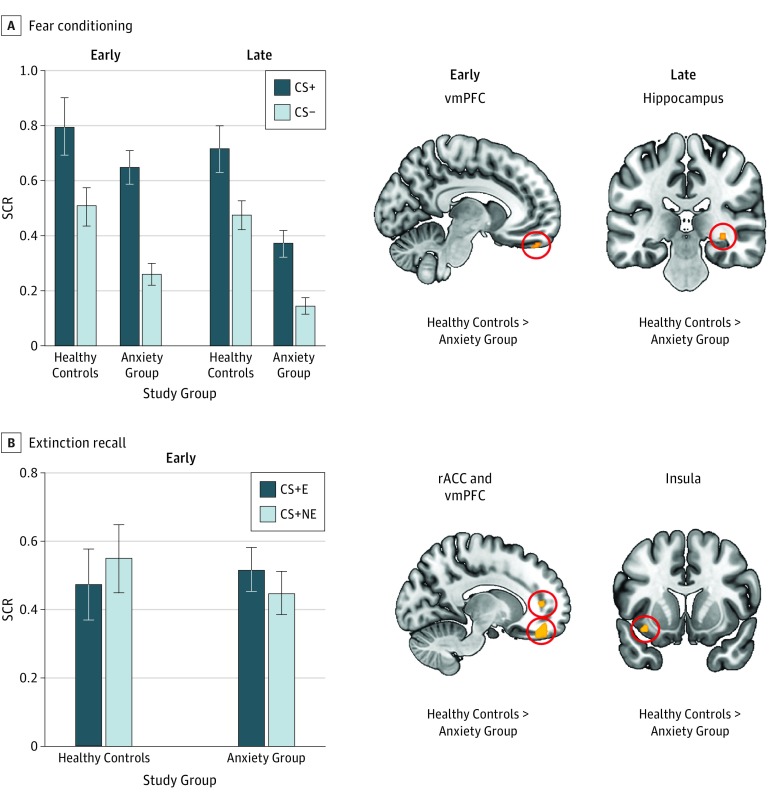
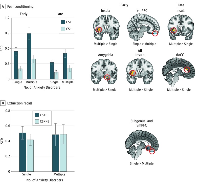
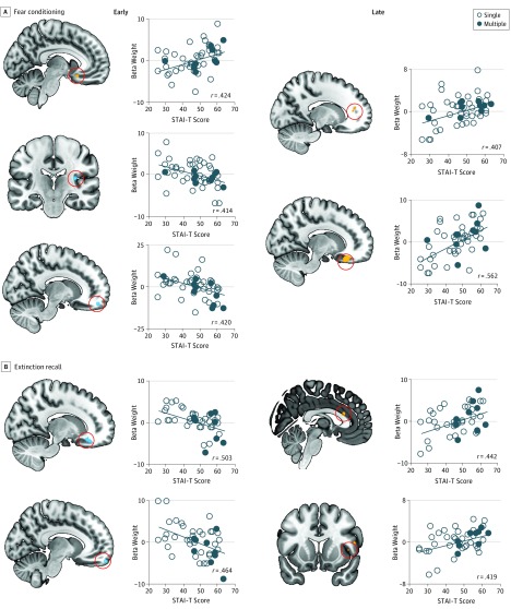
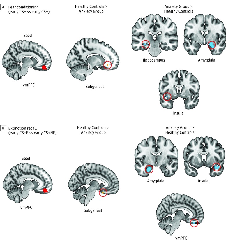
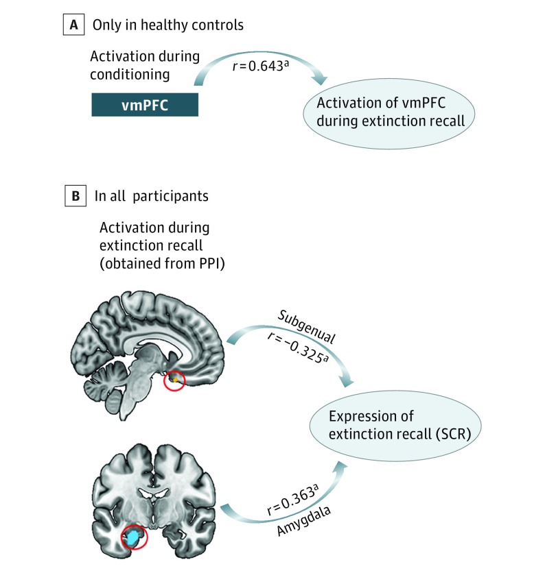
Results: This study included 21 healthy controls (10 women) and 61 individuals with anxiety disorders (36 women). P values reported for the neuroimaging results are all familywise error corrected. Skin conductance responses during extinction recall did not differ between individuals with anxiety disorders and healthy controls (ηp2 = 0.001, P = .79), where ηp2 is partial eta squared. The anxiety group had lower activation of the ventromedial prefrontal cortex (vmPFC) during extinction recall (ηp2 = 0.178, P = .02). A similar hypoactive pattern was found during early conditioning (ηp2 = 0.106, P = .009). The vmPFC hypoactivation was associated with anxiety symptom severity (r = -0.420, P = .01 for conditioning and r = -0.464, P = .004 for extinction recall) and the number of co-occuring anxiety disorders diagnosed (ηp2 = 0.137, P = .009 for conditioning and ηp2 = 0.227, P = .004 for extinction recall). Psychophysiological interaction analyses revealed that the fear network connectivity differed between healthy controls and the anxiety group during fear learning (ηp2 range between 0.088 and 0.176 and P range between 0.02 and 0.003) and extinction recall (ηp2 range between 0.111 and 0.235 and P range between 0.02 and 0.002).

Conclusions and relevance: Despite no skin conductance response group differences during extinction recall, brain activation patterns between anxious and healthy individuals differed. These findings encourage future studies to examine the conditions longitudinally and in the context of treatment trials to improve and guide therapeutics via advanced neurobiological understanding of each disorder.


Conflict of interest statement

Conflict of Interest Disclosures: None reported.

Figures

Figure 1.
Figure 1.. Comparison of Healthy Controls and Individuals With Anxiety Disorders During Fear Conditioning and Extinction Recall
Shown are skin conductance response (SCR) and voxelwise analyses contrasting healthy controls with individuals with anxiety disorders. A, On the left, SCR is shown during early and late conditioning as a function of conditioned stimuli (CS) type and group. On the right, significant group differences are shown in terms of brain activation during the early CS+ vs CS− contrast and late CS+ vs CS− contrast from fear conditioning. B, On the left, SCR is shown during early extinction recall as a function of CS type and group. On the right, brain regions are shown for which the 2 groups differed during extinction recall for the CS + E vs CS + NE contrast. Hot colors indicate greater activation in healthy controls relative to the anxiety group. P = .005 is used for all results. Error bars are SEM. CS+ indicates cues that were partially reinforced with a shock; CS−, cue that was never reinforced with a shock; CS + E, extinguished CS+; CS + NE, nonextinguished CS+; rACC, rostral anterior cingulate cortex; and vmPFC, ventromedial prefrontal cortex.
Figure 2.
Figure 2.. Comparison of One Anxiety Disorder With Multiple Anxiety Disorders During Fear Conditioning and Extinction Recall
Shown are skin conductance response (SCR) and voxelwise analyses contrasting individuals diagnosed as having one anxiety disorder with those diagnosed as having multiple anxiety disorders. A, On the left, SCR is shown during early and late conditioning as a function of conditioned stimuli (CS) type and group. On the right, significant group differences are shown in terms of brain activation during the early CS+ vs CS− contrast, late CS+ vs CS− contrast, and all CS+ vs CS− contrast from fear conditioning. B, On the left, SCR is shown during early extinction recall as a function of CS type and group. On the right, the brain region is shown for which the 2 groups differed during extinction recall for the CS + E vs CS + NE contrast. Hot colors indicate greater activation in the multiple disorders group relative to the single disorder group. Cold colors indicate greater activation in the single disorder group relative to the multiple disorders group. P = .005 is used for all results. Error bars are SEM. CS+ indicates cues that were partially reinforced with a shock; CS−, cue that was never reinforced with a shock; CS + E, extinguished CS+; CS + NE, nonextinguished CS+; dACC, dorsal anterior cingulate cortex; and vmPFC, ventromedial prefrontal cortex.
Figure 3.
Figure 3.. Trait Anxiety Prediction of Brain Activation During Fear Conditioning and Extinction Recall in the Anxiety Group
Shown are voxelwise analyses performed in the anxiety group only with trait anxiety scores as the regressor. A, The regions of the fear network are shown that demonstrated significant association with trait anxiety scores during early and late fear conditioning. B, The regions of the fear network are shown that demonstrated significant associations with trait anxiety scores during early extinction recall. STAI-T indicates State Trait Anxiety Inventory–Trait Form.
Figure 4.
Figure 4.. Psychophysiological Interactions With Ventromedial Prefrontal Cortex (vmPFC) as the Seed During Fear Conditioning and Extinction Recall
Shown are between-group psychophysiological interaction analyses contrasting healthy controls with individuals with anxiety disorders during early fear conditioning (A) and extinction recall (B). Hot colors indicate that the healthy controls exhibited greater connectivity than the anxiety group between the seed and the given region. Cold colors indicate that the anxiety group exhibited greater connectivity than the healthy controls between the seed and the given region. P = .005 is used for all results. CS indicates conditioned stimuli; CS+ indicates cues that were partially reinforced with a shock; CS−, the cue that was never reinforced with a shock; CS + E, extinguished CS+; and CS + NE, nonextinguished CS+.
Figure 5.
Figure 5.. Exploratory Analyses
A, Activation in the ventromedial prefrontal cortex (vmPFC) during early fear conditioning is associated with vmPFC activation during extinction recall. This association was only significant in healthy controls and failed to reach significance in the anxiety group. B, Shown are significant associations between brain regions demonstrating significant psychophysiological interaction (PPI) between-group differences during extinction recall and skin conductance response (SCR) during extinction recall. Expression of extinction recall is the computation of SCR to the first 4 trials of extinguished conditioned stimuli minus SCR to the first 4 trials of nonextinguished conditioned stimuli.
aP < .05.

Similar articles

Cited by

References

    1. Shin LM, Liberzon I. The neurocircuitry of fear, stress, and anxiety disorders. Neuropsychopharmacology. 2010;35(1):169-191. - PMC - PubMed
    1. VanElzakker MB, Dahlgren MK, Davis FC, Dubois S, Shin LM. From Pavlov to PTSD: the extinction of conditioned fear in rodents, humans, and anxiety disorders. Neurobiol Learn Mem. 2014;113:3-18. - PMC - PubMed
    1. Milad MR, Quirk GJ. Fear extinction as a model for translational neuroscience: ten years of progress. Annu Rev Psychol. 2012;63:129-151. - PMC - PubMed
    1. Yehuda R, LeDoux J. Response variation following trauma: a translational neuroscience approach to understanding PTSD. Neuron. 2007;56(1):19-32. - PubMed
    1. Pitman RK, Rasmusson AM, Koenen KC, et al. . Biological studies of post-traumatic stress disorder. Nat Rev Neurosci. 2012;13(11):769-787. - PMC - PubMed

Publication types

MeSH terms