Review
. 2018 Feb;24(1):73-83.
doi: 10.1177/1073858417703033. Epub 2017 Apr 12.

Mechanisms Mediating Functional Hyperemia in the Brain

Affiliations
Review

Mechanisms Mediating Functional Hyperemia in the Brain

Amy R Nippert et al. Neuroscientist. 2018 Feb.

Abstract

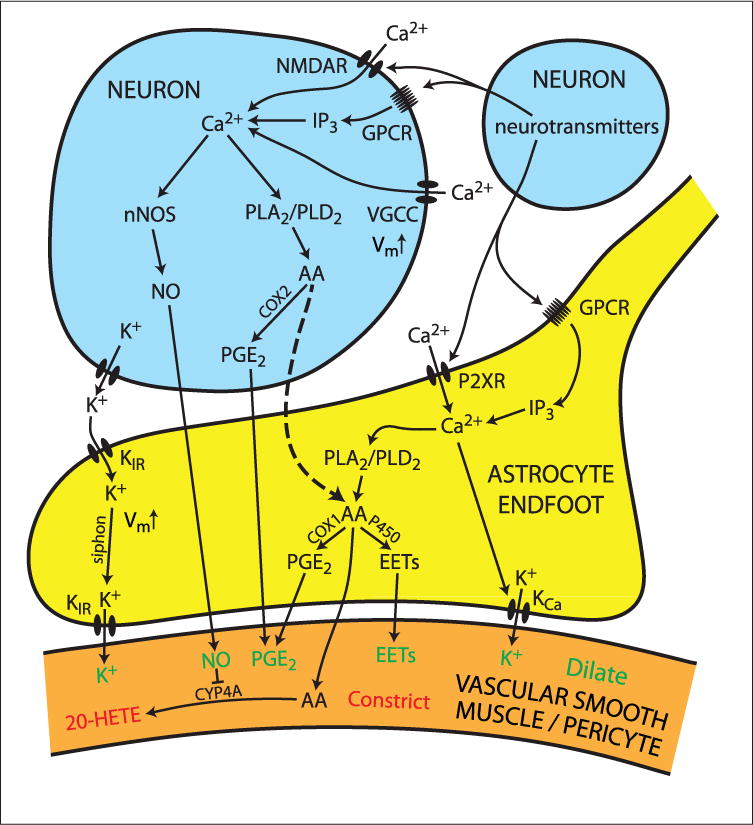
Neuronal activity within the brain evokes local increases in blood flow, a response termed functional hyperemia. This response ensures that active neurons receive sufficient oxygen and nutrients to maintain tissue function and health. In this review, we discuss the functions of functional hyperemia, the types of vessels that generate the response, and the signaling mechanisms that mediate neurovascular coupling, the communication between neurons and blood vessels. Neurovascular coupling signaling is mediated primarily by the vasoactive metabolites of arachidonic acid (AA), by nitric oxide, and by K+. While much is known about these pathways, many contentious issues remain. We highlight two controversies, the role of glial cell Ca2+ signaling in mediating neurovascular coupling and the importance of capillaries in generating functional hyperemia. We propose signaling pathways that resolve these controversies. In this scheme, capillary dilations are generated by Ca2+ increases in astrocyte endfeet, leading to production of AA metabolites. In contrast, arteriole dilations are generated by Ca2+ increases in neurons, resulting in production of nitric oxide and AA metabolites. Arachidonic acid from neurons also diffuses into astrocyte endfeet where it is converted into additional vasoactive metabolites. While this scheme resolves several discrepancies in the field, many unresolved challenges remain and are discussed in the final section of the review.

Keywords: EETs; PGE2; arachidonic acid; astrocyte; cerebral blood flow; functional hyperemia; neurovascular coupling.


Conflict of interest statement

Declaration of Conflicting Interests

The author(s) declared no potential conflicts of interest with respect to the research, authorship, and/or publication of this article.

Figures

Figure 1
Figure 1
Interactions between cells of the neurovascular unit regulate blood flow in the brain. The diameter of arterioles and arteries is controlled by the contractile state of smooth muscle cells, which form one or more continuous layers around the vessels. Capillary diameter is controlled by pericytes, whose longitudinal and circumferential contractile processes envelop capillaries. Astrocyte endfeet almost completely surround arterioles and capillaries and their contractile cells. Chemicals released from both astrocyte endfeet and neurons control the contractile state of smooth muscle cells and pericytes.
Figure 2
Figure 2
Astrocytes are well suited to mediate signaling from neurons to blood vessels. This 1899 drawing by Santiago Ramon y Cajal illustrates that astrocytes, the darkly colored cells (A, B), form extensive contacts with neurons, the lightly colored cells (C, D), and with blood vessels (F). Astrocyte processes surround many synapses in the brain and generate intracellular Ca2+ increases in response to transmitter release from these synapses. Astrocyte endfeet envelope capillaries and arterioles. Astrocyte Ca2+ increases trigger the synthesis and release of vasoactive agents from the cell endfeet onto capillaries, inducing vessel dilation. Drawing courtesy of the Cajal Institute-CSIC, Madrid, Spain.
Figure 3
Figure 3
Metabolites of arachidonic acid dilate and constrict blood vessels. Calcium increases in both astrocytes and neurons activate phospholipase A2 and D2, leading to the conversion of membrane phospholipids to arachidonic acid. Arachidonic acid, in turn, is converted to the vasodilators EETs and PGE2. Arachidonic acid is also converted to the vasoconstrictor 20-HETE. Nitric oxide (NO) inhibits the synthesis of 20-HETE and EETs.
Figure 4
Figure 4
Proposed neurovascular coupling pathways mediating vasodilation of capillaries and arterioles. At capillaries, neurotransmitters released from active neurons evoke Ca2+ increases in astrocyte endfeet by activation of metabotropic (GPCR) and ionotropic (P2XR) receptors. These Ca2+ increases result in the synthesis and release of vasodilating PGE2 and EETs onto pericytes surrounding capillaries. Calcium increases may also open Ca2+-activated K+ channels (KCa), leading to the release of vasodilating K+. At arterioles, Ca2+ increases are evoked in neurons though activation of GPCRs, NMDA receptors (NMDAR) and though depolarization and activation of voltage-gated Ca2+ channels (VGCC). These Ca2+ increases result in the synthesis and release of nitric oxide (NO) through activation of neuronal nitric oxide synthase (nNOS) and PGE2. These vasodilators diffuse through astrocyte endfeet onto vascular smooth muscle cells surrounding arterioles. Nitric oxide also inhibits the synthesis of vasoconstricting 20-HETE within vascular smooth muscle cells, leading to vasodilation. Arachidonic acid (AA) produced in neurons also diffuses into astrocyte endfeet (dashed line) where it will be converted into PGE2 and EETs. In this way neuronal Ca2+ increases trigger the synthesis and release of vasodilators from astrocyte endfeet. Dilation of both capillaries and arterioles may also be mediated by a siphoning mechanism, where the release of K+ from active neurons depolarizes astrocytes, leading to the release of K+ onto vessels.

References

    1. Akgoren N, Dalgaard P, Lauritzen M. Cerebral blood flow increases evoked by electrical stimulation of rat cerebellar cortex: relation to excitatory synaptic activity and nitric oxide synthesis. Brain Res. 1996;710:204–14. - PubMed
    1. Alkayed NJ, Birks EK, Hudetz AG, Roman RJ, Henderson L, Harder DR. Inhibition of brain P-450 arachidonic acid epoxygenase decreases baseline cerebral blood flow. Am J Physiol. 1996a;271:H1541–6. - PubMed
    1. Alkayed NJ, Narayanan J, Gebremedhin D, Medhora M, Roman RJ, Harder DR. Molecular characterization of an arachidonic acid epoxygenase in rat brain astrocytes. Stroke. 1996b;27:971–9. - PubMed
    1. Anderson AW, Marois R, Colson ER, Peterson BS, Duncan CC, Ehrenkranz RA, et al. Neonatal auditory activation detected by functional magnetic resonance imaging. Magn Reson Imaging. 2001;19:1–5. - PubMed
    1. Anenberg E, Chan AW, Xie Y, LeDue JM, Murphy TH. Optogenetic stimulation of GABA neurons can decrease local neuronal activity while increasing cortical blood flow. J Cereb Blood Flow Metab. 2015;35:1579–86. - PMC - PubMed

Publication types