

A Dalbergia odorifera extract improves the survival of endotoxemia model mice by inhibiting HMGB1 release
- PMID: 28403838
- PMCID: PMC5389052
- DOI: 10.1186/s12906-017-1725-0
A Dalbergia odorifera extract improves the survival of endotoxemia model mice by inhibiting HMGB1 release
Abstract
Background: Dalbergia odorifera T. Chen (Leguminosae) is an indigenous medicinal herb that is widely used as a popular remedy in northern and eastern Asia. However, the cellular mechanisms underlying the biological activity of D. odorifera are not fully elucidated.
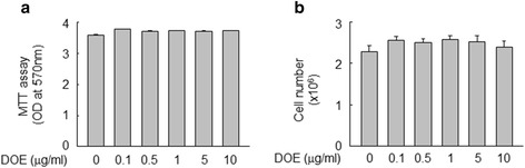
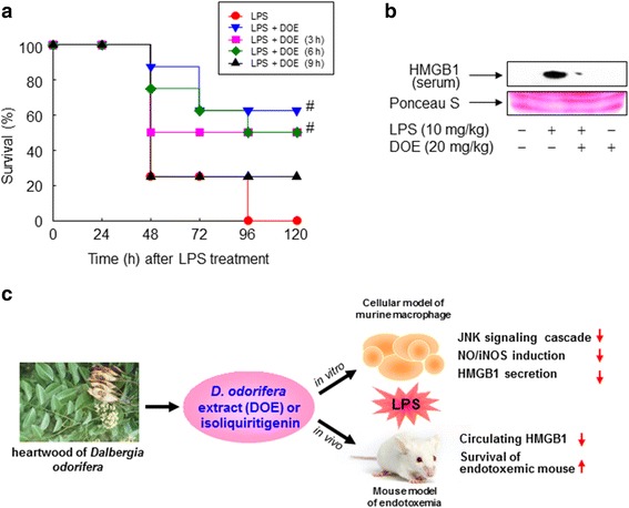
Methods: Anti-inflammatory effect of D. odorifera extract (DOE) was determined through intraperitoneal injection in a mouse model of endotoxemia induced by lipopolysaccharide (LPS). RAW 264.7 cells, a murine macrophage, were also treated with LPS to generate a cellular model of inflammation, and investigated the anti-inflammatory activity and underlying mechanisms of DOE and its constituent isoliquiritigenin.
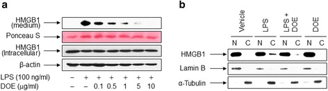
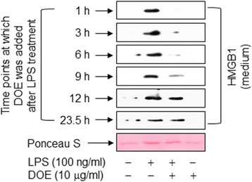
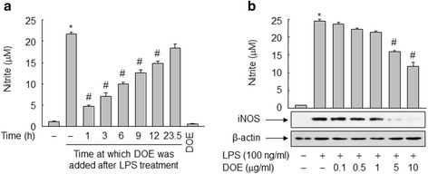
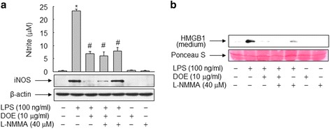
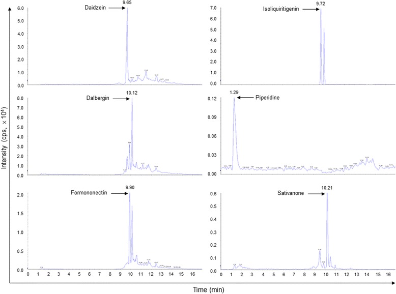
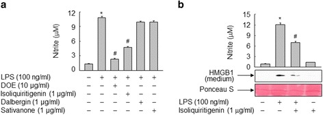
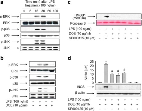
Results: DOE dose-dependently inhibited LPS-induced release of high mobility group box 1 (HMGB1), a late proinflammatory cytokine, and decreased cytosolic translocation of HMGB1 in RAW264.7 cells. This inhibitory effect of DOE on HMGB1 release was observed in cells treated with DOE before or after LPS treatment, suggesting that DOE is effective for both treatment and prevention. In addition, DOE significantly inhibited LPS-induced formation of nitric oxide (NO) and expression of inducible NO synthase (iNOS) in a dose-dependent manner. These effects of DOE were accompanied by suppression of HMGB1 release triggered by LPS, suggesting a possible mechanism by which DOE modulates HMGB1 release through NO signaling. Isoriquiritigenin, a constituent of DOE, also attenuated LPS-triggered NO formation and HMGB1 release in RAW264.7 cells, indicating that isoriquiritigenin is an indexing molecule for the anti-inflammatory properties of DOE. Furthermore, c-Jun N-terminal kinase, but not extracellular signal-regulated kinase and p38, mediated DOE-dependent inhibition of HMGB1 release and NO/iNOS induction in RAW 264.7 cells exposed to LPS. Notably, administration of DOE ameliorated survival rates in a mouse model of endotoxemia induced by LPS, where decreased level of circulating HMGB1 was observed.
Conclusion: These results suggest that DOE confers resistance to LPS-triggered inflammation through NO-mediated inhibitory effects on HMGB1 release.
Keywords: Dalbergia Odorifera; Endotoxemia; HMGB1; Inflammation; Nitric oxide.
Figures
Similar articles
-
A Review on the Medicinal Plant Dalbergia odorifera Species: Phytochemistry and Biological Activity.Evid Based Complement Alternat Med. 2017;2017:7142370. doi: 10.1155/2017/7142370. Epub 2017 Dec 4. Evid Based Complement Alternat Med. 2017. PMID: 29348771 Free PMC article. Review.
-
Ginseng leaf extract ameliorates the survival of endotoxemic mice by inhibiting the release of high mobility group box 1.J Food Biochem. 2021 Jul;45(7):e13805. doi: 10.1111/jfbc.13805. Epub 2021 Jun 6. J Food Biochem. 2021. PMID: 34096077
-
[Anti-inflammation of flavonoid compounds from Dalbergia odorifera T. Chen in lipopolysaccharide stimulated RAW264.7 macrophages].Xi Bao Yu Fen Zi Mian Yi Xue Za Zhi. 2013 Jul;29(7):681-4. Xi Bao Yu Fen Zi Mian Yi Xue Za Zhi. 2013. PMID: 23837974 Chinese.
-
Subcritical water-hydrolyzed fish collagen ameliorates survival of endotoxemic mice by inhibiting HMGB1 release in a HO-1-dependent manner.Biomed Pharmacother. 2017 Sep;93:923-930. doi: 10.1016/j.biopha.2017.07.041. Epub 2017 Jul 13. Biomed Pharmacother. 2017. PMID: 28715873
-
Extracellular HMGB1 as a proinflammatory cytokine.J Interferon Cytokine Res. 2004 Jun;24(6):329-33. doi: 10.1089/107999004323142187. J Interferon Cytokine Res. 2004. PMID: 15212706 Review.
Cited by
-
Function of the R2R3-MYB Transcription Factors in Dalbergia odorifera and Their Relationship with Heartwood Formation.Int J Mol Sci. 2023 Aug 4;24(15):12430. doi: 10.3390/ijms241512430. Int J Mol Sci. 2023. PMID: 37569814 Free PMC article.
-
Genetic Diversity and Population Structure Analysis of DalbergiaOdorifera Germplasm and Development of a Core Collection Using Microsatellite Markers.Genes (Basel). 2019 Apr 6;10(4):281. doi: 10.3390/genes10040281. Genes (Basel). 2019. PMID: 30959931 Free PMC article.
-
A Review on the Medicinal Plant Dalbergia odorifera Species: Phytochemistry and Biological Activity.Evid Based Complement Alternat Med. 2017;2017:7142370. doi: 10.1155/2017/7142370. Epub 2017 Dec 4. Evid Based Complement Alternat Med. 2017. PMID: 29348771 Free PMC article. Review.
-
Heartwood of Dalbergia cochinchinensis: 4,7,2'-Trihydroxy-4'-methoxyisoflavanol and 6,4'-Dihydroxy-7-methoxyflavane Reduce Cytokine and Chemokine Expression In Vitro.Molecules. 2022 Feb 15;27(4):1321. doi: 10.3390/molecules27041321. Molecules. 2022. PMID: 35209110 Free PMC article.
References
-
- Wang H, Bloom O, Zhang M, Vishnubhakat JM, Ombrellino M, Che J, Frazier A, Yang H, Ivanova S, Borovikova L, Manogue KR, Faist E, Abraham E, Andersson J, Andersson U, Molina PE, Abumrad NN, Sama A, Tracey KJ. HMG-1 as a late mediator of endotoxin lethality in mice. Science. 1999;285:248–251. doi: 10.1126/science.285.5425.248. - DOI - PubMed
-
- Yang H, Ochani M, Li J, Qiang X, Tanovic M, Harris HE, Susarla SM, Ulloa L, Wang H, DiRaimo R, Czura CJ, Wang H, Roth J, Warren HS, Fink MP, Fenton MJ, Andersson U, Tracey KJ. Reversing established sepsis with antagonists of endogenous high- mobility group box 1. Proc Natl Acad Sci U S A. 2004;101:296–301. doi: 10.1073/pnas.2434651100. - DOI - PMC - PubMed
MeSH terms
Substances
LinkOut - more resources
Full Text Sources
Other Literature Sources
Medical
Research Materials
Miscellaneous
