

Pathobiological investigation of naturally infected canine rabies cases from Sri Lanka
- PMID: 28403882
- PMCID: PMC5389160
- DOI: 10.1186/s12917-017-1024-5
Pathobiological investigation of naturally infected canine rabies cases from Sri Lanka
Abstract
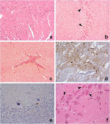
Background: The recommended screening of rabies in 'suspect' animal cases involves testing fresh brain tissue. The preservation of fresh tissue however can be difficult under field conditions and formalin fixation provides a simple alternative that may allow a confirmatory diagnosis. The occurrence and location of histopathological changes and immunohistochemical (IHC) labelling for rabies in formalin fixed paraffin embedded (FFPE) canine brain is described in samples from 57 rabies suspect cases from Sri-Lanka. The presence of Negri bodies and immunohistochemical detection of rabies virus antigen were evaluated in the cortex, hippocampus, cerebellum and brainstem. The effect of autolysis and artefactual degeneration of the tissue was also assessed.
Results: Rabies was confirmed in 53 of 57 (93%) cases by IHC. IHC labelling was statistically more abundant in the brainstem. Negri bodies were observed in 32 of 53 (60.4%) of the positive cases. Although tissue degradation had no effect on IHC diagnosis, it was associated with an inability to detect Negri bodies. In 13 cases, a confirmatory Polymerase chain reaction (PCR) testing for rabies virus RNA was undertaken by extracting RNA from fresh frozen tissue, and also attempted using FFPE samples. PCR detection using fresh frozen samples was in agreement with the IHC results. The PCR method from FFPE tissues was suitable for control material but unsuccessful in our field cases.
Conclusions: Histopathological examination of the brain is essential to define the differential diagnoses of behaviour modifying conditions in rabies virus negative cases, but it is unreliable as the sole method for rabies diagnosis, particularly where artefactual change has occurred. Formalin fixation and paraffin embedding does not prevent detection of rabies virus via IHC labelling even where artefactual degeneration has occurred. This could represent a pragmatic secondary assay for rabies diagnosis in the field because formalin fixation can prevent sample degeneration. The brain stem was shown to be the site with most viral immunoreactivity; supporting recommended sampling protocols in favour of improved necropsy safety in the field. PCR testing of formalin fixed tissue may be successful in certain circumstances as an alternative test.
Keywords: Rabies canine histopathology immunohistochemistry hemi-nested reverse transcription polymerase chain reaction.
Figures
Similar articles
-
A simple method for detection of rabies viral sequences in 16-year old archival brain specimens with one-week fixation in formalin.J Virol Methods. 2006 Jun;134(1-2):267-71. doi: 10.1016/j.jviromet.2006.02.002. Epub 2006 Mar 9. J Virol Methods. 2006. PMID: 16529825
-
Pathological, immunological and molecular diagnosis of rabies in clinically suspected animals of different species using four detection techniques in Jordan.Transbound Emerg Dis. 2012 Apr;59(2):154-64. doi: 10.1111/j.1865-1682.2011.01255.x. Epub 2011 Aug 21. Transbound Emerg Dis. 2012. PMID: 22390575
-
Rabies in apparently healthy dogs: histological and immunohistochemical studies.Niger Postgrad Med J. 2006 Jun;13(2):128-34. Niger Postgrad Med J. 2006. PMID: 16794650
-
Neuropathology and diagnostic features of rabies in a litter of piglets, with a brief review of the literature.J Vet Diagn Invest. 2020 Jan;32(1):166-168. doi: 10.1177/1040638719898687. Epub 2020 Jan 9. J Vet Diagn Invest. 2020. PMID: 31916501 Free PMC article. Review.
-
Clinical laboratory advances in the detection of rabies virus.Clin Chim Acta. 2005 Jan;351(1-2):49-63. doi: 10.1016/j.cccn.2004.09.018. Clin Chim Acta. 2005. PMID: 15563871 Review.
Cited by
-
Lateral flow devices for samples collected by straw sampling method for postmortem canine rabies diagnosis.PLoS Negl Trop Dis. 2021 Dec 9;15(12):e0009891. doi: 10.1371/journal.pntd.0009891. eCollection 2021 Dec. PLoS Negl Trop Dis. 2021. PMID: 34882672 Free PMC article.
-
Rabies-induced behavioural changes are key to rabies persistence in dog populations: Investigation using a network-based model.PLoS Negl Trop Dis. 2019 Sep 23;13(9):e0007739. doi: 10.1371/journal.pntd.0007739. eCollection 2019 Sep. PLoS Negl Trop Dis. 2019. PMID: 31545810 Free PMC article.
-
Meningeal Lymphatic and Glymphatic Structures in a Pelagic Delphinid (Delphinus delphis).Animals (Basel). 2025 Mar 4;15(5):729. doi: 10.3390/ani15050729. Animals (Basel). 2025. PMID: 40076012 Free PMC article.
-
Histopathological and immunohistochemical examination of the brains of rabid dogs in the Philippines.J Vet Med Sci. 2024 Dec 1;86(12):1243-1251. doi: 10.1292/jvms.24-0249. Epub 2024 Oct 8. J Vet Med Sci. 2024. PMID: 39384379 Free PMC article.
-
Tracking lethal threat: in-depth review of rabies.Open Vet J. 2023 Nov;13(11):1385-1399. doi: 10.5455/OVJ.2023.v13.i11.1. Epub 2023 Nov 30. Open Vet J. 2023. PMID: 38107233 Free PMC article. Review.
References
MeSH terms
Substances
LinkOut - more resources
Full Text Sources
Other Literature Sources
Medical
