. 2017 Apr 13;16(1):78.
doi: 10.1186/s12943-017-0639-2.

DC - SIGNR by influencing the lncRNA HNRNPKP2 upregulates the expression of CXCR4 in gastric cancer liver metastasis

Affiliations

DC - SIGNR by influencing the lncRNA HNRNPKP2 upregulates the expression of CXCR4 in gastric cancer liver metastasis

Yu Zhang et al. Mol Cancer. .

Abstract

Background: Profiling evidences of selectin demonstrate that they play an crucial role in cancer progression and metastasis. However, DC-SIGNR as a family member of selectin participates in gastric cancer liver metastasis remains unknown.

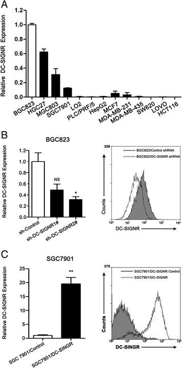
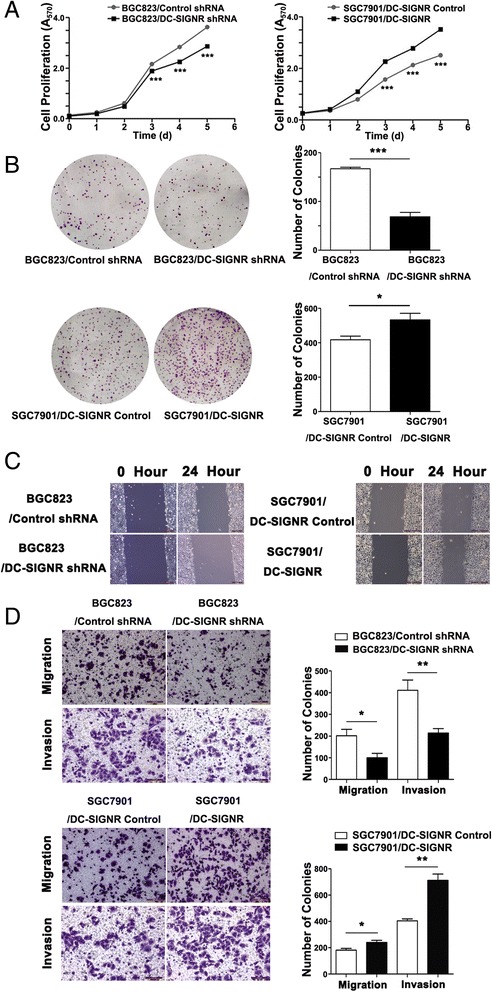
Methods: The serum level of DC-SIGNR was evaluated in gastric cancer patients by ELISA. Manipulation DC-SIGNR expression in BGC823 and SGC7901 cell lines was mediated by lentivirus. Investigation the biological effects of DC-SIGNR were verified by MTT, wounding and transwell in vitro and experiments on animals to confirm gastric cancer liver metastasis by IVIS. Insights of the mechanism were employed microarray and bioinformatic analysis. Further to confirm the results were conducted by qRT-PCR, western blot and by flow cytometry.

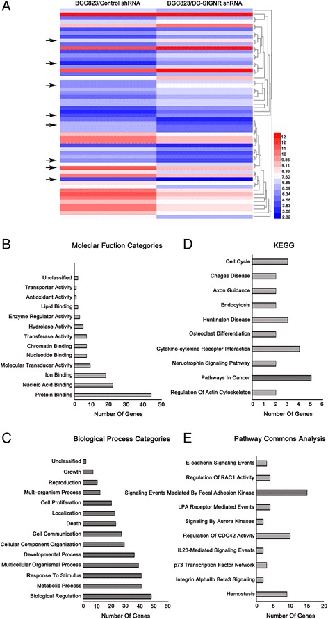
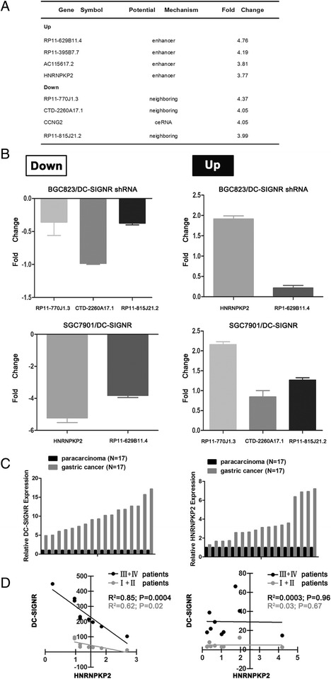
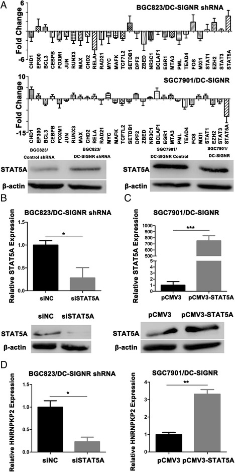
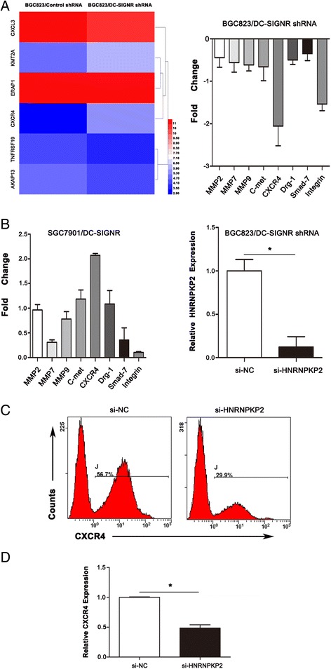
Results: DC-SIGNR serum level was significantly increased in gastric cancer patients compared with healthy group. Additionally, DC-SIGNR level was associated with an advanced pathological stage in gastric cancer patients. DC-SIGNR knockdown inhibited the proliferation, migration and invasion of gastric cancer cells in vitro and suppressed the liver metastasis in vivo. While, DC-SIGNR overexpression promoted cell proliferation, migration and invasion. In mechanism, HNRNPKP2 as a lncRNA was upregulated after DC-SIGNR knockdown. Importantly, STAT5A promoted HNRNPKP2 expression after knockdown DC-SIGNR. Furthermore after HNRNPKP2 depletion, the downstream target gene CXCR4 was downregulated.

Conclusions: DC-SIGNR promoted gastric cancer liver metastasis mediated with HNRNPKP2 which expression was regulated by STAT5A. And HNRNPKP2 decreased the expression of downstream target gene CXCR4. These findings indicated potential therapeutic candidates for gastric cancer liver metastasis.

Keywords: CXCR4; DC-SIGNR; Gastric cancer; Liver metastasis; STAT5A; lncRNA HNRNPKP2.


Figures

Fig. 1
Fig. 1
The level of DC-SIGNR in gastric cancer patients and its clinical significance. a, the level of DC-SIGNR in gastric cancer patients serum (n = 207) compared with healthy group (n = 197). DC-SIGNR level was examined by ELISA. b, examination of the correlation between DC-SIGNR level and advanced pathological stage. c, DC-SIGNR in healthy group (n = 197); patients with no metastasis (n = 151), liver metastasis (n = 43), or lung, ovary and colon metastasis (n = 17)
Fig. 2
Fig. 2
The expression of DC-SIGNR in various cell lines and manipulate the level of DC-SIGNR expression in gastric cancer cell lines. a, qRT-PCR was performed to demonstrate DC-SIGNR expression in various cell lines. b qRT-PCR (left panel) and flow cytometry assay (right panel) were used to confirm the knockdown efficiency of BGC823 infected with sh-DC-SIGNR. c qRT-PCR (left panel) and flow cytometry assay (right panel) were used to confirm the overexpression efficiency of SGC7901 infected with DC-SIGNR mediated by lentivirus
Fig. 3
Fig. 3
The function of DC-SIGNR in vitro. a, cell proliferation was measured by MTT assay. BGC823 cells were infected with sh-DC-SIGNR. SGC7901 cells were infected with DC-SIGNR mediated by lentivirus. b colony-forming growth assays were performed to determine the proliferation of sh-DC-SIGNR infected BGC823 cells and DC-SIGNR mediated by lentivirus infected SGC7901 cells. Colonies were counted and captured. c wound-healing assays were conducted to determine the rate of migration by measuring the distance from one edge of the wound to the other side. d transwell assays were conducted to investigate changes in BGC823 cells and SGC7901 cells migration and invasion. All experiments were performed in biologic triplicates with three technical replicates; *P < 0.05, **P < 0.01 and ***P < 0.001
Fig. 4
Fig. 4
The effects of DC-SIGNR knockdown on gastric cancer liver metastasis cascade in vivo. a images of GFP signals of mice 15 days after subcutaneous injection with different concentrations of BGC823 cells infected negative control (left panel). The relationship between tumour volume and fluorescence intensity (right panel). b representative images of GFP signals of mice in each group after intrasplenic injection with 5 × 106 BGC823 cells after knockdown DC-SIGNR and photographs of tumours after dissection. c average volume of tumours on liver and spleen derived from BGC823 cells after infected DC-SIGNR and negative control (left panel) and average volume of tumours on spleen derived from BGC823 cells after infected DC-SIGNR and negative control (right panel). d H&E, CK7, CK20 and Ki-67 staining of sections from liver tumours and spleen tumours were performed (scale bar = 50 μm)
Fig. 5
Fig. 5
DC-SIGNR knockdown leads to cancer-related lncRNA expression profiling. a, the hierarchical cluster heatmap demonstrated lncRNAs expression with change fold > 2 from microarray data. b-c, bar graphs analyzed differentially expressed genes based on GO Slim with molecular function and biological processes. d-e KEGG and Pathway commons analysis revealed enrichment of genes related to various events
Fig. 6
Fig. 6
Five gene expression profiling with DC-SIGNR level change and correlation between DC-SIGNR and HNRNPKP2. a top 8 genes significantly upregulated or downregulated in BGC823 cells after DC-SIGNR knockdown. b real-time PCR confirmed the 8 genes expression using the gene specific primers. c relative expression of DC-SIGNR and HNRNPKP2 in gastric FFPET (n = 17) and paracarcinoma FFPET (n = 17) was detected by real-time PCR. d Correlation DC-SIGNR expression and HNRNPKP2 expression inI,IIgastric FFPET and III,IVgastric FFPET (left). And in I,II paracarcinoma FFPET and III,IVparacarcinoma FFPET (right). FFPET, formalin-fixed paraffin-embeded tissue
Fig. 7
Fig. 7
STAT5A promotes HNRNPKP2 expression. a, The levels of 29 transcription factors in BGC823 cells after DC-SIGNR knockdown by real-time PCR (up). The levels of 29 transcription factors in SGC7901 cells after DC-SIGNR overexpression by real-time PCR (middle). Western blot confirmed the protein level of STAT5A in BGC823 cells after DC-SIGNR knockdown and in SGC7901 cells after DC-SIGNR overexpression (down). b-c Real-time PCR confirmed the STAT5A expression and western blot analysis of STAT5A protein level in BGC823 cells after DC-SIGNR knockdown with si-STAT5A and in SGC7901 cells after DC-SIGNR overexpression with pCMV3-STAT5A. D, HNRNPKP2 expression in BGC823 cells after DC-SIGNR with si-STAT5A and in SGC7901 cells after DC-SIGNR overexpression with pCMV3-STAT5A is analyzed by real-time PCR
Fig. 8
Fig. 8
CXCR4 is significantly disregulated after DC-SIGNR knockdown. a a heatmap of target genes (CXCL3, KMT2A, ERAP1, CXCR4, TNFRSF19 and AKAP13) after DC-SIGNR knockdown (left panel). The levels of metastasis and invasion markers in BGC823 cells after DC-SIGNR knockdown by qRT-PCR (right panel). b,the levels of metastasis and invasion markers in SGC7901 cells after DC-SIGNR overexpression by qRT-PCR. And HNRNPKP2 expression in BGC823 cells after DC-SIGNR and HNRNPKP2 knockdown was analyzed by qRT-PCR. c flow cytometry indicated CXCR4 in BGC823 cells after DC-SIGNR and HNRNPKP2 knockdown. d CXCR4 expression in BGC823 cells after DC-SIGNR and HNRNPKP2 knockdown was analyzed by qRT-PCR

References

    1. Torre LA, Bray F, Siegel RL, Ferlay J, Lortet-Tieulent J, Jemal A. Global cancer statistics, 2012. CA Cancer J Clin. 2015;65:87–108. doi: 10.3322/caac.21262. - DOI - PubMed
    1. Ferro A, Peleteiro B, Malvezzi M, et al. Worldwide trends in gastric cancer mortality (1980–2011), with predictions to 2015, and incidence by subtype. Eur J Cancer. 2014;50:1330–1344. doi: 10.1016/j.ejca.2014.01.029. - DOI - PubMed
    1. Sakamoto Y, Ohyama S, Yamamoto J, et al. Surgical resection of liver metastases of gastric cancer: an analysis of a 17-year experience with 22 patients. Surgery. 2003;133:507–511. doi: 10.1067/msy.2003.147. - DOI - PubMed
    1. Koga R, Yamamoto J, Ohyama S, et al. Liver resection for metastatic gastric cancer: experience with 42 patients including eight long-term survivors. Jpn J Clin Oncol. 2007;37:836–842. doi: 10.1093/jjco/hym113. - DOI - PubMed
    1. Macdonald JS, Smalley SR, Benedetti J, et al. Chemoradiotherapy after surgery compared with surgery alone for adenocarcinoma of the stomach or gastroesophageal junction. N Engl J Med. 2001;345:725–730. doi: 10.1056/NEJMoa010187. - DOI - PubMed

Publication types

MeSH terms