. 2017 Jul:82:105-117.
doi: 10.1016/j.mcn.2017.04.001. Epub 2017 Apr 9.

Paclitaxel inhibits the activity and membrane localization of PKCα and PKCβI/II to elicit a decrease in stimulated calcitonin gene-related peptide release from cultured sensory neurons

Affiliations

Paclitaxel inhibits the activity and membrane localization of PKCα and PKCβI/II to elicit a decrease in stimulated calcitonin gene-related peptide release from cultured sensory neurons

Lisa M Darby et al. Mol Cell Neurosci. 2017 Jul.

Abstract

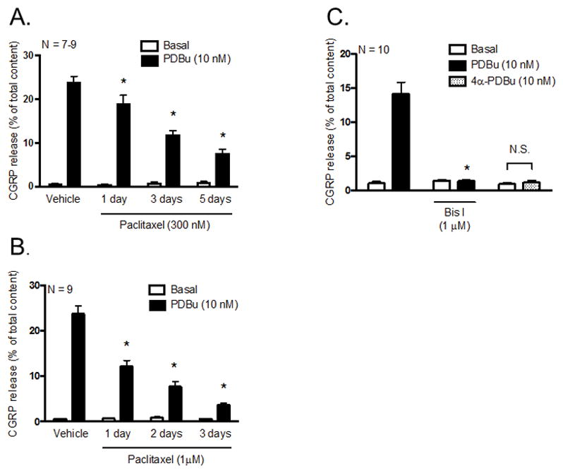
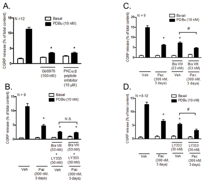
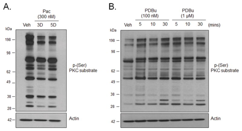
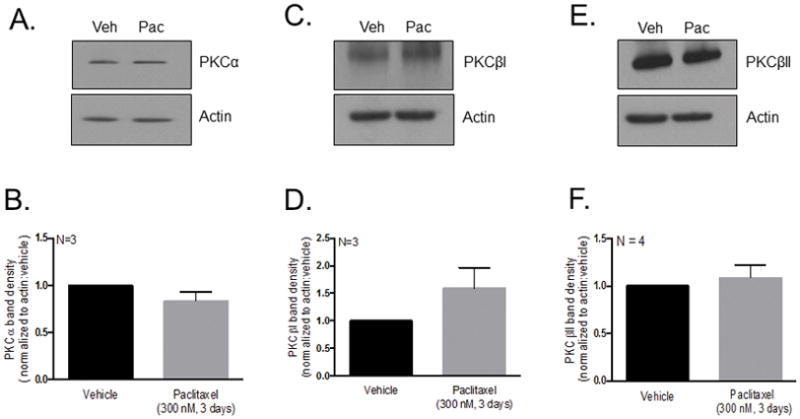
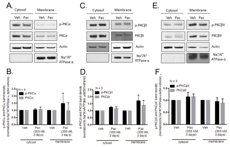
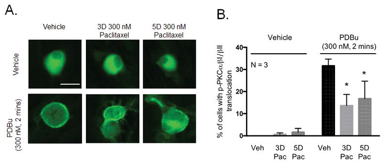
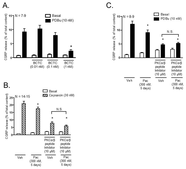
Peripheral neuropathy is a dose-limiting and debilitating side effect of the chemotherapeutic drug, paclitaxel. Consequently, elucidating the mechanisms by which this drug alters sensory neuronal function is essential for the development of successful therapeutics for peripheral neuropathy. We previously demonstrated that chronic treatment with paclitaxel (3-5days) reduces neuropeptide release stimulated by agonists of TRPV1. Because the activity of TRPV1 channels is modulated by conventional and novel PKC isozymes (c/nPKC), we investigated whether c/nPKC mediate the loss of neuropeptide release following chronic treatment with paclitaxel (300nM; 3 and 5days). Release of the neuropeptide, calcitonin gene-related peptide (CGRP), was measured as an index of neuronal sensitivity. Following paclitaxel treatment, cultured dorsal root ganglia sensory neurons were stimulated with a c/nPKC activator, phorbol 12,13-dibutyrate (PDBu), or a TRPV1 agonist, capsaicin, in the absence and presence of selective inhibitors of conventional PKCα and PKCβI/II isozymes (cPKC). Paclitaxel (300nM; 3days and 5days) attenuated both PDBu- and capsaicin-stimulated release in a cPKC-dependent manner. Under basal conditions, there were no changes in the protein expression, phosphorylation or membrane localization of PKC α, βI or βII, however, paclitaxel decreased cPKC activity as indicated by a reduction in the phosphorylation of cPKC substrates. Under stimulatory conditions, paclitaxel attenuated the membrane translocation of phosphorylated PKC α, βI and βII, providing a rationale for the attenuation in PDBu- and capsaicin-stimulated release. Our findings suggest that a decrease in cPKC activity and membrane localization are responsible for the reduction in stimulated peptide release following chronic treatment with paclitaxel in sensory neurons.

Keywords: CGRP; Dorsal root ganglia; PKC; Paclitaxel; Peripheral neuropathy; TRPV1.


Figures

Figure 1
Figure 1
Paclitaxel decreases PDBu-stimulated CGRP release in cultured sensory neurons. Each column represents the mean ± SEM of basal (white columns) or stimulated (black columns) CGRP release expressed as % of total content. Cultures were exposed to (A) 300 nM or (B) 1 μM paclitaxel for 1, 2, 3 or 5 days prior to stimulation with PDBu (10 nM). An * indicates a significant decrease in PDBu-stimulated release in paclitaxel-treated (300 nM and 1 μM) neurons compared to vehicle-treated neurons (p < 0.05, N = 7–9). (C) Naïve cultures were pre-treated with Bis I (1 μM) for 10 minutes prior to stimulation with PDBu. As a control experiment, naïve cultures were also incubated with 10 nM 4α-PDBu (dotted columns). An * indicates a significant decrease in PDBu-stimulated release in neurons pre-treated with Bis I (p < 0.05, N = 10). Significance was determined using a two-way ANOVA with Tukey’s post-hoc test. N.S. – not significant.
Figure 2
Figure 2
Inhibition of PKCα and PKCβI/II activity mediates the decrease in PDBu-stimulated CGRP release following chronic exposure to paclitaxel in cultured sensory neurons. Each column represents the mean ± SEM of basal (white columns) or PDBu-stimulated (black columns) CGRP release expressed as % of total content in the absence and presence of PKCα and PKCβI/II inhibitors. (A) Naïve cultures were pre-treated with Gö6976 (100 nM) or a myristoylated PKCα/β peptide inhibitor (10 μM) for 10 minutes prior to stimulation with PDBu (10 nM). An * indicates a significant decrease in PDBu-stimulated release in neurons pre-treated with Gö6976 or the myristoylated PKCα/β peptide inhibitor (p < 0.05, N = 12). Cultures were exposed to 300 nM paclitaxel for 3 days prior to stimulation with PDBu (10 nM) in the presence and absence of the (B) PKCα inhibitor (Bis VIII; 53 nM) + PKCβI/II inhibitor (LY333531; 30 nM), (C) Bis VIII or (D) LY333531. An * indicates a significant decrease in PDBu-stimulated release in paclitaxel-only treated neurons, vehicle- and paclitaxel-treated neurons pre-treated with (B) Bis VIII + LY333531, (C) Bis VIII or (D) LY333531 compared to vehicle-only treated neurons and a # indicates a significant decrease in PDBu-stimulated release in paclitaxel-treated neurons compared to vehicle-treated neurons pre-treated with (C) Bis VIII or (D) LY333531. Significance was determined using a two-way ANOVA with Tukey’s post-hoc test (p < 0.05, N = 8–12). Veh - Vehicle; Pac - Paclitaxel; LY333 – LY333531; N.S. – not significant.
Figure 3
Figure 3
Chronic treatment with paclitaxel decreases the basal phosphorylation of PKC α/β substrates in cultured sensory neurons. (A) Representative western blot image showing immunoreactivity for phosphorylation of PKC α, βI and βII substrates in whole cell lysates in neurons treated with vehicle and paclitaxel (300 nM, 3 and 5 days). (B) Representative western blot image showing immunoreactivity for phosphorylation of PKC α, βI and βII substrates in neurons treated with PDBu (100 nM and 1 μM; 5, 10 or 30 minutes). Veh - Vehicle; Pac – Paclitaxel; 3D – 3 days; 5D – 5 days.
Figure 4
Figure 4
Chronic treatment with paclitaxel does not reduce basal PKC α, βI or βII, immunoreactivity in cultured sensory neurons. (A, C, E) Representative western blot images showing immunoreactivity for PKC α, βI and βII in whole cell lysates in neurons treated with vehicle and paclitaxel (300 nM, 3 days). (B, D, F) Densitometry analysis showing no significant differences in PKC immunoreactivity normalized to actin in paclitaxel-treated (300 nM, 3 days) neurons compared to vehicle-treated neurons using a t-test (N = 3–4, a.u. ± SEM). Veh - Vehicle; Pac -Paclitaxel.
Figure 5
Figure 5
Chronic treatment with paclitaxel does not attenuate the basal phosphorylation level or membrane localization of PKC α, βI or βII in cultured sensory neurons. (A, C, E) Representative western blot images showing immunoreactivity for PKC α, βI and βII in cytosolic and membrane fractions of neurons treated with vehicle and paclitaxel (300 nM, 3 days). (B, D, F) Densitometry analysis showing no significant differences in PKC immunoreactivity in paclitaxel-treated (300 nM, 3 days) neurons compared to vehicle-treated neurons in cytosolic and membrane fractions when normalized to actin and Na+/K+ ATPase α, respectively. Significance was determined using a two-way ANOVA with Tukey’s post-hoc test (N = 3–4, a.u. ± SEM). Veh - Vehicle; Pac -Paclitaxel.
Figure 6
Figure 6
Chronic treatment with paclitaxel decreases PDBu-stimulated membrane localization of PKC α, βI and βII in cultured sensory neurons. (A) Representative fluorescent images showing membrane localization of immunoreactive phosphorylated PKC α, βI and βII in neurons treated with paclitaxel (300 nM, 3 days or 5 days) following stimulation with PDBu (300 nM, 2 minutes). (B) Graphical analysis showing the number of neuronal cell bodies demonstrating translocation of phosphorylated PKC α, βI and βII in paclitaxel-treated neurons (300 nM, 3 and 5 days) following stimulation with PDBu (300 nM, 2 minutes) expressed as % of phospho- PKC α/βI/II translocation. An * indicates a significant decrease in the number of cells with PKC α, βI and βII translocation. Significance was determined using a two-way ANOVA with Dunnett’s post-hoc test (p < 0.05, N = 3). Scale bar = 20 μm. Veh - Vehicle; Pac –Paclitaxel, 3D – 3 days, 5D – 5 days.
Figure 7
Figure 7
Inhibition of PKCα and PKCβI/II activity mediates the decrease in capsaicin-stimulated CGRP release following chronic exposure to paclitaxel in cultured sensory neurons. Each column represents the mean ± SEM of basal (white columns), capsaicin-stimulated (striped columns) or PDBu-stimulated (black columns) CGRP release expressed as % of total content. (A) Naïve cultures were pre-treated with the TRPV1 antagonist, BCTC, for 10 minutes prior to stimulation with capsaicin (30 nM). An * indicates a significant decrease in capsaicin-stimulated release in neurons pre-treated with BCTC compared to control neurons (p < 0.05, N = 7–9). Cultures were exposed to 300 nM paclitaxel for 3 days and pre-treated with a myristolyated PKCα/β peptide inhibitor (10 μM) for 10 minutes prior to stimulation with (B) 30 nM capsaicin or (C) 10 nM PDBu. An * indicates a decrease in (B) capsaicin-stimulated and (C) PDBu-stimulated release in paclitaxel-only treated neurons, vehicle- and paclitaxel treated neurons pre-treated with the myristolyated PKCα/β peptide inhibitor compared to vehicle-only treated neurons. Significance was determined using a two-way ANOVA with Tukey’s post-hoc test (p < 0.05, N = 8–15). N.S. – not significant.

Similar articles

Cited by

References

    1. Authier N, Gillet JP, Fialip J, Eschalier A, Coudore F. Description of a short-term Taxol-induced nociceptive neuropathy in rats. Brain research. 2000;887:239–249. - PubMed
    1. Barber LA, Vasko MR. Activation of protein kinase C augments peptide release from rat sensory neurons. Journal of neurochemistry. 1996;67:72–80. - PubMed
    1. Betz A, Ashery U, Rickmann M, Augustin I, Neher E, Sudhof TC, Rettig J, Brose N. Munc13-1 is a presynaptic phorbol ester receptor that enhances neurotransmitter release. Neuron. 1998;21:123–136. - PubMed
    1. Bhave G, Hu HJ, Glauner KS, Zhu W, Wang H, Brasier DJ, Oxford GS, Gereau RWt. Protein kinase C phosphorylation sensitizes but does not activate the capsaicin receptor transient receptor potential vanilloid 1 (TRPV1) Proc Natl Acad Sci U S A. 2003;100:12480–12485. - PMC - PubMed
    1. Bradford MM. A rapid and sensitive method for the quantitation of microgram quantities of protein utilizing the principle of protein-dye binding. Analytical biochemistry. 1976;72:248–254. - PubMed

MeSH terms