Review
. 2017 May;18(5):693-711.
doi: 10.15252/embr.201744045. Epub 2017 Apr 12.

Local translation in neuronal compartments: how local is local?

Affiliations
Review

Local translation in neuronal compartments: how local is local?

Vidhya Rangaraju et al. EMBO Rep. 2017 May.

Abstract

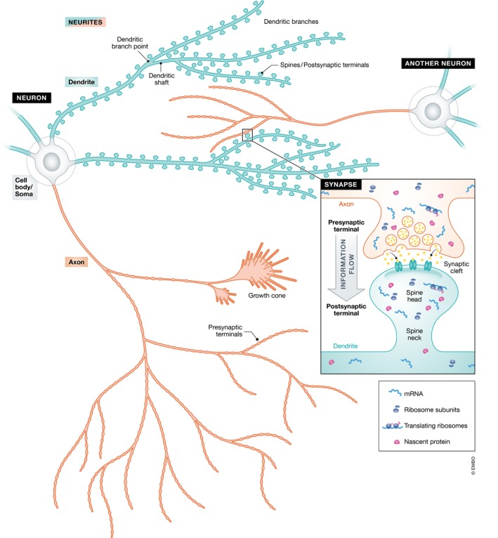
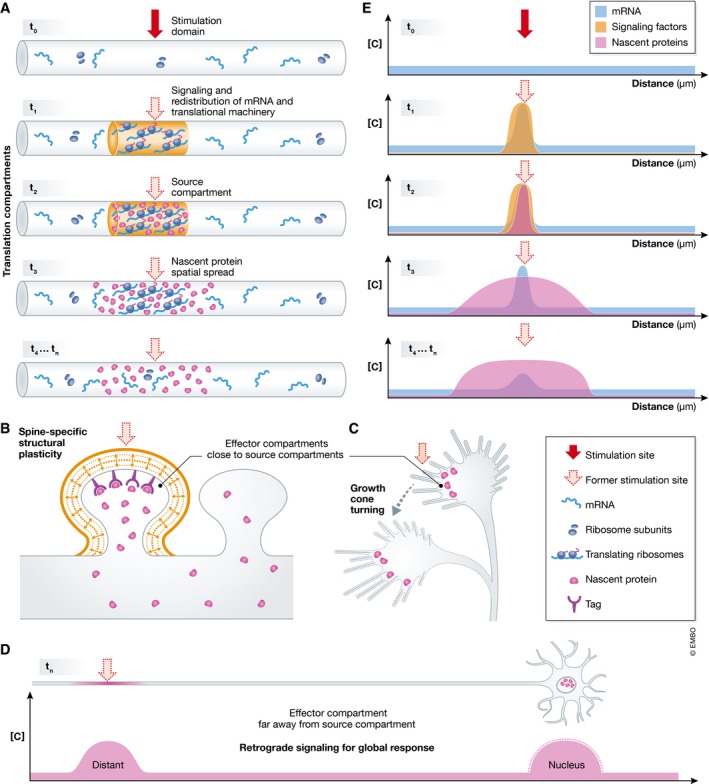
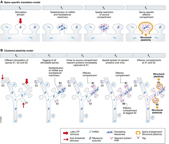
Efficient neuronal function depends on the continued modulation of the local neuronal proteome. Local protein synthesis plays a central role in tuning the neuronal proteome at specific neuronal regions. Various aspects of translation such as the localization of translational machinery, spatial spread of the newly translated proteins, and their site of action are carried out in specialized neuronal subcompartments to result in a localized functional outcome. In this review, we focus on the various aspects of these local translation compartments such as size, biochemical and organelle composition, structural boundaries, and temporal dynamics. We also discuss the apparent absence of definitive components of translation in these local compartments and the emerging state-of-the-art tools that could help dissecting these conundrums in greater detail in the future.

Keywords: compartments; local translation; nascent protein; plasticity; spatial spread.


Figures

Figure 1
Figure 1. Neuron and its structural compartments
Morphology of the neuron showing its cell body (gray) and neurites–composed of dendrites (blue) and axons (red). The inset shows a synapse formed between the presynaptic terminal of one neuron (red) and the postsynaptic terminal of another neuron (blue).
Figure 2
Figure 2. Local translation compartments
(A) On receiving a focal stimulus (red filled arrow)—stimulation domain (t 0)—mRNA and translational machinery are redistributed to the stimulation site (t 1). This local redistribution of mRNA and translational machinery, on overlap with signaling events (orange) form the site of synthesis for nascent proteins (magenta)—source compartment (t 2)—near the former stimulation site (red dotted arrow). The nascent proteins quickly spread over time. This nascent protein spatial spread (t 3) gradually increases and might reach a compartment of stable size defined by unknown factors (t 4, t n). The site of action of the nascent proteins—effector compartment—is restricted within a smaller region of the nascent protein spatial spread and can be either close to the source compartment as in a spine (B) and the growth cone (C) or hundreds of microns apart as in the nucleus (D). All these translation compartments operate in unison to elicit a functional outcome, for example, spine‐specific structural plasticity (B), growth cone turning (C), and retrograde signaling for global response (D). (E) Graphical representation showing the concentration ([C]) of mRNA (blue), signaling factors (orange), and nascent proteins (magenta) plotted at various time points t 0, t 1, t 2, t 3, t 4, t n.
Figure 3
Figure 3. Subcompartments in dendrites
(A) Concept of spine‐specific translation: A spine‐specific stimulation (t 0) could result in redistribution of mRNA and translational machinery to the stimulated spine (t 1). On overlapping with spine‐specific signaling events, this would result in the synthesis of nascent proteins—source compartment—whose spatial spread is restricted within the spine, due to diffusional restriction by the spine neck (t 2). This would lead to a spine‐specific effector compartment and subsequent functional outcome—structural plasticity (t n). (B) Clustered plasticity model: Spine S1 receives a late‐LTP stimulus, and spines S2 and S3 receive a subthreshold stimulus (t 0). All three spines get tagged but mRNA and translational machinery redistribute only close to spine S1 that received the late‐LTP stimulus (t 1). The newly translated proteins—source compartment—are instantly captured at spine S1 (t 2) and with time, the spatial spread of the nascent proteins increases allowing for its additional capture at adjacent tagged spine S2 (t 3). Both the tagged spines S1 and S2 that capture nascent proteins—effector compartment—undergo spine‐specific structural plasticity—functional outcome (t n). However, only tagged spines clustered within a nascent protein spatial spread of ~50 μm show this functional outcome—tagged spine (S3) present beyond this spatial spread does not.

References

    1. Hoagland MB, Keller EB, Zamecnik PC (1956) Enzymatic carboxyl activation of amino acids. J Biol Chem 218: 345–358 - PubMed
    1. Hoagland MB, Stephenson ML, Scott JF, Hecht LI, Zamecnik PC (1958) A soluble ribonucleic acid intermediate in protein synthesis. J Biol Chem 231: 241–257 - PubMed
    1. Holt CE, Schuman EM (2013) The central dogma decentralized: new perspectives on RNA function and local translation in neurons. Neuron 80: 648–657 - PMC - PubMed
    1. Garner CC, Matus A (1988) Different forms of microtubule‐associated protein 2 are encoded by separate mRNA transcripts. J Cell Biol 106: 779–783 - PMC - PubMed
    1. Burgin KE, Waxham MN, Rickling S, Westgate SA, Mobley WC, Kelly PT (1990) In situ hybridization histochemistry of Ca2+/calmodulin‐dependent protein kinase in developing rat brain. J Neurosci 10: 1788–1798 - PMC - PubMed

LinkOut - more resources