. 2017 Jun 23;121(1):56-70.
doi: 10.1161/CIRCRESAHA.117.310870. Epub 2017 Apr 12.

Myeloperoxidase Mediates Postischemic Arrhythmogenic Ventricular Remodeling

Affiliations

Myeloperoxidase Mediates Postischemic Arrhythmogenic Ventricular Remodeling

Martin Mollenhauer et al. Circ Res. .

Abstract

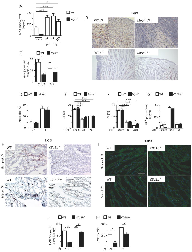
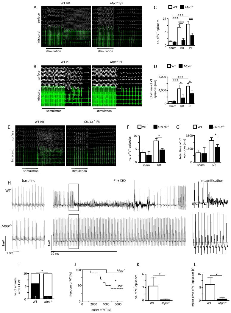
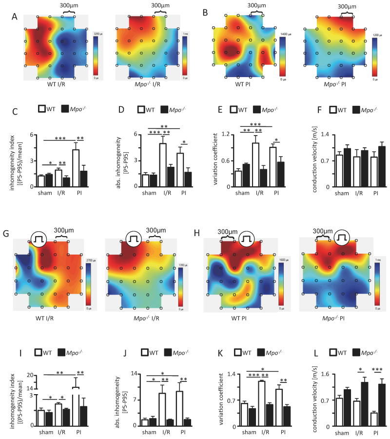
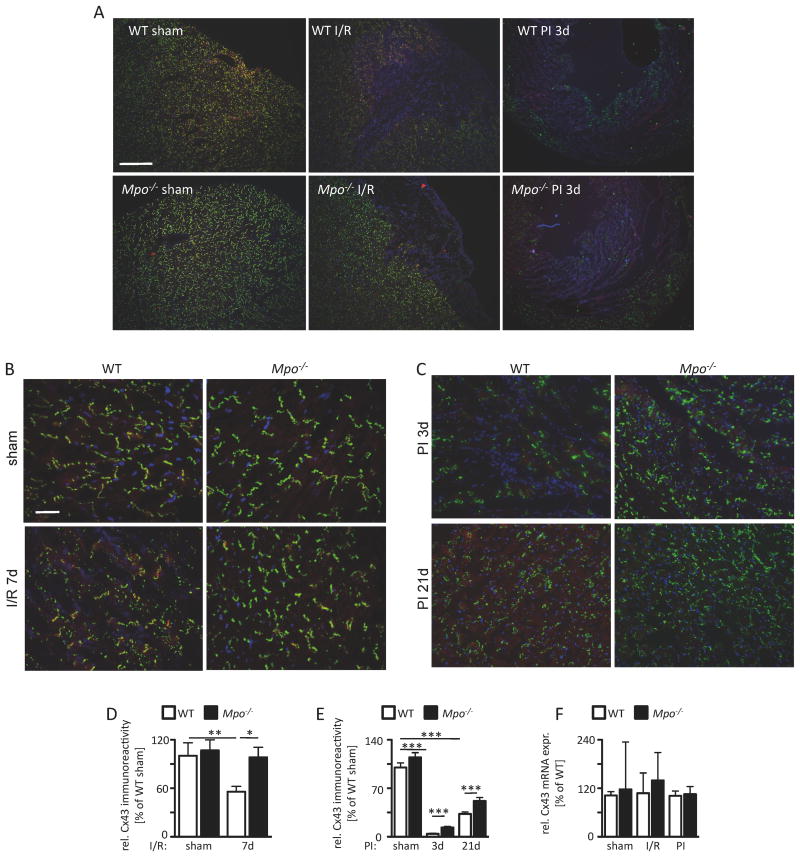
Rationale: Ventricular arrhythmias remain the leading cause of death in patients suffering myocardial ischemia. Myeloperoxidase, a heme enzyme released by polymorphonuclear neutrophils, accumulates within ischemic myocardium and has been linked to adverse left ventricular remodeling.

Objective: To reveal the role of myeloperoxidase for the development of ventricular arrhythmias.

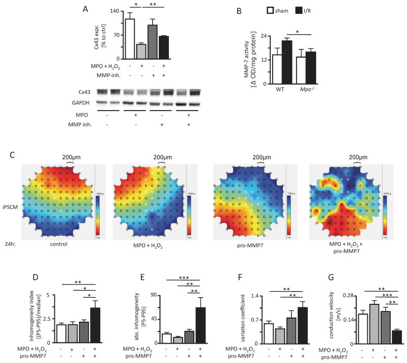
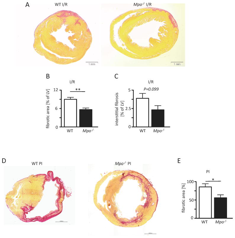
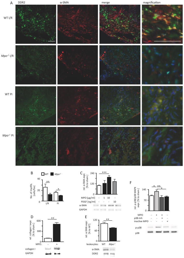
Methods and results: In different murine models of myocardial ischemia, myeloperoxidase deficiency profoundly decreased vulnerability for ventricular tachycardia on programmed right ventricular and burst stimulation and spontaneously as assessed by ECG telemetry after isoproterenol injection. Experiments using CD11b/CD18 integrin-deficient (CD11b-/-) mice and intravenous myeloperoxidase infusion revealed that neutrophil infiltration is a prerequisite for myocardial myeloperoxidase accumulation. Ventricles from myeloperoxidase-deficient (Mpo-/-) mice showed less pronounced slowing and decreased heterogeneity of electric conduction in the peri-infarct zone than wild-type mice. Expression of the redox-sensitive gap junctional protein Cx43 (Connexin 43) was reduced in the peri-infarct area of wild-type compared with Mpo-/- mice. In isolated wild-type cardiomyocytes, Cx43 protein content decreased on myeloperoxidase/H2O2 incubation. Mapping of induced pluripotent stem cell-derived cardiomyocyte networks and in vivo investigations linked Cx43 breakdown to myeloperoxidase-dependent activation of matrix metalloproteinase 7. Moreover, Mpo-/- mice showed decreased ventricular postischemic fibrosis reflecting reduced accumulation of myofibroblasts. Ex vivo, myeloperoxidase was demonstrated to induce fibroblast-to-myofibroblast transdifferentiation by activation of p38 mitogen-activated protein kinases resulting in upregulated collagen generation. In support of our experimental findings, baseline myeloperoxidase plasma levels were independently associated with a history of ventricular arrhythmias, sudden cardiac death, or implantable cardioverter-defibrillator implantation in a cohort of 2622 stable patients with an ejection fraction >35% undergoing elective diagnostic cardiac evaluation.

Conclusions: Myeloperoxidase emerges as a crucial mediator of postischemic myocardial remodeling and may evolve as a novel pharmacological target for secondary disease prevention after myocardial ischemia.

Keywords: Connexin 43; fibrosis; infarction; inflammation; myocardial ischemia; tachycardia.


Figures

Figure 1
Figure 1
A) MPO plasma level of sham WT and Mpo-/- mice or WT mice after left ventricular ischemia and 7 days of reperfusion (I/R) and 5 and 21 days upon permanent ischemia (PI) as assessed by ELISA. WT sham n=28, Mpo-/- sham n=8, WT I/R n=30, WT 5 days PI n=5, WT 21 days n=7. Infiltration of PMN. (B) Immunohistochemical Ly6G stainings (brown) of WT and Mpo-/- hearts subjected to I/R or PI. (C) Quantitative analysis of Ly6G+ cells into the infarct and periinfarct zones of WT and Mpo-/- hearts after I/R and PI. Scale bar = 200 μm. WT I/R / Mpo-/- I/R n=7/13; WT PI / Mpo-/- PI n=6/7. Infarct size and left ventricular function. (D) Infarct size in WT and Mpo-/- hearts after 7 days I/R and 21 days upon PI. WT / Mpo-/- I/R n=6/5; WT / Mpo-/- PI n=6/6. Ejection fraction (EF) after (E) 3 and 7 days I/R or (F) 3 days and 21 days PI of WT and Mpo-/- animals. E: WT / Mpo-/- sham n=4, WT / Mpo-/- 3d I/R n=5/4, WT / Mpo-/- 7d I/R n=8/5; F: WT / Mpo-/- sham n=4, WT / Mpo-/- 3d PI n=5/3, WT / Mpo-/- 21d PI n=4/3. Infiltration of PMN and accumulation of MPO. (G) MPO plasma levels of WT and CD11b-/- mice subjected to ischemia following 6 hours or 2 days of reperfusion. (H, J) Immunohistochemical Ly6G stainings (brown) and quantitative analysis of Ly6G+ cells into the infarct and periinfarct zones of WT and CD11b-/- hearts and (I, K) immunofluorescence MPO stainings (light green) and quantitative analysis of MPO immunoreactivity in WT and CD11b-/- hearts subjected to ischemia following 6 hours or 2 days of reperfusion. G: WT / CD11b-/- sham n=5/5, WT / CD11b-/- 6h I/R n=5/5, WT / CD11b-/- 2d I/R n=3/7; J: WT / CD11b-/- 6h I/R n=3/4, WT / CD11b-/- 2d I/R n=5/5; K: WT / CD11b-/- 6h I/R n=5/5, WT / CD11b-/- 2d I/R n=5/7. *=P<0.05, ***=P<0.001, Kruskal Wallis test followed by Bonferroni post hoc test. Mean ± SEM is shown.
Figure 2
Figure 2. Vulnerability to ventricular arrhythmias: Representative surface (top panel) and intracardiac (bottom panel)
ECG recordings of electrical ventricular stimulation of WT and Mpo-/- mice after (A) ischemia and 7 days of reperfusion (I/R) and (B) 21 days upon permanent ischemia (PI). (C) Number of episodes of ventricular tachycardia (VT) and (D) total time of VT after I/R and PI. (E) Representative ECG recordings of WT and CD11b-/- mice after ischemia following 2 days of reperfusion. (F) Number of VT episodes and (G) total time of VT in WT and CD11b-/- mice after ischemia following 2 days of reperfusion. sham: n=4; C, D: WT / Mpo-/- I/R n=8/9; WT / Mpo-/- PI n=6/11; F, G: sham n=11/4, WT / CD11b-/- I/R n=9/8. Analysis of ventricular tachycardia 24 hrs after LAD-ligation: (H) Representative ECG-traces recorded by telemetry investigations (for 2 hrs) before (left panel) and 24 hrs after LAD-ligation and two Isoproterenol injections (2 mg/kg BW) (middle panel) in WT and Mpo-/- animals. Magnification of the highlighted section is shown in the right panel. Analyses of (I) VT probability, (J) onset of VT, (K) mean number of VT episodes and (L) mean time of VT episodes in WT and Mpo-/- animals. n=10/10. *=P<0.05, **=P<0.01, ***=P<0.001, by Kruskal Wallis test followed by Bonferroni post hoc test. (I) Chi-square test. (J) Log-rank test.
Figure 3
Figure 3. Epicardial mapping analyses. Representative maps of spontaneous conduction in the periinfarct region of WT and Mpo-/- hearts
(A) after left ventricular ischemia and 7 days of reperfusion (I/R) and (B) 21 days upon permanent ischemia PI (from dark red to dark blue, bars indicate total time from first to last measurement within one heartbeat). (C) Inhomogeneity index, (D) absolute inhomogeneity, (E) variation coefficient of conduction and (F) mean conduction velocity of WT and Mpo-/- hearts. sham n=5; WT I/R / Mpo-/- I/R n=6/6; WT PI / Mpo-/- PI n=7/5. Representative conduction maps of stimulated WT and Mpo-/- hearts subjected to (G) I/R or (H) PI with indicated pacing position. (I) Inhomogeneity index, (J) absolute inhomogeneity, (K) variation coefficient and of conduction and (L) mean conduction velocity in WT and Mpo-/- hearts subjected to I/R or PI. Mean ± SEM is shown; sham WT / Mpo-/- n=3/4; WT / Mpo-/- I/R n=5/6; WT / Mpo-/- PI n=5/7. *=P<0.05, **=P<0.01, ***=P<0.001, by Kruskal Wallis test followed by Bonferroni post hoc test.
Figure 4
Figure 4. Connexin43 (Cx43) integrity. (A) Immunoreactivity for Cx43 in cardiac sections of WT and Mpo-/- mice after left ventricular ischemia and 7 days of reperfusion (I/R) and 3 and 21 days upon PI (green=Cx43, red=N-cadherin, blue=DAPI; scale bar=200 μm)
Immunoreactivity for Cx43 and N-cadherin in the cardiac periinfarct zone of WT and Mpo-/- animals subjected to (B) sham-operation and 7 days I/R or (C) 3 and 21 days PI (red=N-cadherin, green=Cx43, blue=DAPI; scale bar=30 μm). (D) Quantitative analysis of Cx43 immunoreactivity in the periinfarct zone of WT- and Mpo-/- mice upon I/R or (E) 3 and 21 days PI (C: WT / Mpo-/- sham n=9/4, WT / Mpo-/- 7d I/R n=7/11; D: WT / Mpo-/- sham n=6/11, WT / Mpo-/- 3d PI n=6/7, WT / Mpo-/- 21d PI n=6/7. (F) Analysis of left ventricular Cx43 mRNA expression of sham, I/R or PI hearts of WT and Mpo-/- mice. n=3 for all groups. *=P<0.05, **=P<0.01, ***=P<0.001, by Kruskal Wallis test followed by Bonferroni post hoc test. Mean ± SEM is shown.
Figure 5
Figure 5. MPO-dependent effect of MMP-7 on Cx43 and electrical homogeneity. (A) Immunoblot analyses of Cx43 in isolated cardiomyocytes upon MPO treatment with and without additional MMP-inhibitor treatment (sham: n=4; MPO: n=4; MMP-inhibitor: n=4; MPO/MMP-inhibitor: n=3, the original immunoblot is shown in Online Figure VIII)
(B) MMP-7 activity in the periinfarct region of I/R WT and Mpo-/- hearts (WT / Mpo-/- sham n=5/5, WT / Mpo-/- I/R n=10/8). (C) Representative maps of spontaneous conduction of iPSCM treated with NaCl (control), MPO/H2O2, pro-MMP-7 or MPO/H2O2/pro-MMP-7 with analysis of (D) inhomogeneity index, (E) enhanced absolute inhomogeneity, (F) variation coefficient of conduction and (G) conduction velocity. Control: n=11, MPO/H2O2: n=5, pro-MMP-7: n=5, MPO/H2O2/pro-MMP-7: n=7. *=P<0.05, **=P<0.01, by Kruskal Wallis test followed by Bonferroni post hoc test. Mean ± SEM is shown.
Figure 6
Figure 6. MPO-dependent fibrotic remodeling. Picrosirius red stained cardiac sections of WT and Mpo-/- mice
(A) upon ischemia and 7 days of reperfusion (I/R) or (D) 21 days of permanent ischemia (PI) with red areas indicating collage deposition and analysis of (B) total left ventricular fibrotic area and (C) interstitial fibrosis; B,C: WT / Mpo-/- 7d I/R n=6/7 and (E) total left ventricular fibrosis after PI of WT and Mpo-/- hearts. Mean ± SEM is shown; WT / Mpo-/- 21d PI n=7/9. Scale bar = 1mm. *=P<0.05, **=P<0.01, by unpaired Students's t-test.
Figure 7
Figure 7. MPO-dependent fibroblast transdifferentiation
(A) Representative immunofluorescence stainings for fibroblast marker DDR2 (green) and myofibroblast marker α-SMA (red) within the infarct area of WT and Mpo-/- mice after left ventricular ischemia and 7 days of reperfusion (I/R) and 5 days of permanent ischemia (PI) (blue=DAPI; scale bar=200 μm). (B) Quantitative analysis of myofibroblasts within the infarct and periinfarct region after 7 days of I/R and 5 days PI; WT / Mpo-/- 7d I/R n=11/15, WT / Mpo-/- 5d PI n=5/5. (C) Relative α-SMA protein expression of isolated cardiac fibroblasts after 8 hours of MPO and PDGF treatment. (D) Relative collagen I expression of isolated cardiac fibroblasts after 36 hours of MPO treatment. (E) Relative α-SMA protein expression of isolated cardiac fibroblasts after co-culture with WT or Mpo-/- leukocytes. (F) Relative phosphorylation of p38 MAP-kinase (p-p38/p38 MAPK) in isolated fibroblasts upon 15 minutes of MPO treatment. Original immunoblots are shown in Online Figure IX-XII. Mean ± SEM is shown. C, D, E, F: n=4 independent experiments. *=P<0.05, **=P<0.01, ***=P<0.001, by unpaired Students's t-test for B, D, E and by Kruskal Wallis test followed by Bonferroni post hoc test for C, F.

Comment in

References

    1. Zipes DP, Camm AJ, Borggrefe M, Buxton AE, Chaitman B, Fromer M, Gregoratos G, Klein G, Moss AJ, Myerburg RJ, Priori SG, Quinones MA, Roden DM, Silka MJ, Tracy C. ACC/AHA/ESC 2006 Guidelines for Management of Patients With Ventricular Arrhythmias and the Prevention of Sudden Cardiac Death. Circulation. 2006;114 - PubMed
    1. Lau D, Baldus S. Myeloperoxidase and its contributory role in inflammatory vascular disease. Pharmacol Ther. 2006;111:16–26. - PubMed
    1. Abu-Soud HM, Hazen SL. Nitric oxide is a physiological substrate for mammalian peroxidases. J Biol Chem. 2000;275:37524–32. - PubMed
    1. Vasilyev N, Williams T, Brennan M-L, Unzek S, Zhou X, Heinecke JW, Spitz DR, Topol EJ, Hazen SL, Penn MS. Myeloperoxidase-generated oxidants modulate left ventricular remodeling but not infarct size after myocardial infarction. Circulation. 2005;112:2812–20. - PubMed
    1. Askari AT, Brennan M-L, Zhou X, Drinko J, Morehead A, Thomas JD, Topol EJ, Hazen SL, Penn MS. Myeloperoxidase and plasminogen activator inhibitor 1 play a central role in ventricular remodeling after myocardial infarction. J Exp Med. 2003;197:615–24. - PMC - PubMed