. 2017 Jun;58(6):1055-1066.
doi: 10.1194/jlr.M071506. Epub 2017 Apr 12.

Surfactant inhibits ATP-induced release of interleukin-1β via nicotinic acetylcholine receptors

Affiliations

Surfactant inhibits ATP-induced release of interleukin-1β via nicotinic acetylcholine receptors

Sören Backhaus et al. J Lipid Res. 2017 Jun.

Abstract

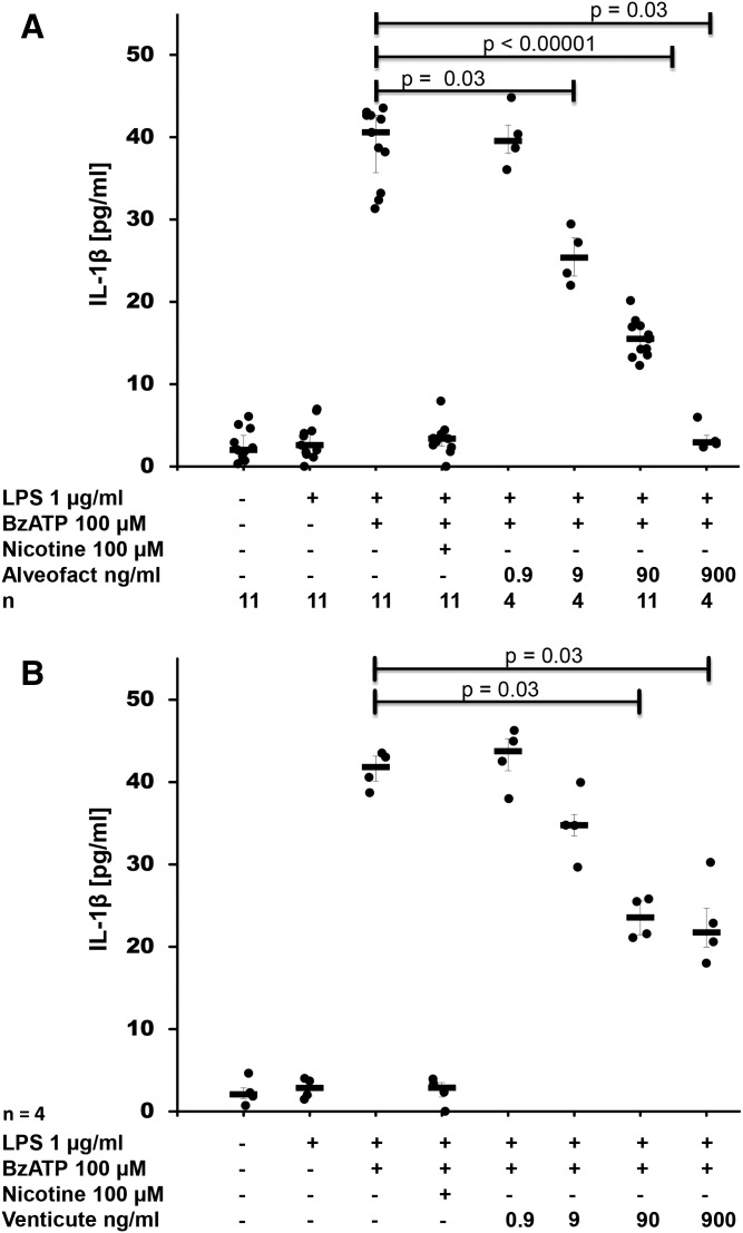
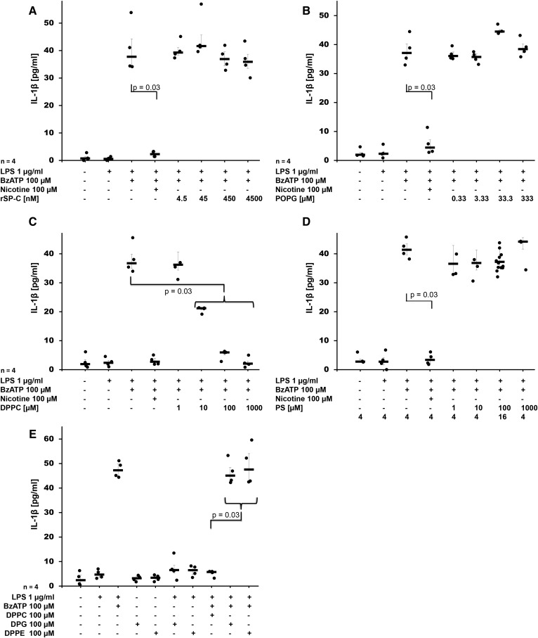
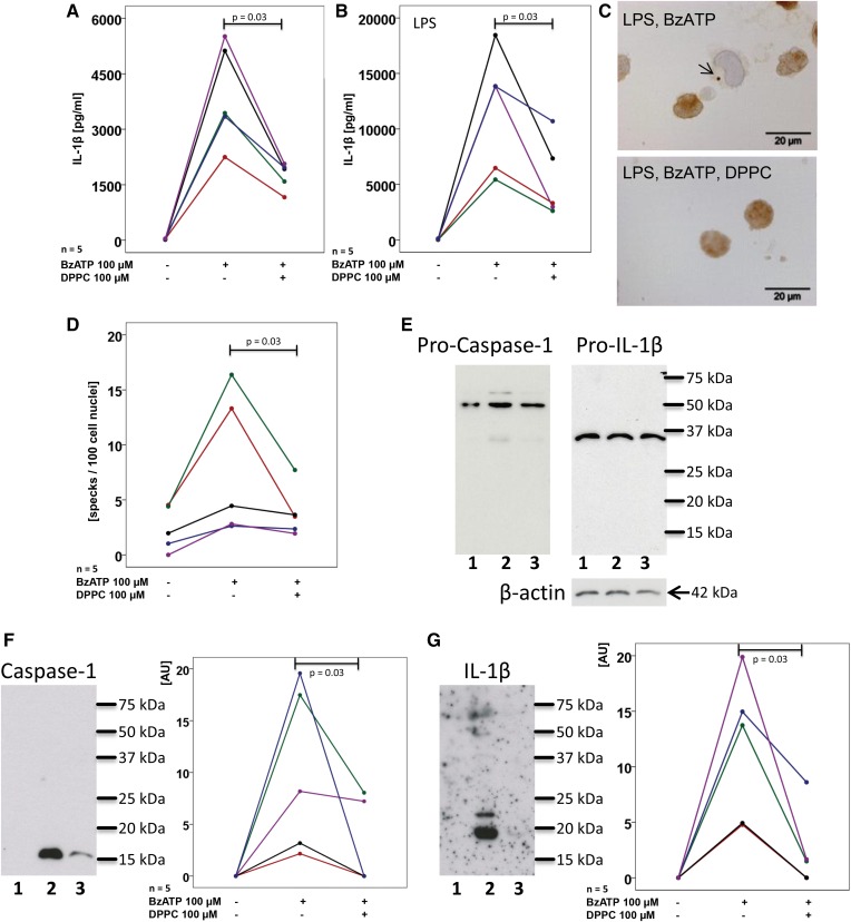
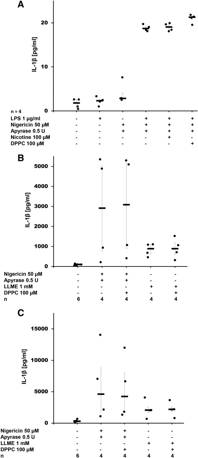
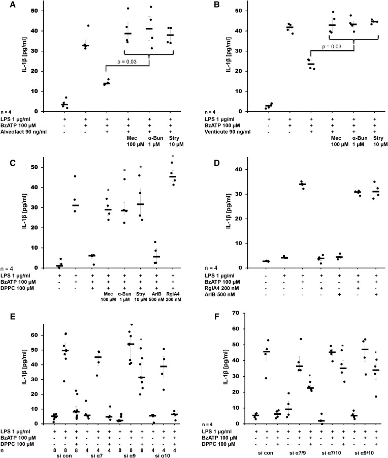
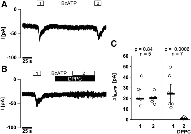
Interleukin (IL)-1β is a potent pro-inflammatory cytokine of innate immunity involved in host defense. High systemic IL-1β levels, however, cause life-threatening inflammatory diseases, including systemic inflammatory response syndrome. In response to various danger signals, the pro-form of IL-1β is synthesized and stays in the cytoplasm unless a second signal, such as extracellular ATP, activates the inflammasome, which enables processing and release of mature IL-1β. As pulmonary surfactant is known for its anti-inflammatory properties, we hypothesize that surfactant inhibits ATP-induced release of IL-1β. Lipopolysaccharide-primed monocytic U937 cells were stimulated with an ATP analog in the presence of natural or synthetic surfactant composed of recombinant surfactant protein (rSP)-C, palmitoylphosphatidylglycerol, and dipalmitoylphosphatidylcholine (DPPC). Both surfactant preparations dose-dependently inhibited IL-1β release from U937 cells. DPPC was the active constituent of surfactant, whereas rSP-C and palmitoylphosphatidylglycerol were inactive. DPPC was also effective in primary mononuclear leukocytes isolated from human blood. Experiments with nicotinic antagonists, siRNA technology, and patch-clamp experiments suggested that stimulation of nicotinic acetylcholine receptors (nAChRs) containing subunit α9 results in a complete inhibition of the ion channel function of ATP receptor, P2X7. In conclusion, the surfactant constituent, DPPC, efficiently inhibits ATP-induced inflammasome activation and maturation of IL-1β in human monocytes by a mechanism involving nAChRs.

Keywords: CHRNA10; CHRNA7; CHRNA9; dipalmitoylphosphatidylcholine; inflammasome; monocyte; phosphatidylcholine; purinergic receptor P2X 7.


Figures

Fig. 1.
Fig. 1.
Surfactant dose-dependently inhibits BzATP-mediated release of IL-1β. Different concentrations of the natural surfactant preparation, Alveofact® (A), and the synthetic surfactant, Venticute® (B), were added to LPS-primed U937 cells together with BzATP. IL-1β levels were measured 30 min thereafter in cell culture supernatants. Nicotine was included as a known inhibitor of BzATP-dependent IL-1β release. A Kruskal-Wallis test was followed by Mann-Whitney rank-sum test; data are presented as individual data points; bars represent median; whiskers represent percentiles 25 and 75.
Fig. 2.
Fig. 2.
DPPC is the active component of surfactant that dose-dependently inhibits BzATP-mediated release of IL-1β. Different concentrations of rSP-C (A), POPG (B), DPPC (C), PS (D), DPPE (E), or DPG (E) were added to LPS-primed U937 cells together with BzATP. IL-1β levels were measured 30 min thereafter in cell culture supernatants. Nicotine was included as a known inhibitor of BzATP-dependent IL-1β release. A Kruskal-Wallis test was followed by Mann-Whitney rank-sum test; data are presented as individual data points; bars represent median; whiskers represent percentiles 25 and 75.
Fig. 3.
Fig. 3.
DPPC inhibits BzATP-mediated inflammasome activation in human PBMCs. PBMCs were isolated from healthy human donors, left untreated (A) or pulsed with LPS (B), cultured for 3 h and stimulated with BzATP in the presence or absence of DPPC. IL-1β levels were measured 30 min thereafter in cell culture supernatants. C, D: ASC specks were detected by immunocytochemistry in LPS-pulsed PBMC and stimulated with BzATP in the presence or absence of DPPC. C: Micrographs of PBMC: ASC immunopositive material is stained in brown, cell nuclei were lightly counterstained with hemalum, and the arrow is pointing to an ASC speck. D: The number of specks per 100 cell nuclei is depicted. E–G: PBMCs were pulsed with LPS (lane 1) and stimulated with BzATP in the absence (lane 2) or presence (lane 3) of DPPC and Western blots were performed with antibodies directed to human caspase-1, IL-1β, and β-actin. E: In cell lysates, exclusively, the pro-forms, pro-caspase-1 and pro-IL-1β, were detected; whereas, in concentrated cell culture supernatants, mature caspase-1 (F) and IL-1β (G) were detected. F, G: Immunopositive signals were quantified by densitometry and expressed as arbitrary units (AU). Data points obtained from individual blood donors are depicted and connected by lines. Data from the same individuals are coded by the same color in (A) and (B) as well as in (F) and (G), but in (A) and (B) a cohort of volunteers was investigated that was different than in (F) and (G). Wilcoxon sign rank test.
Fig. 4.
Fig. 4.
DPPC does not inhibit nigericin- or LLME-induced release of IL-1β. A: DPPC was added to LPS-primed U937 cells together with nigericin. IL-1β levels were measured 30 min after application of nigericin in cell culture supernatants. Nicotine was included as a known inhibitor of BzATP-dependent IL-1β release. B, C: Human PBMCs were isolated from healthy human donors, left untreated (B) or pulsed with LPS (C), cultured for 3 h and stimulated with nigericin or LLME in the presence or absence of DPPC. IL-1β levels were measured 30 min thereafter in cell culture supernatants. Apyrase was included (A–C) to hydrolyze ATP that might have been released by U937 cells. A Kruskal-Wallis test was followed by Mann-Whitney rank-sum test; data are presented as individual data points; bars represent median; whiskers represent percentiles 25 and 75.
Fig. 5.
Fig. 5.
The inhibitory effect of surfactant and DPPC on the BzATP-mediated release of IL-1β by LPS-primed U937 cells is mediated by nAChR subunits α7, α9, and α10. Natural surfactant Alveofact® (A), synthetic surfactant Venticute® (B), or DPPC (C) were added to LPS-primed U937 cells together with BzATP in the presence and absence of the nicotinic antagonists mecamylamine (Mec), α-bungarotoxin (α-Bun), strychnine (Stry), RgIA4, or ArIB[V11L,V16D] (ArIB). IL-1β levels were measured 30 min thereafter in cell culture supernatants. In LPS-primed U937 cells that were transfected with control siRNA (si con) the BzATP-stimulated IL-1β release was inhibited by DPPC Conotoxins RgIA4 or ArIB did neither induce the release of IL-1beta by LPS-primed U937 cells nor impair the effects of BzATP. (E, F). E: After transfection of siRNA to nAChR subunit α9, the effect of DPPC was blunted, whereas silencing of subunit α7 and α10 or the use of control siRNA (si con) did not provoke any effect. F: When double knock-down experiments were performed, the inhibitory effect of DPPC was impaired in all siRNA combinations investigated (α7/9, α7/10, α9/10). A Kruskal-Wallis test was followed by a Mann-Whitney rank-sum test; data are presented as individual data points; bars represent median; whiskers represent percentiles 25 and 75; *P ≥ 0.03 compared with respective experiments performed on cells treated with si con.
Fig. 6.
Fig. 6.
DPPC inhibits BzATP-induced ion currents in LPS-primed U937 cells. Whole cell patch-clamp recordings of U937 cells are depicted in (A, B), and changes in ion currents in response to BzATP (ΔIBzATP) are summarized in (C). Two consecutive BzATP applications (1 and 2) resulted in repeatable ion current changes that were abolished in the presence of DPPC (100 μM). Data are presented as individual data points; bars indicate median; whiskers represent percentiles 25 and 75.

References

    1. Hiemstra P. S. 2007. The role of epithelial beta-defensins and cathelicidins in host defense of the lung. Exp. Lung Res. 33: 537–542. - PubMed
    1. Alkhouri H., Poppinga W. J., Tania N. P., Ammit A., and Schuliga M.. 2014. Regulation of pulmonary inflammation by mesenchymal cells. Pulm. Pharmacol. Ther. 29: 156–165. - PubMed
    1. Bhattacharya J., and Westphalen K.. 2016. Macrophage-epithelial interactions in pulmonary alveoli. Semin. Immunopathol. 38: 461–469. - PMC - PubMed
    1. Robb C. T., Regan K. H., Dorward D. A., and Rossi A. G.. 2016. Key mechanisms governing resolution of lung inflammation. Semin. Immunopathol. 38: 425–448. - PMC - PubMed
    1. Chen G. Y., and Nuñez G.. 2010. Sterile inflammation: sensing and reacting to damage. Nat. Rev. Immunol. 10: 826–837. - PMC - PubMed

Publication types

MeSH terms

LinkOut - more resources