. 2017 May 1;216(5):1357-1369.
doi: 10.1083/jcb.201609022. Epub 2017 Apr 12.

Preprotein mature domains contain translocase targeting signals that are essential for secretion

Affiliations

Preprotein mature domains contain translocase targeting signals that are essential for secretion

Katerina E Chatzi et al. J Cell Biol. .

Abstract

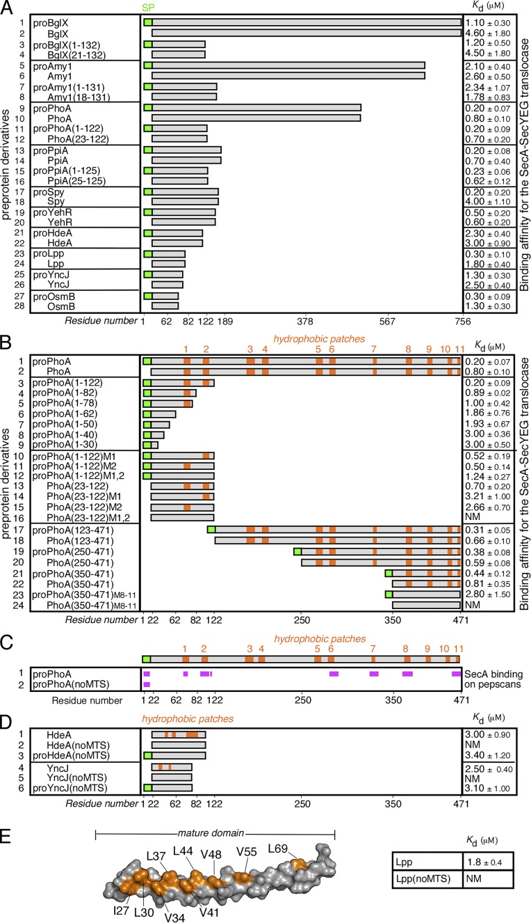
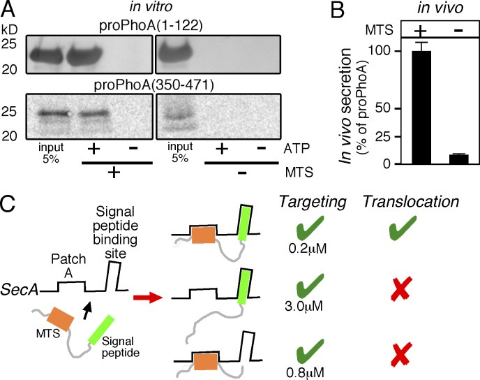
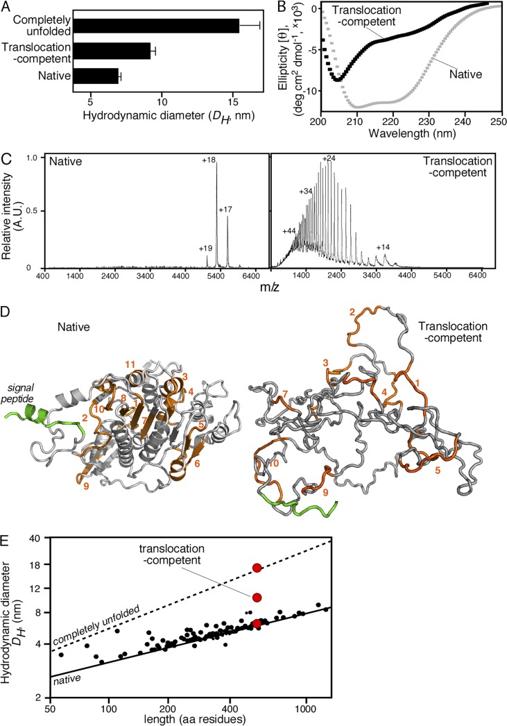
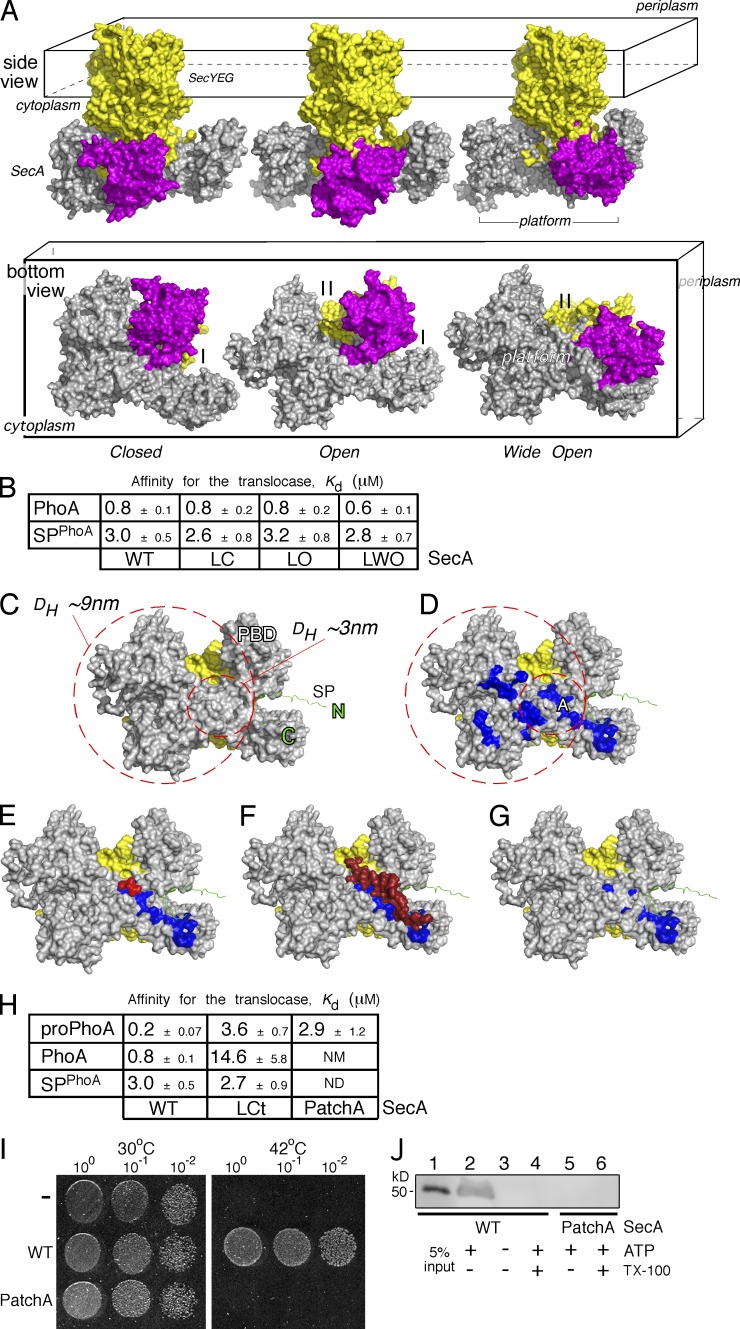
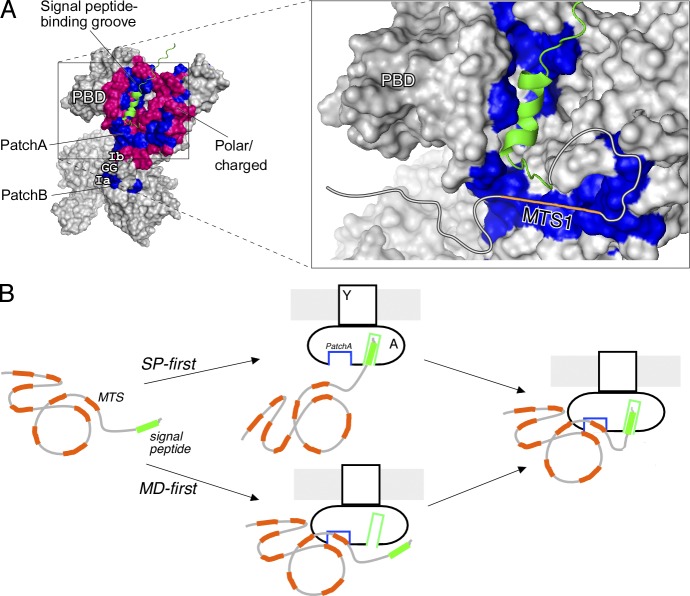
Secretory proteins are only temporary cytoplasmic residents. They are typically synthesized as preproteins, carrying signal peptides N-terminally fused to their mature domains. In bacteria secretion largely occurs posttranslationally through the membrane-embedded SecA-SecYEG translocase. Upon crossing the plasma membrane, signal peptides are cleaved off and mature domains reach their destinations and fold. Targeting to the translocase is mediated by signal peptides. The role of mature domains in targeting and secretion is unclear. We now reveal that mature domains harbor their own independent targeting signals (mature domain targeting signals [MTSs]). These are multiple, degenerate, interchangeable, linear or 3D hydrophobic stretches that become available because of the unstructured states of targeting-competent preproteins. Their receptor site on the cytoplasmic face of the SecYEG-bound SecA is also of hydrophobic nature and is located adjacent to the signal peptide cleft. Both the preprotein MTSs and their receptor site on SecA are essential for protein secretion. Evidently, mature domains have their own previously unsuspected distinct roles in preprotein targeting and secretion.


Figures

Figure 1.
Figure 1.
MTSs in preproteins. (A) Equilibrium dissociation constants (Kd; micromoles; right) of the indicated preproteins, their mature domains and truncated analogues for the wild-type SecA-SecYEG translocase. No detectable binding of proPhoA or derivatives occurs to the SecYEG-inverted membrane vesicles in the absence of SecA (for detailed analysis, see Gouridis et al., 2009). n = 3–9. x axis indicates preprotein length in residues. SP, signal peptide. (B) Hydrophobic patches (HPs; orange) in the mature domain of PhoA (see also Fig. S1 A) and their contribution to targeting. The Kd of the indicated protein derivatives for the translocase were determined; n = 3–9. M1,2 and M8–11, hydrophobicity-reducing mutations in the indicated HPs of PhoA; the mutated residues are detailed in Fig. S1 A. x axis: proPhoA residues. (C) Binding experiments of soluble SecA onto proPhoA or proPhoA(noMTS) peptide arrays are summarized; n = 6 (see also Fig. S2 A). HPs are indicated; x axis: proPhoA residues. (D) HPs (orange) in the secretory proteins HdeA and YncJ (see also Fig. S1 A) and their contribution to targeting. The Kd of the indicated protein derivatives for the translocase were determined; n = 3–6. noMTS, hydrophobicity-reducing mutations in all HPs; the mutated residues are detailed in Fig. S1 A. (E) Left: 3D surface representation of the Lpp structure (PDB: 1EQ7; a single protomer is shown). The residues that were mutated in Lpp(noMTS), shown in orange, are detailed in Fig. S1 B. The Kd of Lpp(noMTS) for the translocase was determined (right); n = 3. NM in C–E, nonmeasurable binding for the translocase (i.e., >20 µM). Affinity values in A, B, D, and E represent means ± SEM.
Figure 2.
Figure 2.
MTSs are essential for protein secretion. (A) Representative, in vitro, SecA-dependent translocation assays of proPhoA(1–122)M1,2 compared with proPhoA(1–122) (top) and proPhoA(350–471)M8-11 compared with proPhoA(350–471) (bottom) into the lumen of SecYEG containing inverted membrane vesicles; n = 3. 5% of the input is indicated. (B) In vivo secretion of proYncJ-PhoA (left) and proYncJ(noMTS)-PhoA (right) was compared with that of proPhoA (considered as 100%) under identical conditions (MC4100 cells; OD600 = 0.2; 0.002% wt/vol arabinose; 30min; 30°C). In all cases, the measured PhoA enzymatic activity (Gouridis et al., 2009) was normalized to the amount of PhoA or fusion-PhoA protein produced. This provided a means to quantitate the in vivo secretion of all three proteins. The alkaline phosphatase units per microgram PhoA for cells expressing proPhoA was considered 100%; proYncJ-PhoA and proYncJ(noMTS)-PhoA values were expressed as a percentage of this value. n = 5. Values are expressed as means ± SEM. (C) Schematic summary of requirements for preprotein targeting and translocation (as indicated). Preproteins are bivalent ligands with distinct binding sites on SecA. Targeting to the translocase is efficiently achieved by either targeting element, the signal peptide (middle) or one/more MTSs (bottom), independently. However, preprotein translocation requires binding of both (top).
Figure 3.
Figure 3.
Biophysical characterization of translocation-competent proPhoA. (A) Hydrodynamic diameter (DH, nanometers; x axis) of native (no urea; no DTT), translocation-competent (no urea; 1 mM DTT) and completely unfolded proPhoA (8M urea; 1 mM DTT) as determined by quasielastic laser light scattering that was performed online after gel permeation chromatography on a Superdex HR200 (see also Fig. S3 A); n = 6–15. Values represent means ± SD. For the native species, natively purified proPhoA was diluted and chromatographed in buffer L. For the translocation-competent and the completely unfolded species, urea-purified proPhoA (0.5 mM; 6 M urea), preincubated with DTT (10mM; 30min; ice), was diluted and chromatographed in buffer L supplemented with the indicated urea and DTT concentration. (B) Representative circular dichroism spectra of natively folded (no DTT) and translocation-competent (1 mM DTT) proPhoA; n > 3. x axis: wavelength (nanometers); y axis: mean residue molar ellipticity ([θ]MRW). For the translocation-competent species, urea-purified proPhoA was preincubated with 10 mM DTT (30 min; ice) and dialyzed in buffer U supplemented with 8M urea and 1 mM DTT. The natively purified proPhoA was dialyzed in 5 liters buffer U (15 h; 4°C). Spectra for both were recorded in buffer U supplemented with 1 mM EDTA, 0.2 M urea, and DTT (as indicated). Natively folded proPhoA exhibits two minima (208 and 222 nm) typical of folded, predominantly α-helical proteins, whereas the translocation-competent proPhoA does not. The urea-purified proPhoA, dialyzed in buffer U in the absence of 5 liters DTT (15 h), folds and gives spectra similar to those of the natively purified proPhoA (not depicted). (C) Representative native nano–electrospray ionization mass spectrometry spectra of native PhoA and translocation-competent proPhoA; n = 3. Translocation-competent proPhoA acquires many charges with broad distribution, typical of unfolded proteins with increased solvent-accessible surface area (Testa et al., 2013), whereas native PhoA acquires few charges with narrow distribution, typical of well-folded, compact proteins, and is a dimer. (D) Ribbon 3D model of folded E. coli PhoA (PDB: 1KHN; a single protomer is shown; left). A signal peptide (green) was modeled. Model of disordered proPhoA derived from the trigger factor–bound structure solved by nuclear magnetic resonance (Saio et al., 2014), with a DH in accordance with the quasielastic laser light scattering measurements of disordered PhoA (right). (E) Predicted and measured hydrodynamic diameters (DH) of SecA-dependent preproteins. Lines show the predicted DH of either folded (solid) or completely unfolded (dotted) preproteins as a function of their length (Wilkins et al., 1999b). Small black dots represent the calculated DH for 40 mature domains with solved structures (Table S8), using Hydropro (García De La Torre et al., 2000). Red circles represent the experimental DH measurements for proPhoA (as indicated; see also A and Fig. S3 B).
Figure 4.
Figure 4.
The mature domain–binding site onto SecA. (A) The E. coli SecA (gray)–SeYEG (yellow) was modeled after the Thermotoga maritima translocase in three conformational states, based on PBD (purple) positioning: closed (left), open (middle), and wide open (right). Side and bottom views are shown (as indicated). I, II: SecA clamps. (B) Kd measurements of PhoA and its signal peptide (SPPhoA) for the wild-type (WT), locked closed (LC), locked open (LO), and locked wide open (LWO) SecA bound to SecYEG-inverted membrane vesicles. proPhoA(1–30) was used as SPPhoA. Affinity values represent means ± SEM; n = 3. (C) Potential space occupied by an incoming preprotein onto the cytoplasmic side (platform) of a SecA(gray)–SecYEG(yellow) translocase; signal peptide is in green. The inner circle represents the minimum area a translocation-competent preprotein would occupy, depicted here by the predicted DH of the smallest known preprotein (proEcnA; ∼3 nm; Table S8). The bigger circle represents the area that the expanded, translocation-competent proPhoA would occupy, based on our experimental measurements (∼7 nm; Figs. 3 A and S3 A). (D) Hydrophobic patches (blue; PatchA is indicated) on SecA’s cytoplasmic platform (the rest as in C). (E–G) Structural models of (E) a tripeptide (red; Zimmer and Rapoport, 2009) bound on PatchA (blue) of SecA (detailed interactions in Fig. S5 B) and (F) the C-tail of SecA (dark red) shielding PatchA (blue; Hunt et al., 2002). The C-tail directly interacts with or shields PatchA residues (detailed interactions in Fig. S5 B) but only partially occludes the signal peptide–binding site on SecA (Gelis et al., 2007; see also Fig. S6 E). Signal peptide–binding-determinant residues (i.e., L306; Fig. S6 E) remain exposed and available for interaction at this state. (G) SecA(PatchA) mutant. Four conserved PatchA amino acids (see also Fig. S5 C) were substituted with alanyl residues (M191A/F193A/I224A/I225A) to disrupt the continuum of its hydrophobic surface. (H) Kd measurements of PhoA, proPhoA, and its signal peptide (SPPhoA) for the wild-type, locked C-tail (LCt), and PatchA SecA bound to SecYEG-inverted membrane vesicles; Affinity values represent means ± SEM; n = 3–12. proPhoA(1–30) was used as SPPhoA. NM: nonmeasurable binding (i.e., >20 µM). (I) Representative, in vivo, genetic complementation assay of the E. coli BL21.19(secAts) strain by either an empty vector (−) or wild-type secA or secA(PatchA) mutant; n = 3. Only wild-type secA allows cell growth at 42°C. An identical plate, grown in parallel, at 30°C is shown; the dilutions of cells that were used are indicated. (J) Representative, in vitro SecA-dependent translocation of proPhoA into wt SecYEG-inverted membrane vesicles using wild-type SecA or SecA(PatchA) mutant under the same conditions; n = 3. 5% of the proPhoA input is indicated. Migration of ovalbumin (prestained protein molecular mass marker; Thermo Fisher Scientific).
Figure 5.
Figure 5.
Preprotein docking on SecA and targeting to the translocase. (A) Structural model for preprotein docking on SecA. Complete view of the SecA features surrounding PatchA and the signal peptide binding groove (left; blue, nonpolar SecA residues; pink, polar/charged SecA residues; green, signal peptide). Zoom-in view of PatchA and the signal peptide–binding groove (right). The signal peptide–binding site on SecA lies within a hydrophobic groove between the PBD and IRA1 (Gelis et al., 2007; for details, see also Fig. S6, C and D). PatchA is a shallow furrow that converges at a 90° angle, forming an “L” shape with the signal peptide groove, and is surrounded by motives (Ia, Ib, and GG) on which helicases bind their nucleic acid substrates (e.g., RNA helicases; Sengoku et al., 2006; Papanikolau et al., 2007). A hypothetical mature domain is drawn as an extension of the signal peptide, bound on PatchA with its MTS1 (orange), based on the mainly hydrophobic nature of the interaction (Fig. 1 and Materials and methods, Bioinformatics approach to define hydrophobic patches on proteins). Additional minor electrostatic or polar contacts might contribute in the association of some mature domains with SecA. (B) Model of preprotein targeting/docking to the Sec translocase initiated by stochastic binding of either the signal peptide (SP-first) or the mature domain (MD-first; see text for details). A, SecA; Y, SecY. Green, signal peptide; orange, mature domain. Only one SecA protomer is shown (the one activated for high-affinity preprotein binding; Gouridis et al., 2013). The second regulatory protomer is omitted for simplicity.

Similar articles

Cited by

References

    1. Bauer B.W., and Rapoport T.A.. 2009. Mapping polypeptide interactions of the SecA ATPase during translocation. Proc. Natl. Acad. Sci. USA. 106:20800–20805. 10.1073/pnas.0910550106 - DOI - PMC - PubMed
    1. Beckwith J. 2013. The Sec-dependent pathway. Res. Microbiol. 164:497–504. 10.1016/j.resmic.2013.03.007 - DOI - PMC - PubMed
    1. Beerten J., Jonckheere W., Rudyak S., Xu J., Wilkinson H., De Smet F., Schymkowitz J., and Rousseau F.. 2012. Aggregation gatekeepers modulate protein homeostasis of aggregating sequences and affect bacterial fitness. Protein Eng. Des. Sel. 25:357–366. 10.1093/protein/gzs031 - DOI - PubMed
    1. Bieker K.L., Phillips G.J., and Silhavy T.J.. 1990. The sec and prl genes of Escherichia coli. J. Bioenerg. Biomembr. 22:291–310. 10.1007/BF00763169 - DOI - PubMed
    1. Blobel G., and Dobberstein B.. 1975. Transfer of proteins across membranes. I. Presence of proteolytically processed and unprocessed nascent immunoglobulin light chains on membrane-bound ribosomes of murine myeloma. J. Cell Biol. 67:835–851. 10.1083/jcb.67.3.835 - DOI - PMC - PubMed

MeSH terms

LinkOut - more resources