Review
. 2017 Jul;30(3):615-645.
doi: 10.1128/CMR.00005-17.

Toxoplasma Effectors Targeting Host Signaling and Transcription

Affiliations
Review

Toxoplasma Effectors Targeting Host Signaling and Transcription

Mohamed-Ali Hakimi et al. Clin Microbiol Rev. 2017 Jul.

Abstract

Early electron microscopy studies revealed the elaborate cellular features that define the unique adaptations of apicomplexan parasites. Among these were bulbous rhoptry (ROP) organelles and small, dense granules (GRAs), both of which are secreted during invasion of host cells. These early morphological studies were followed by the exploration of the cellular contents of these secretory organelles, revealing them to be comprised of highly divergent protein families with few conserved domains or predicted functions. In parallel, studies on host-pathogen interactions identified many host signaling pathways that were mysteriously altered by infection. It was only with the advent of forward and reverse genetic strategies that the connections between individual parasite effectors and the specific host pathways that they targeted finally became clear. The current repertoire of parasite effectors includes ROP kinases and pseudokinases that are secreted during invasion and that block host immune pathways. Similarly, many secretory GRA proteins alter host gene expression by activating host transcription factors, through modification of chromatin, or by inducing small noncoding RNAs. These effectors highlight novel mechanisms by which T. gondii has learned to harness host signaling to favor intracellular survival and will guide future studies designed to uncover the additional complexity of this intricate host-pathogen interaction.

Keywords: chromatin remodeling; epigenetics; immune evasion; innate immunity; intracellular pathogen; serine/threonine kinases; signal transduction; transcription factors.


Figures

FIG 1
FIG 1
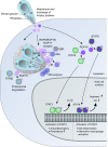
Rhoptry effectors that target host pathways. Following attachment to the host cell, rhoptry (ROP) effector proteins are released into the host cell cytosol prior to the entry of the parasite into the parasitophorous vacuole (PV). ROP proteins are found in the cytosol, traffic to the host nucleus, and also decorate the surface of the vacuole. The secreted kinase ROP18 is assembled on the PV membrane, where it forms complexes with another kinase, ROP17, and the pseudokinase ROP5. By the phosphorylation of IRG monomers/dimers (ROP18) and polymers (ROP17), these kinases prevent the accumulation of IRGs on the PV membrane. The pseudokinase ROP5 binds Irga6 directly and enhances the kinase activity of ROP18. The ROP5/ROP18/ROP17 complex also contains the transmembrane protein GRA7, originating from a parasite organelle named the dense granule. GRA7 can also bind directly to IRG polymers, and it accelerates their turnover. ROP18 has also been shown to phosphorylate the transcription factor ATF6β, marking it for proteosomal degradation. Another secretory rhoptry kinase, ROP16, activates the transcription factors STAT3 and STAT6 by direct phosphorylation, thus altering host transcription.
FIG 2
FIG 2
Secreted T. gondii effectors from dense granules transform host cell signaling pathways. After invasion of the host cell, T. gondii uses a large variety of effector proteins originating from dense granule organelles to manipulate host signaling pathways and gene expression. Some of these proteins translocate to the host nucleus (TgIST, GRA24, and GRA16), while others localize to the PV membrane (GRA15) or only partially interact with the host cell cytosol while residing in the PV (GRA6). Present in all T. gondii type stains, the dense granule protein TgIST globally blocks the interferon (IFN) response by the recruitment of the Mi-2/NuRD repressor complex to STAT1 binding sites in promoter regions of responsive genes. GRA24 bypasses the classical MAPK phosphorylation cascades by forming a complex with p38α, which is able to activate transcription factors such as EGR1 and c-Fos. GRA16 shuttles to the host nucleus while bound to a high-molecular-weight complex, including PP2A-B55 and HAUSP, to control p53 levels. In type 2 strains, GRA15 activates the NF-κB pathway by the activation of TRAF6, which subsequently activates IKK, leading to the phosphorylation and degradation of IκB. GRA6 has a vacuole-restricted location, and its cytosolic region interacts with CAMLG to activate calcineurin and stimulate the transcription factor NFAT4.
FIG 3
FIG 3
Mechanisms of protein export and traffic beyond the PV. The aspartyl protease ASP5 is situated at the Golgi apparatus of the parasite. The cleavage of some (i.e., GRA15, GRA16, and TgIST) but not all (i.e., GRA24) dense granule proteins in their recognition signal by ASP5 is necessary for the export of these proteins. MYR1 is part of a protein complex located at the PV membrane, where it is involved in the export of intrinsically disordered dense granule proteins across the vacuole membrane and into the host cytosol. A number of these export substrates are processed by ASP5. Two additional dense granule proteins, GRA17 and GRA23, which are also located at the PV membrane, are responsible for small-molecule transport between the host cytosol and the vacuole lumen.
None
None
None

Similar articles

Cited by

References

    1. Dubey JP. 2010. Toxoplasmosis of animals and humans. CRC Press, Boca Raton, FL.
    1. Levine ND. 1988. The protozoan phylum Apicomplexa, vol 1 and 2. CRC Press, Boca Raton, FL.
    1. Pawlowski J, Audic S, Adl S, Bass D, Belbahri L, Berney C, Bowser SS, Cepicka I, Decelle J, Dunthorn M, Fiore-Donno AM, Gile GH, Holzmann M, Jahn R, Jirku M, Keeling PJ, Kostka M, Kudryavtsev A, Lara E, Lukes J, Mann DG, Mitchell EA, Nitsche F, Romeralo M, Saunders GW, Simpson AG, Smirnov AV, Spouge JL, Stern RF, Stoeck T, Zimmermann J, Schindel D, de Vargas C. 2012. CBOL protist working group: barcoding eukaryotic richness beyond the animal, plant, and fungal kingdoms. PLoS Biol 10:e1001419. doi:10.1371/journal.pbio.1001419. - DOI - PMC - PubMed
    1. Bartosova-Sojkova P, Oppenheim RD, Soldati-Favre D, Lukes J. 2015. Epicellular apicomplexans: parasites “on the way in.” PLoS Pathog 11:e1005080. doi:10.1371/journal.ppat.1005080. - DOI - PMC - PubMed
    1. Barta JR. 1989. Phylogenetic analysis of the class Sporozoea (phylum Apicomplexan Levine 1970): evidence for the independent evolution of heteroxenous life cycles. J Parasitol 75:195–206. doi:10.2307/3282766. - DOI - PubMed

Publication types