

Prognostic significance of tumor-infiltrating immune cells and PD-L1 expression in esophageal squamous cell carcinoma
- PMID: 28404915
- PMCID: PMC5444735
- DOI: 10.18632/oncotarget.15621
Prognostic significance of tumor-infiltrating immune cells and PD-L1 expression in esophageal squamous cell carcinoma
Abstract
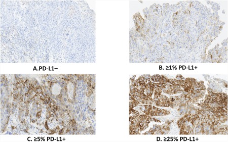
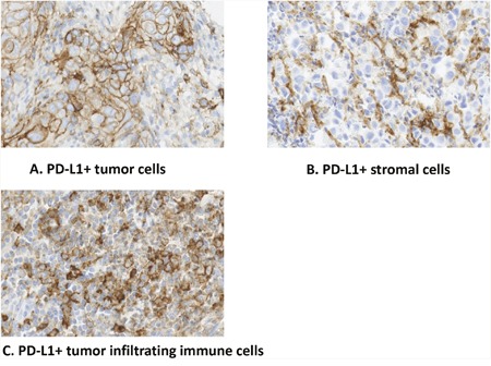
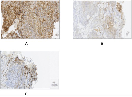
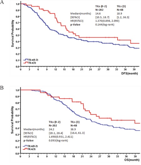
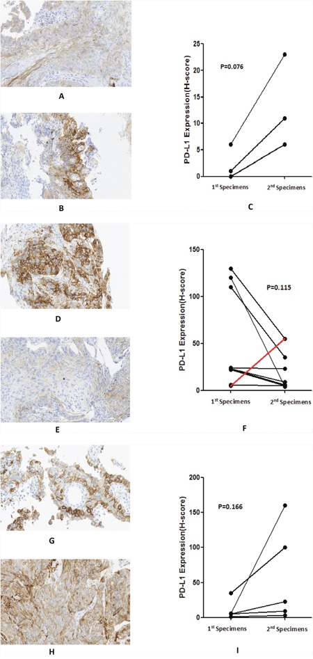
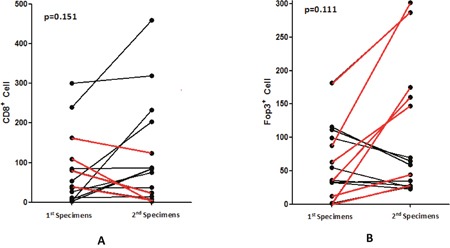
Programmed death-1 receptor (PD-1) and its ligand (PD-L1) play an integral role in regulating the immune response against cancer. This study investigated the prognostic significance of PD-L1 expression on tumor cells and tumor-infiltrating immune cells (TILs) in the tumor microenvironment in Chinese patients with esophageal squamous cell carcinoma (ESCC). Archival formalin-fixed, paraffin-embedded ESCC samples from treatment-naïve patients with ESCC after surgery or by diagnostic endoscopic biopsy were collected between 2004 and 2014. Expression of PD-L1 in ESCC tumor specimens was assessed by immunohistochemistry (IHC), and the degree of TIL infiltration was evaluated by examining hematoxylin and eosin-stained (H&E) specimens. PD-L1+ as defined as ≥1% of tumor cell membranes showing ≥1+ intensity. In 428 patients, specimens from 341 (79.7%) were PD-L1+. In the definitive treatment group (patients who received curative esophagectomy or definitive [chemo-]radiation therapy), PD-L1 positivity was associated with a significantly shorter DFS and OS. In the palliative chemotherapy group exhibited, neither PFS nor OS correlated significantly with PD-L1 expression. PD-L1 expression was positively associated with TIL density. In 17 paired tumor tissues collected before and after treatment, an increase in PD-L1 expression was associated with disease progression, whereas a decrease in PD-L1 expression was associated with response to chemotherapy or disease control. So, PD-L1 expression was associated with a significantly worse prognosis in patients with ESCC. These observations suggest that PD-L1 may play a critical role in ESCC cancer progression and provide a rationale for developing PD-L1 inhibitors for treatment of a subset of ESCC patients.
Keywords: PD-L1; TILs; esophageal squamous cell carcinoma; prognostic factor; tumor microenvironment.
Conflict of interest statement
The Authors do not have any conflicts of interest.
Figures

References
-
- Rustgi AK, El-Serag HB. Esophageal carcinoma. N Engl J Med. 2014;371:2499–509. - PubMed
-
- Hagen P, Hulshof MC, Lanschot JJ, Steyerberg EW, van Berge Henegouwen MI, Wijnhoven BP, Richel DJ, Nieuwenhuijzen GA, Hospers GA, Bonenkamp JJ, Cuesta MA, Blaisse RJ, Busch OR, et al. Preoperative chemoradiotherapy for esophageal or junctional cancer. N Engl J Med. 2012;366:2074–2084. - PubMed
-
- Wheeler J B, Reed C E. Epidemiology of esophageal cancer. SurgClin North Am. 2012;9:1077–1087. - PubMed
-
- Yamasaki M, Miyata H, Tanaka K, Shiraishi O, Motoori M, Peng YF, Yasuda T, Yano M, Shiozaki H, Mori M, Doki Y. Multicenter phase I / II study of docetaxel, cisplatin and fluorouracil combination chemotherapy in patients with advanced or recurrent squamous cell carcinoma of the esophagus. Oncology. 2011;80:307–13. - PubMed
MeSH terms
Substances
LinkOut - more resources
Full Text Sources
Other Literature Sources
Medical
Research Materials
