. 2017 Apr 20;544(7650):367-371.
doi: 10.1038/nature22038. Epub 2017 Apr 12.

Therapeutic reduction of ataxin-2 extends lifespan and reduces pathology in TDP-43 mice

Affiliations

Therapeutic reduction of ataxin-2 extends lifespan and reduces pathology in TDP-43 mice

Lindsay A Becker et al. Nature. .

Abstract

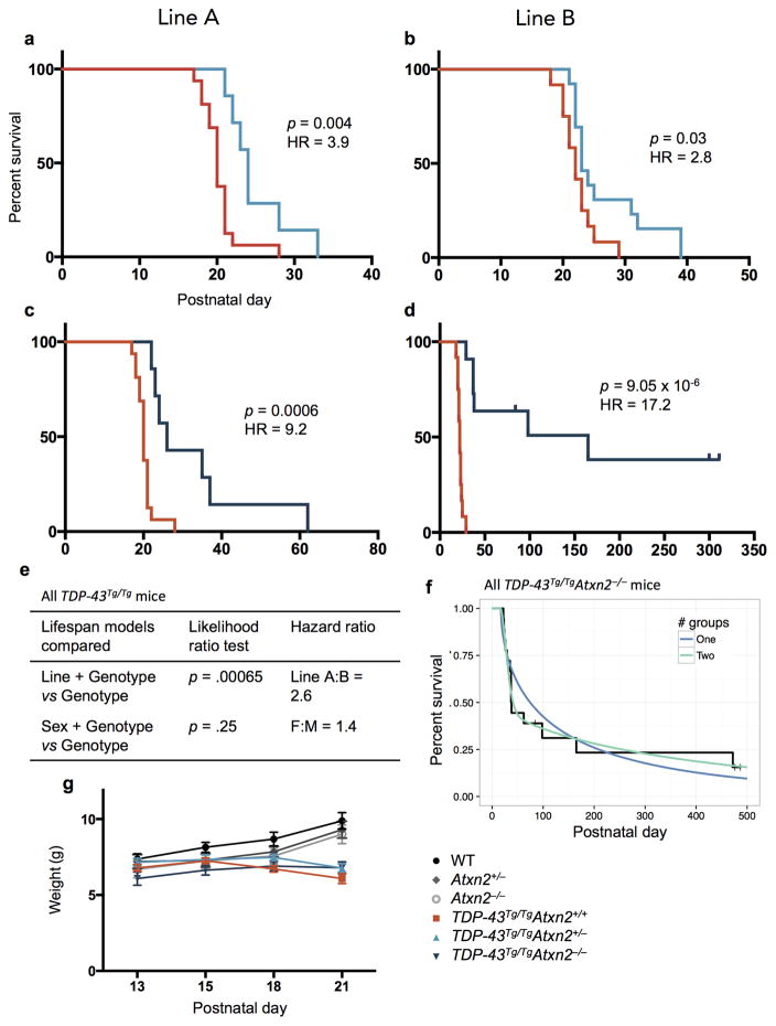
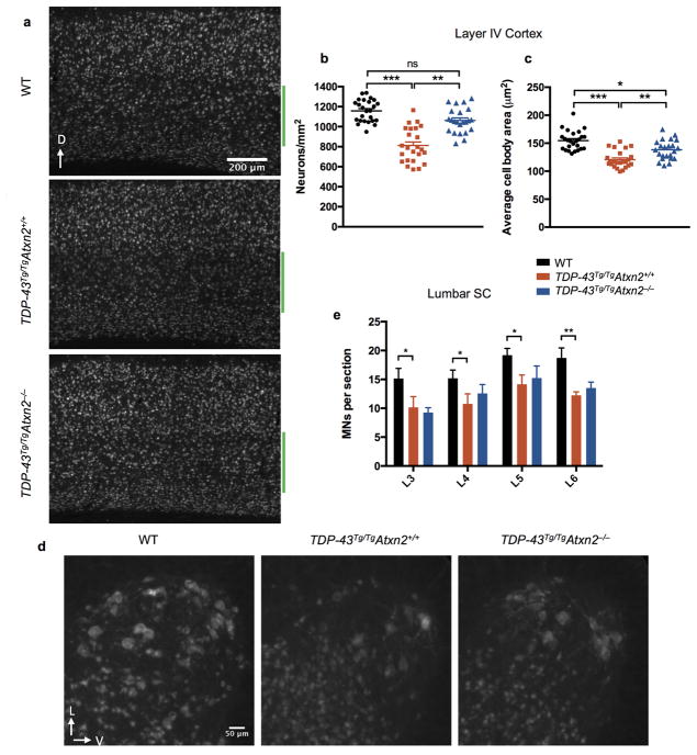
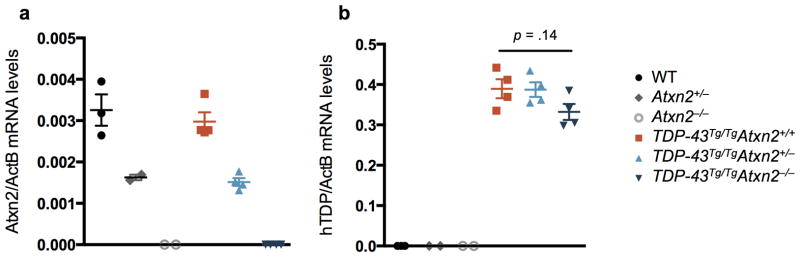
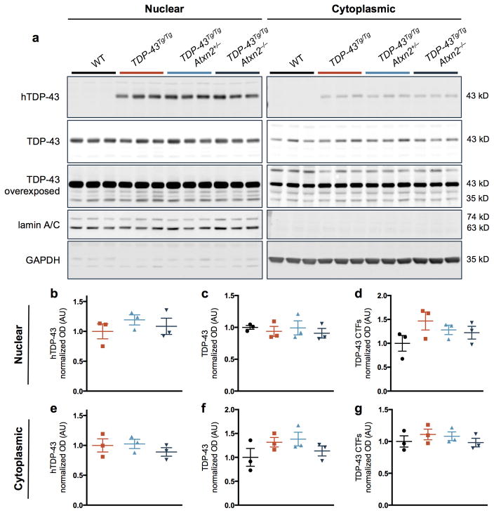
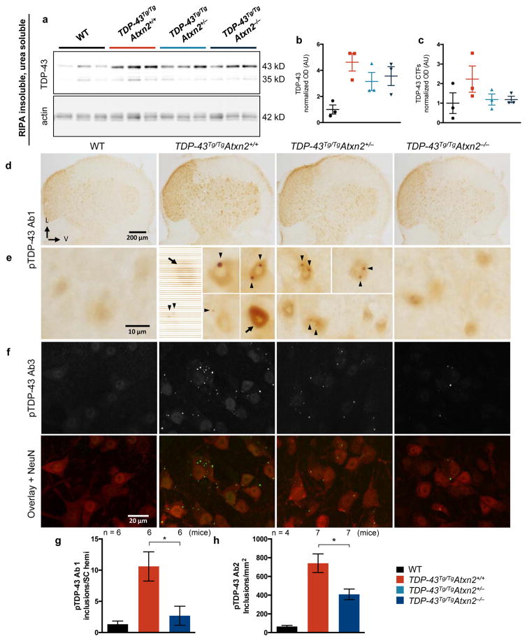
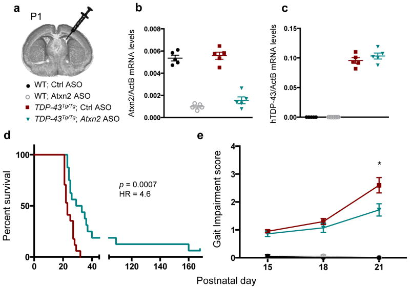
Amyotrophic lateral sclerosis (ALS) is a rapidly progressing neurodegenerative disease that is characterized by motor neuron loss and that leads to paralysis and death 2-5 years after disease onset. Nearly all patients with ALS have aggregates of the RNA-binding protein TDP-43 in their brains and spinal cords, and rare mutations in the gene encoding TDP-43 can cause ALS. There are no effective TDP-43-directed therapies for ALS or related TDP-43 proteinopathies, such as frontotemporal dementia. Antisense oligonucleotides (ASOs) and RNA-interference approaches are emerging as attractive therapeutic strategies in neurological diseases. Indeed, treatment of a rat model of inherited ALS (caused by a mutation in Sod1) with ASOs against Sod1 has been shown to substantially slow disease progression. However, as SOD1 mutations account for only around 2-5% of ALS cases, additional therapeutic strategies are needed. Silencing TDP-43 itself is probably not appropriate, given its critical cellular functions. Here we present a promising alternative therapeutic strategy for ALS that involves targeting ataxin-2. A decrease in ataxin-2 suppresses TDP-43 toxicity in yeast and flies, and intermediate-length polyglutamine expansions in the ataxin-2 gene increase risk of ALS. We used two independent approaches to test whether decreasing ataxin-2 levels could mitigate disease in a mouse model of TDP-43 proteinopathy. First, we crossed ataxin-2 knockout mice with TDP-43 (also known as TARDBP) transgenic mice. The decrease in ataxin-2 reduced aggregation of TDP-43, markedly increased survival and improved motor function. Second, in a more therapeutically applicable approach, we administered ASOs targeting ataxin-2 to the central nervous system of TDP-43 transgenic mice. This single treatment markedly extended survival. Because TDP-43 aggregation is a component of nearly all cases of ALS, targeting ataxin-2 could represent a broadly effective therapeutic strategy.


Conflict of interest statement

P.J.-N., A.S. and F.R. are employed by Ionis Pharmaceuticals, a for-profit company that develops ASO therapies. The other authors declare no competing financial interest.

Figures

Extended Data Figure 1
Extended Data Figure 1. Reduction of ataxin 2 using two independent lines of Atxn2 knockout mice lines extends lifespan of TDP-43 transgenic mice
Lines A and B were generated using Atxn2+/− mice from a congenic C57Bl/6 and a hybrid B6129S background, respectively. Within line A, TDP-43Tg/TgAtxn2+/− (n = 7) (a) and TDP-43Tg/TgAtxn2−/− (n = 7) (c) mice lived significantly longer than TDP-43Tg/TgAtxn2+/+ mice (n = 16). Within line B, TDP-43Tg/TgAtxn2+/− (n = 13) (b) and TDP-43Tg/TgAtxn2−/− (n = 11) (d) mice also lived significantly longer than TDP-43Tg/TgAtxn2+/+ mice (n = 12). Curves were compared by log rank test and effect size estimated by a Cox proportional hazards model (HR = hazard ratio). e) After taking genotype into account, the line that the mice came from, but not the sex of the mice, significantly affected lifespan. A Cox proportional hazards likelihood ratio test was used to compare the null model including only genotype to an alternative model including genotype and line or genotype and sex. f) We found evidence for two groups of responders (strong and weak) in the TDP-43Tg/TgAtxn2−/− population (parametric bootstrap p = 0.02, see online methods). The Kaplan-Meier curve of all TDP-43Tg/TgAtxn2−/− mice from both lines is plotted, and the one and two group models are graphed. g) Knockout of Atxn2 did not affect weight in non-transgenic or TDP-43Tg/Tg adolescents. Means are plotted, and error bars indicate S.E.M.
Extended Data Figure 2
Extended Data Figure 2. Cortical layer V and lower motor neuron loss in TDP-43 transgenic mice
a) Representative NeuN stains of a sagittal sections through cortex. Layer V is marked by a green bar. Layer V neurons were 30% less numerous (b) and had smaller cell bodies (c) in TDP-43Tg/TgAtxn2+/+ mice. These phenotypes were significantly ameliorated in TDP-43Tg/TgAtxn2−/− mice. Four mice were quantified per genotype, and the values for individual brain sections are plotted. Genotype groups were compared using linear mixed models with a random effect to appropriately account for the multiple measurements per mouse (see online methods). d) Representative NeuN stains of L5 lumbar ventral horn showing large lower motor neurons. e) Quantification of motor neuron cell bodies present in the ventral horn of the lumbar enlargement at levels L3-L6. There was an 27% decrease in motor neurons on average in TDP-43Tg/TgAtxn2+/+ mice compared to WT. Six P23 animals were used per genotype. Two-tailed t-tests were performed between groups of interest. Means are plotted, and error bars indicate S.E.M. *p < 0.05, **p < 0.01.
Extended Data Figure 3
Extended Data Figure 3. Lowering ataxin 2 levels does not affect expression of the human TDP-43 transgene
a) Atxn2 mRNA levels were decreased in Atxn2+/− mouse brain by ~50% and completely absent in Atxn2−/− mice. b) Among TDP-43Tg/Tg mice, Atxn2 reduction did not significantly affect levels of the human TDP-43 (hTDP-43) transgene by ordinary one-way ANOVA. Samples were collected at P21. Error bars indicate S.E.M.
Extended Data Figure 4
Extended Data Figure 4. Protein levels of TDP-43 are not significantly affected by ataxin 2 reduction in TDP-43Tg/Tg mice
a) Nucleocytoplasmic fractioning of mouse brain tissue segregated the nuclear marker lamin A/C from cytoplasmic marker GAPDH. Nuclear levels of human TDP-43 (b) or total full-length TDP-43 (c) were not altered among the genotypes, though nuclear TDP-43 C-terminal fragments (CTFs) were slightly elevated in TDP-43Tg/TgAtxn2+/+ mice (d). e–g) Cytoplasmic hTDP-43 (e) and total TDP-43 CTFs (g) were also unaltered among the genotypes, though cytoplasmic full-length TDP-43 (f) seemed slightly elevated in TDP-43Tg/TgAtxn2+/+ mice. Samples were collected at P21. Error bars indicate S.E.M. Gel source data in SI Fig 1.
Extended Data Figure 5
Extended Data Figure 5. Stress granules contain phosphorylated TDP-43
Two different phosphorylation-specific TDP-43 antibodies (a, b) and a C-terminal epitope TDP-43 antibody (c) readily stain stress granules, indicated by the stress granule markers EIF3η and ataxin 2.
Extended Data Figure 6
Extended Data Figure 6. Two different types of inclusions are recognized in TDP-43Tg/Tg mice. a–d,m–p)
None of the TDP-43 antibodies tested recognized inclusions in WT mice. One of the three phosphorylated TDP-43 (pTDP-43) specific antibodies (e) and TDP-43 antibodies not phosphorylation-specific (f,h,q–t) recognized spherical, predominantly nuclear inclusions. The other two pTDP-43 specific antibodies (only one is shown) recognized smaller cytoplasmic and nuclear inclusions (g). i,j,l) The first type of inclusion was very rare in TDP-43Tg/TgAtxn2−/− mice. k) The second type of inclusion appeared smaller and reduced in number in TDP-43Tg/TgAtxn2−/− mice. q–t) Nuclear inclusions were effectively stained with total TDP-43 (tTDP-43), human-specific TDP-43 (hTDP-43), and mouse-specific TDP-43 (mTDP-43) antibodies. s) Diffuse mTDP-43 is greatly decreased in TDP-43Tg/Tg mice, an expected outcome of TDP-43 autoregulation. u) Levels of diffuse nuclear tTDP-43 were quantified by IF in WT neurons or TDP-43Tg/Tg neurons with or without inclusions. These 3 groups were compared in a pairwise fashion using linear mixed models with a term to appropriately account for multiple measurements per mouse (n = 3 mice per genotype, see online methods). Median and min to max are plotted. Images were taken in cervical spinal cord. Samples were collected at P21. Scale bars are 10 μm.
Extended Data Figure 7
Extended Data Figure 7. An antisense oligonucleotide (ASO) targeting Atxn2 is able to successfully reduce mRNA levels throughout the central nervous system
a) ICV injection at P1 of an ASO targeting Atxn2 was able to successfully reduce levels of Atxn2 mRNA in the spinal cord by ~75% when assessed at P28. Atxn2 reduction was also seen in the cortex (b) and cerebellum (c). d) Grip strength of WT mice was not effected by Ctrl or Atxn2 ASO injection (n = 16 per treatment). Mean and SEM plotted. Genetic markers of gliosis, Aif1 and Gfap, were not altered in the spinal cord (e, f) or cortex (g, h) after ASO injection. i) Using a Luminex 38-plex assay, we could not detect a significant difference in inflammatory markers among uninjected WT mice (n = 5) and WT mice treated with the Atxn2 (n = 4) or Ctrl (n = 5) ASOs (two-way ANOVA treatment group factor p = .32). However, the ASO treated animals had a small increase in 1 of the 38 markers, VEGF. Multiplicity-adjusted pairwise tests revealed that this difference was not significant for Atxn2 ASO treated mice (p = .17), but was for Ctrl ASO treated mice (p = .006). Min to max plotted. a–c,e–h) Biological replicates and means are plotted.
Figure 1
Figure 1. Reducing levels of ataxin 2 extends lifespan, improves motor function, and slows the rate of disease progression in TDP-43 transgenic mice
a,b) Kaplan-Meier survival curves comparing survival of TDP-43Tg/TgAtxn2+/+ (n = 28), TDP-43Tg/TgAtxn2+/− (n = 20) and TDP-43Tg/TgAtxn2−/− (n = 18) mice. Curves were compared by log rank test, and effect size was estimated by a Cox proportional hazards model (HR = hazard ratio). c) The gait impairment score of TDP-43Tg/Tg mice worsened with age, but this was attenuated by reducing ataxin 2. d) A line was fit to each mouse’s gait scores over time and the slope of that line was plotted, grouping animals by genotype. The error bars are min to max, and the three groups were compared by ordinary one-way ANOVA. e,f) Tremor and kyphosis scores were also decreased by lowering ataxin 2 in TDP-43Tg/Tg mice. c,e,f) At each age, the three genotypes were compared by ordinary one-way ANOVA. Means are plotted, and error bars indicate S.E.M. *p < .05, **p < .01, ***p < .001.
Figure 2
Figure 2. Knockdown of ataxin 2 delays the maturation of stress granules and decreases recruitment of TDP-43 to stress granules
a) An EIF3η antibody was used to visualize stress granules (SGs) in U2OS cells. During continued stress, stress granules fuse to form larger structures. ATXN2 siRNA inhibited maturation of stress granules, resulting in smaller, more numerous stress granules at each time point (quantified in b–c). d) Western blot of ataxin 2 knockdown (source data in SI Fig. 1). e, f) Ataxin 2 siRNA treatment caused fewer cells to have TDP-43-positive stress granules at 60 minutes of arsenite exposure. b,c,f) The mean of 3 separate wells (116–146 cells per well) is plotted for each data point. Two-tailed t-tests were used to compare treatment groups. Means are plotted, and error bars represent SEM. *p < .05, **p < .01, ***p < .001.
Figure 3
Figure 3. Lowering ataxin 2 levels decreases TDP-43 pathology
a) RIPA-insoluble, urea soluble full-length and CTF TDP-43 are elevated in TDP-43Tg/TgAtxn2+/+ mice (quantified b–c), and appeared moderately reduced in TDP-43Tg/TgAtxn2−/− mice (source data in SI Fig. 1). d) Lumbar spinal cord hemispheres (SC hemi) stained with pTDP-43 Ab1. e) Magnification to the cellular level reveals weak, diffuse nuclear staining in WT mice, and pTDP-43 inclusions (arrow heads) and strong diffuse nuclear pTDP-43 (arrows) in TDP-43Tg/TgAtxn2+/+ mice. Fluorescent stains (Extended Data Fig. 6e) demonstrate that most pTDP-43 Ab1 inclusions are nuclear. f) pTDP-43 Ab3 reveals inclusions that are much more numerous and usually cytoplasmic (Extended Data Fig. 6g). Lowering ataxin 2 decreased pTDP-43 Ab1 inclusions at P21 (g), and pTDP-43 Ab2 inclusions at P23 (h) in lumbar spinal cord. Two-tailed t-tests were performed between groups of interest. Means are plotted, and error bars indicate S.E.M. *p < 0.05
Figure 4
Figure 4. ASOs targeting ataxin 2 extend lifespan and improve motor performance in TDP-43 transgenic mice
a) P1 mouse pups were treated with Ctrl or Atxn2 ASO via ICV injection. At P21, mRNA levels of Atxn2 (b) were decreased by 77% in the Atxn2 ASO-injected mouse brains without affecting mRNA levels of the human TDP-43 transgene (c). d) Lifespan was significantly extended by Atxn2 ASO treatment (n = 16) vs Ctrl ASO (n = 17). Curves were compared by log rank test, and effect size was estimated using a Cox proportional hazards model. e) Gait impairment score was also improved in Atxn2 ASO treated mice (n = 14) by P21 compared to Ctrl ASO (n = 20). Two-tailed t-tests were used to compare the two treatment groups at each age. Means are indicated, and error bars indicate S.E.M. *p < 0.05

Comment in

References

    1. Taylor JP, Brown RH, Jr, Cleveland DW. Decoding ALS: from genes to mechanism. Nature. 2016;539:197–206. - PMC - PubMed
    1. Neumann M, et al. Ubiquitinated TDP-43 in frontotemporal lobar degeneration and amyotrophic lateral sclerosis. Science. 2006;314:130–133. - PubMed
    1. Lagier-Tourenne C, Cleveland DW. Rethinking ALS: the FUS about TDP-43. Cell. 2009;136:1001–1004. - PMC - PubMed
    1. Southwell AL, Skotte NH, Bennett CF, Hayden MR. Antisense oligonucleotide therapeutics for inherited neurodegenerative diseases. Trends Mol Med. 2012;18:634–643. - PubMed
    1. Smith RA, et al. Antisense oligonucleotide therapy for neurodegenerative disease. J Clin Invest. 2006;116:2290–2296. - PMC - PubMed

MeSH terms