. 2017 Apr 20;544(7650):362-366.
doi: 10.1038/nature22044. Epub 2017 Apr 12.

Antisense oligonucleotide therapy for spinocerebellar ataxia type 2

Affiliations

Antisense oligonucleotide therapy for spinocerebellar ataxia type 2

Daniel R Scoles et al. Nature. .

Abstract

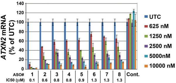
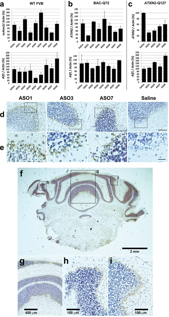
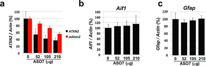
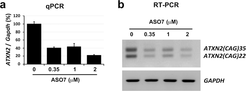
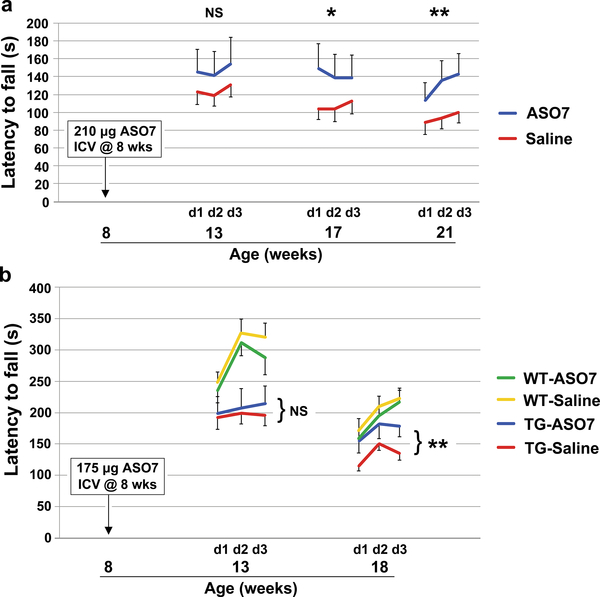
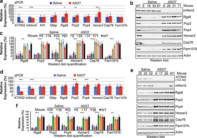
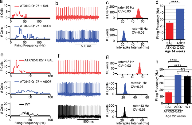
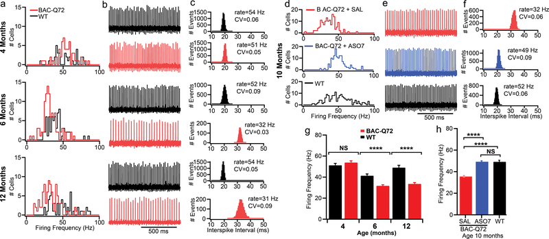
There are no disease-modifying treatments for adult human neurodegenerative diseases. Here we test RNA-targeted therapies in two mouse models of spinocerebellar ataxia type 2 (SCA2), an autosomal dominant polyglutamine disease. Both models recreate the progressive adult-onset dysfunction and degeneration of a neuronal network that are seen in patients, including decreased firing frequency of cerebellar Purkinje cells and a decline in motor function. We developed a potential therapy directed at the ATXN2 gene by screening 152 antisense oligonucleotides (ASOs). The most promising oligonucleotide, ASO7, downregulated ATXN2 mRNA and protein, which resulted in delayed onset of the SCA2 phenotype. After delivery by intracerebroventricular injection to ATXN2-Q127 mice, ASO7 localized to Purkinje cells, reduced cerebellar ATXN2 expression below 75% for more than 10 weeks without microglial activation, and reduced the levels of cerebellar ATXN2. Treatment of symptomatic mice with ASO7 improved motor function compared to saline-treated mice. ASO7 had a similar effect in the BAC-Q72 SCA2 mouse model, and in both mouse models it normalized protein levels of several SCA2-related proteins expressed in Purkinje cells, including Rgs8, Pcp2, Pcp4, Homer3, Cep76 and Fam107b. Notably, the firing frequency of Purkinje cells returned to normal even when treatment was initiated more than 12 weeks after the onset of the motor phenotype in BAC-Q72 mice. These findings support ASOs as a promising approach for treating some human neurodegenerative diseases.


Conflict of interest statement

The authors declare no competing financial interests. S.M.P. is a consultant for Progenitor Life Sciences and Ataxion Pharmaceuticals. T.S.O. is an employee of F. Hoffmann-La Roche, Ltd. G.H., F.R., and C.F.B are employed by Ionis Pharmaceuticals, which supplied the ASOs used in the study.

Figures

Extended Data Figure 1
In vitro screen for ATXN2 ASOs by qPCR. A total of 152 ASOs were delivered at 4.5 μM to HepG2 cells by electroporation in two 384-well plates. ATXN2 expression was evaluated by qPCR (n=3 wells per ASO). Shown is the evaluation of the eight best positive hit ASOs for IC50 determination. Values are means ± SD of ATXN2 quantity relative to total RNA. Cont. = scrambled control ASO.
Extended Data Figure 2
Positive hit ASOs evaluated in vivo. a-c, 250 μg of the indicated ASOs in a total of 7 μl was delivered by ICV injection. After 7 days treatment the expression of mouse Atxn2 or human ATXN2 and Aif1 was determined by qPCR relative to actin. a, Wildtype FVB mice. b, BAC-Q72 mice. c, ATXN2-Q127 mice. Values are means ± SD relative to those determined from normal saline treated mice. The n number of mice (left-to-right in each chart) was as follows: a, 2,2,2,3,2,2,2,2,1. b, 2,1,1,2,2,2,1,2. c, 2,3,1,1,1. d & e, ASOs localized to the cerebellar Purkinje cell layer of treated mice. Mice were treated by ICV injection into the right lateral ventricle of the indicated lead ASOs for 7 days in BAC-Q72 mice (ASO3 and ASO7 used at 250 μg) or 10 wks in ATXN2-Q127 mice (ASO1 used at 200 μg). ASOs were localized in paraffin embedded sections by immunohistochemical peroxidase staining using anti-ASO antibody. Saline= ATXN2-Q127 mice treated by ICV injection of 7 μl saline for 10 wks. d, 10x objective. e, 3x digital zoom of a region of the PC layer in the corresponding 10x image, indicated by the box. Bar = 100 μm (d), 25 μm (e). f-i, Distribution of ASO7 in the cerebellum. f, ASO7 was distributed in Purkinje cell layers throughout the cerebellum (2x objective). Higher power images at regions indicated in f demonstrated ASO7 localization in Purkinje cells across the cerebellum: g, 10x objective; h & i, 40x objective.
Extended Data Figure 3
Effects of ASO7 on ATXN2 expression in vivo by dose and time. a-c, Dosewise effect of ASO7 on ATXN2 expression in BAC-Q72 mice treated with ASO7 by ICV injection for 14 days. a, Expression of cerebellar human ATXN2 and mouse Atxn2 determined by qPCR. b, Cerebellar Aif1 expression determined by qPCR. c, Cerebellar Gfap expression determined by qPCR. The n # of mice for the saline, 52 μg, 105 μg, and 210 μg treatments was 3, 2, 3 and 3, respectively.
Extended Data Figure 4
Weights of mice before and after rotarod testing. a, Rotarod test of ATXN2-Q127 mice treated with a single ICV dose of 210 μg ASO7. b, Rotarod test of BAC-Q72 mice treated with a single ICV dose of 175 μg ASO7. Weeks of ASO treatments are indicated on the X-axes. Significance tests demonstrated significant differences between weights of BAC-Q72 mice compared to wildtype littermates (p<0.001 for any age group, Student’s t-test). Comments on the relevance of mouse weights on motor phenotype testing are provided in Supplementary Discussion.
Extended Data Figure 5
ASO7 lowered expression of wildtype and mutant ATXN2 in cultured SCA2 patient derived fibroblasts. a, Patient derived SCA2(CAG35) fibroblasts were transfected with the indicated quantities of ASO7. After 72 hours RNAs were prepared and total ATXN2 expression was determined by qPCR. b, To determine ASO7 effect on the expression of non-mutant (CAG22) and mutant (CAG35) ATXN2, RT-PCR reactions were evaluated by agarose gel electrophoresis, with loading controlled by GAPDH.
Figure 1
Figure 1
Effect of ASO7 on motor phenotypes. a, Eight wk old ATXN2-Q127 mice were treated with 210 μg ASO7 or saline ICV and rotorod tested at the indicated ages. n=15 mice per group. b, Eight wk old BAC-Q72 mice and wildtype littermates were treated with 175 μg ASO7 or saline ICV and rotarod tested at the indicated ages. n= 13, 13, 11, 14 for WT-ASO, WT-Saline, TG-ASO, and TG-Saline, respectively. Values shown are mean±SE. Probabilities of significance were determined using the method of generalized estimating equations (GEE). NS, nonsignificant, *=p<0.05, **=p<0.01.
Figure 2
Figure 2
Cerebellar gene expression for ASO treated SCA2 mice following rotatod tests. a-c, Expression in ATXN2-Q127 mice. a, ATXN2-Q127 mice were treated as in Fig. 1a and the cerebellar expression of the indicated genes was determined at 22 wks of age. The n number of mice for saline and ASO7 treatments, respectively, were: ATXN2, 11, 11; Atxn2, 11, 11; Aif1, 7, 8; Gfap, 11, 11; Rgs8, 4, 4; Pcp2, 11, 11; Pcp4, 10, 11; Cep76, 10, 11; Homer3, 10, 11; Fam107b, 10, 10. b & c, Cerebellar expression in ATXN2-Q127 mice for ATXN2, mouse Atxn2, Rgs8, Pcp2, Pcp4, Cep76, Homer3, Fam107b, and Actin (b), with abundances expressed as a percent relative to actin, determined densitometrically (c). d-f, Expression in BAC-Q72 mice. d, BAC-Q72 mice were treated as in Fig. 1b and the cerebellar expression of the indicated genes was determined at 19 wks of age. The n per group in qPCR analyses was 14 saline treated mice and 11 ASO7 treated mice for all genes except Rgs8 and Fam107b where the n was 14 saline treated and 10 ASO treated mice. Values are mean±SD. NS, nonsignificant, *=p<0.05, **=p<0.01, ***=p<0.001 by Student’s t-test. e, Cerebellar expression in BAC-Q72 mice of expanded ATXN2 detected with anti-1C2 antibody, and expression of mouse Atxn2, Rgs8, Pcp2, Pcp4, Cep76, Homer3, Fam107b and Actin (e), with abundances expressed as a percent relative to actin, determined densitometrically (f). Values shown in c and f are mean±SD from three replicate blots.
Figure 3
Figure 3
Slow ATXN2-Q127 mouse PC firing frequency (FF) was restored by ASO7. a-d, ATXN2-Q127 mice were treated with 210 μg ASO7 or saline by ICV injection at 8 wks, and PC FF was evaluated at 14 wks of age. a, Mean FF distribution for PCs from saline and ASO7 treated mice. b, Representative PC traces of 1s duration, and (c) interspike-interval histogram of the same cell as in (b) calculated from 2 min duration, with coefficients of variation (CV) shown. d, Mean FF (±SEM) were 25±2 Hz for the saline treated mouse (n=50 neurons, mice=1) and 46±2 Hz for the ASO7 treated mice (n=88 neurons, mice=2) (****, p<0.0001, Student’s t-test). e-h, ATXN2-Q127 mice were treated with 210 μg ASO7 or saline by ICV injection at 8 wks. PC FF was evaluated at 22 weeks of age. e, Mean FFs of all PCs measured from saline and ASO7 treated mice and age-matched WT mice. f, Representative PC traces of 1s duration, and (g) interspike-interval histogram of the same cell as in (f) calculated from 2 min duration, with CV values shown. h, Mean FF (± SEM) were 16±1 Hz for the saline treated ATXN2-Q127 mice (n=107 neurons, mice=2), 42±2 Hz for the ASO7 treated ATXN2-Q127 mice (n=102 neurons, mice=2), and 40±1 Hz for an age-matched WT mouse (n=50 neurons, mice=1). (****, p<0.0001, Student’s t-test). All recordings were measured at 34.5±1 °C.
Figure 4
Figure 4
Slow BAC-Q72 mouse PC FF was restored by ASO7. a-c, FFs of BAC-Q72 mice at indicated ages. a, FF distributions for WT and BAC-Q72 PCs. The n number of PCs were 33 WT, 59 BAC-Q72 (4 mo); 49 WT, 101 BAC-Q72 (6 mo); 55 WT, 92 BAC-Q72 (12 mo). b, Example recordings from PCs for WT and BAC-Q72 mice. c, Interspike intervals of the same cell over 2 min with mean FF and CV indicated. d-f, The PC FF was restored in 10 mo. old BAC-Q72 mice treated with 210 μg ASO7 ICV for 10 wks. d, PC FF in BAC-Q72 mice, BAC-Q72 mice treated with ASO7, and WT littermates. e, Representative PC 1s trace and (f) interspike intervals of the same cell over 2 min in saline treated BAC-Q72 mice, ASO treated BAC-Q72, and WT littermates with PC FF and CV indicated. g-h, Mean PC FFs in (a) and (d). g, Age-dependent PC FF in BAC-Q72 mice. At age 4 mo the mean FFs in Hz were 51±2 for WT (n=33 PCs, mice=1) and 54±2 for BAC-Q72 (n= 59 PCs, mice=1). At 6 mo the values were 41±2 for WT (n=49 PCs, mice=2) and 32±1 for BAC-Q72 (n=101 PCs, mice=2). At age 12 mo they were 49±2 for WT (n=55 PCs, mice=1) and 34±1 for BAC-Q72 (n=92 PCs, n=1). h, ASO7 treatment restored the PC FF of BAC-Q72 mice. The mean PC FF for 10 mo old BAC-Q72 mice treated with saline was 36±1 Hz (n=148 PCs, mice=3) while the mean FF of BAC-Q72 mice treated with ASO7 was 49±1 (n=134 PCs, mice=4), similar to that of WT mice (49±1, n=155 PCs, mice=3). (****, p<0.0001, ANOVA followed by Tukey’s multiple comparison posthoc testing).

Comment in

References

    1. Pulst SM, Nechiporuk A & Starkman S Anticipation in spinocerebellar ataxia type 2. Nat Genet 5, 8–10, doi: 10.1038/ng0993-8c (1993). - DOI - PubMed
    1. Rigo F, Seth PP & Bennett CF Antisense oligonucleotide-based therapies for diseases caused by pre-mRNA processing defects. Adv. Exp. Med. Biol 825, 303–352, doi: 10.1007/978-1-4939-1221-6_9 (2014). - DOI - PubMed
    1. Dansithong W et al. Ataxin-2 regulates RGS8 translation in a new BAC-SCA2 transgenic mouse model. PLoS Genet 11, e1005182, doi: 10.1371/journal.pgen.1005182 (2015). - DOI - PMC - PubMed
    1. Hansen ST, Meera P, Otis TS & Pulst SM Changes in Purkinje cell firing and gene expression precede behavioral pathology in a mouse model of SCA2. Hum Mol Genet 22, 271–283, doi: 10.1093/hmg/dds427 (2013). - DOI - PMC - PubMed
    1. Pulst SM Degenerative ataxias, from genes to therapies: The 2015 Cotzias Lecture. Neurology 86, 2284–2290, doi: 10.1212/WNL.0000000000002777 (2016). - DOI - PMC - PubMed

Publication types

MeSH terms