. 2017 Mar 28;23(12):2175-2184.
doi: 10.3748/wjg.v23.i12.2175.

Acanthopanax senticosus polysaccharides-induced intestinal tight junction injury alleviation via inhibition of NF-κB/MLCK pathway in a mouse endotoxemia model

Affiliations

Acanthopanax senticosus polysaccharides-induced intestinal tight junction injury alleviation via inhibition of NF-κB/MLCK pathway in a mouse endotoxemia model

Jie Han et al. World J Gastroenterol. .

Abstract

Aim: To examine the effects of Acanthopanax senticosus polysaccharides (ASPS) on intestinal tight junction (TJ) disruption and nuclear factor-kappa B (NF-κB)/myosin light chain kinase (MLCK) activation in endotoxemia.

Methods: BALB/C mice (6-8-weeks-old) received continuous intragastric gavage of ASPS for 7 d before injection of lipopolysaccharide (LPS), or received ASPS once after LPS injection. Blood and intestinal mucosal samples were collected 6 h after LPS challenge. Clinical symptoms, histological injury, intestinal permeability, TJ ultrastructure, and TJ protein expression were determined.

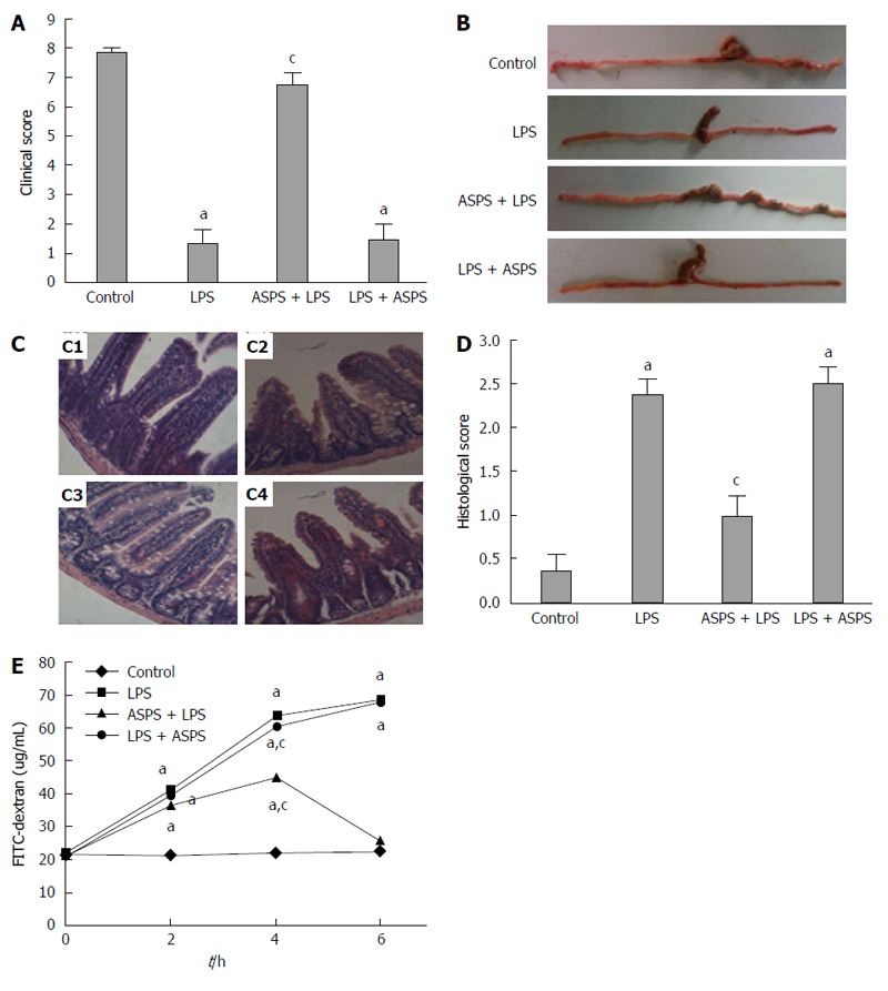
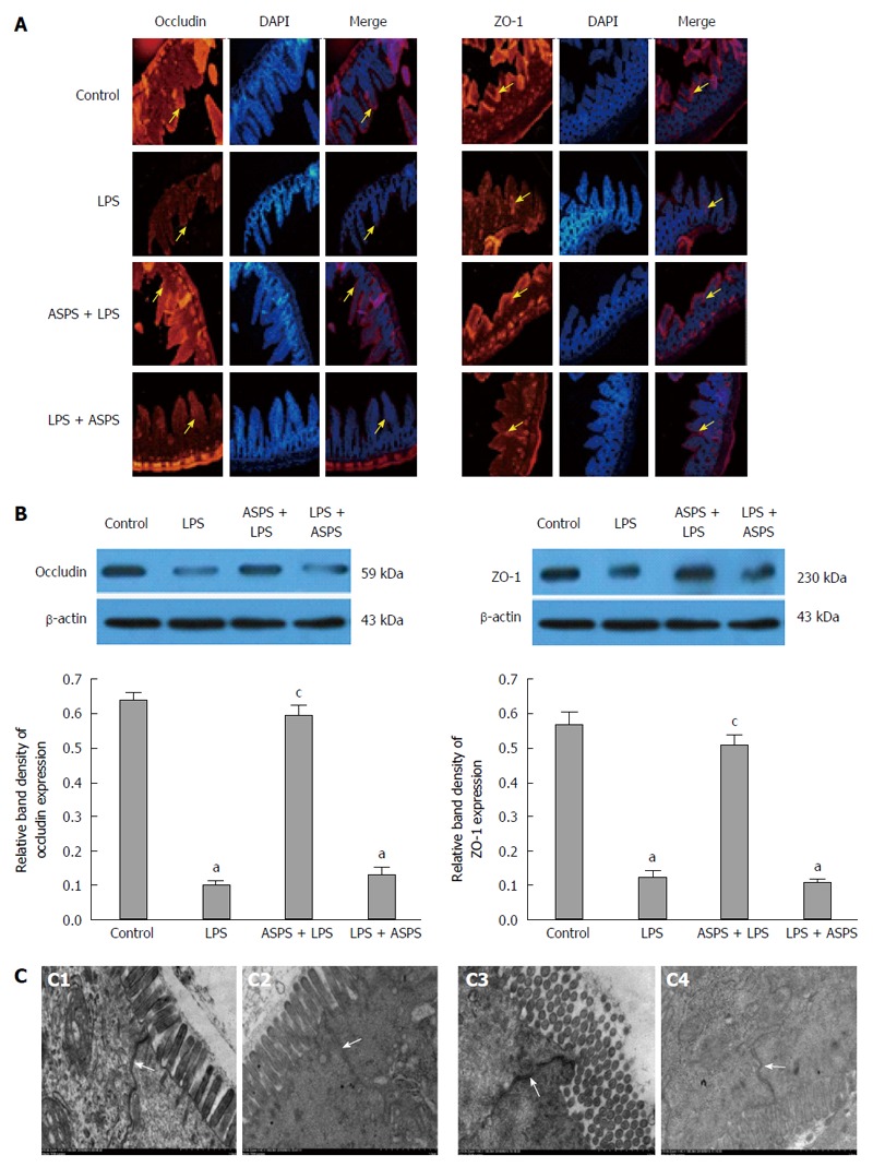
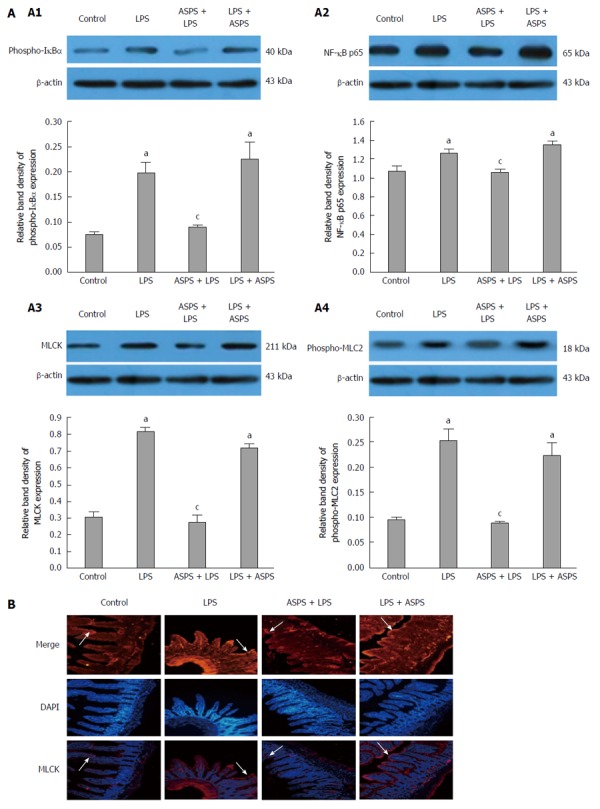
Results: Compared with mice in the LPS group, pretreatment with ASPS improved clinical and histological scores by 390.9% (P < 0.05) and 57.89% (P < 0.05), respectively, and gut permeability change in endotoxemic mice was shown by a 61.93% reduction in reduced leakage of fluorescein isothiocyanate-dextran 6 h after LPS injection (P < 0.05). ASPS pretreatment also prevented LPS-induced TJ ultrastructure breakdown supported by increased electron dense materials between adjoining cells, sustained redistribution and expression of occludin (0.597 ± 0.027 vs 0.103 ± 0.009, P < 0.05) and zonula occludens-1 (0.507 ± 0.032 vs 0.125 ± 0.019, P < 0.05), and suppressed activation of the NF-κB/MLCK pathway indicated by reduced expression of NF-κB, phospho-inhibitor kappa B-alpha, MLCK and phospho-myosin light-chain-2 by 16.06% (P < 0.05), 54.31% (P < 0.05), 66.10% (P < 0.05) and 64.82% (P < 0.05), respectively.

Conclusion: ASPS pretreatment may be associated with inhibition of the NF-κB/MLCK pathway and concomitant amelioration of LPS-induced TJ dysfunction of intestinal epithelium in endotoxemia.

Keywords: Acanthopanax senticosus polysaccharide; Intestinal permeability; Myosin light chain kinase; Nuclear factor-kappa B; Tight junction.


Conflict of interest statement

Conflict-of-interest statement: The authors declare that there are no conflicts of interest related to this study.

Figures

Figure 1
Figure 1
Effects of acanthopanax senticosus polysaccharides on clinical score, macroscopic features of distal ileum and colon, histological appearance and score of distal ileum in lipopolysaccharide-induced mice. A: Mice were assessed for clinical score at designated time points after lipopolysaccharide (LPS) challenge (n = 8); B: Representative photographs of the distal ileums and colons at 6 h after LPS injection (n = 8); C: Effects of acanthopanax senticosus polysaccharides (ASPS) on LPS-induced intestinal histopathologic changes. Ileum was processed for morphological and histopathologic evaluation at 6 h after LPS induction (n = 3). The representative photomicrographs of ileal segments stained with hematoxylin and eosin at 200 × magnification of C1, control group; C2, LPS group; C3, ASPS + LPS group; and C4, LPS + ASPS group; D: Intestinal histopathologic score was determined at 6 h after LPS challenge (n = 3); E: Effects of ASPS on LPS-induced increase in iliac mucosal permeability. The intestinal permeability of 4 kDa fluorescein isothiocyanate (FITC)-dextran in ileal pouch was measured at 2, 4 and 6 h after LPS administration (n = 8). aP < 0.05, vs the control group; cP < 0.05, vs the LPS group.
Figure 2
Figure 2
Localization and expression of tight junction proteins, and tight junction proteins ultrastructure in ileum were evaluated 6 h after lipopolysaccharide administration in mice of four groups. A: Effects of Acanthopanax senticosus polysaccharides (ASPS) on distribution of occludin and ZO-1. Staining of both proteins along the villous epithelium at a 200 × magnification (red fluorescence) were observed by immunofluorescence. Nuclei were stained by DAPI (blue fluorescence). Arrows indicate the location of tight junction (TJ) proteins staining; B: Effects of ASPS on intestinal TJ proteins expression of occludin and ZO-1 (n = 3). Protein samples were analyzed by western blotting, and β-actin was used as an internal control. The values are presented as mean ± SE. aP < 0.05, vs the control group; cP < 0.05, vs the LPS group; C: Effects of ASPS on intestinal TJ ultrastructure in ileum viewed under transmission electron microscope of C1, control group; C2, LPS group; C3, ASPS + LPS group; and C4, LPS + ASPS group. Arrows indicate the location of the TJ (scale bar = 1 μm).
Figure 3
Figure 3
Protein expression of phospho-IκBα, nuclear factor-kappa B p65, myosin light chain kinase and phospho-MLC2 (A) and MLCK localization (B) in ileum epithelium. A: Protein expression of phospho-IκBα (A1), nuclear factor-kappa B (NF-κB) p65 (A2), myosin light chain kinase (MLCK) (A3) and phospho-MLC2 (A4) were analyzed by western blotting at 6 h after lipopolysaccharide (LPS) induction, and β-actin was used as internal control (n = 3). Data are shown as mean ± SE (n = 3). aP < 0.05, vs the control group; cP < 0.05, vs the LPS group; B: MLCK location was observed by immunofluorescence at 6 h after LPS administration at 200 × magnification (red fluorescence) (n = 3). Nuclei were stained by DAPI (blue fluorescence).

References

    1. Angus DC, Wax RS. Epidemiology of sepsis: an update. Crit Care Med. 2001;29:S109–S116. - PubMed
    1. Polderman KH, Girbes AR. Drug intervention trials in sepsis: divergent results. Lancet. 2004;363:1721–1723. - PubMed
    1. Hassoun HT, Kone BC, Mercer DW, Moody FG, Weisbrodt NW, Moore FA. Post-injury multiple organ failure: the role of the gut. Shock. 2001;15:1–10. - PubMed
    1. Lee SH. Intestinal permeability regulation by tight junction: implication on inflammatory bowel diseases. Intest Res. 2015;13:11–18. - PMC - PubMed
    1. De-Souza DA, Greene LJ. Intestinal permeability and systemic infections in critically ill patients: effect of glutamine. Crit Care Med. 2005;33:1125–1135. - PubMed

LinkOut - more resources