

Breast cancer metastasizing to the stomach mimicking primary gastric cancer: A case report
- PMID: 28405154
- PMCID: PMC5374138
- DOI: 10.3748/wjg.v23.i12.2251
Breast cancer metastasizing to the stomach mimicking primary gastric cancer: A case report
Abstract
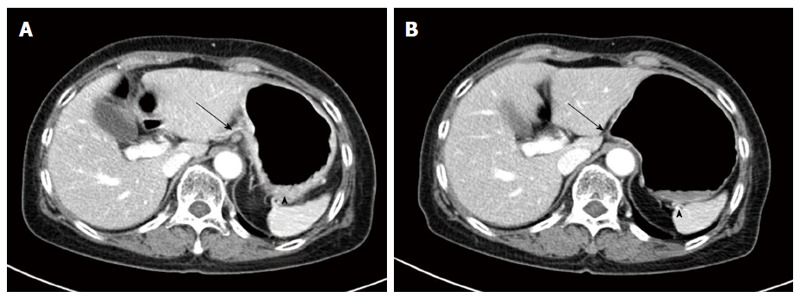
Breast cancer with stomach metastasis rare with an incidence of 1% or less among metastatic breast cancer patients. We experienced a case of breast cancer metastasizing to the stomach in 65-year-old female patient. She experienced dyspepsia and poor oral intake before visiting the clinic. Diffuse infiltration with nodular mucosal thickening of the stomach wall was observed, suggesting advanced gastric cancer based on gross endoscopic finding. Spread of poorly cohesive tumor cells in the gastric mucosa observed upon hematoxylin and eosin stain resembled signet ring cell carcinoma, but diffuse positive staining for GATA3 in immunohistochemical stain allowed for a conclusive diagnosis of breast cancer metastasizing to the stomach. Based on the final diagnosis, systemic chemotherapy was administered instead of primary surgical resection. After 2 cycles of docetaxel administration, she showed a partial response based on abdominal computed tomography scan. This case is an unusual presentation of breast cancer metastasizing to the gastrointestinal tract.
Keywords: Breast cancer; GATA3; GCDFP-15; Gastric cancer; Immunohistochemical stain; Metastasis.
Conflict of interest statement
Conflict-of-interest statement: All authors have no personal, financial, or other conflicts of interest to declare.
Figures


Similar articles
-
Breast cancer metastasis to the stomach may mimic primary gastric cancer: report of two cases and review of literature.World J Surg Oncol. 2007 Jul 9;5:75. doi: 10.1186/1477-7819-5-75. World J Surg Oncol. 2007. PMID: 17620117 Free PMC article. Review.
-
Gastric metastasis of mammary signet ring cell carcinoma--a differential diagnosis with primary gastric signet ring cell carcinoma.J Korean Med Sci. 1997 Jun;12(3):256-61. doi: 10.3346/jkms.1997.12.3.256. J Korean Med Sci. 1997. PMID: 9250925 Free PMC article.
-
Gastric Metastasis as the First Presentation One Year Before Diagnosis of Primary Breast Cancer.Am J Case Rep. 2018 Mar 26;19:354-359. doi: 10.12659/ajcr.908039. Am J Case Rep. 2018. PMID: 29576606 Free PMC article.
-
[A case of gastric metastasis of breast carcinoma resembling early gastric cancer].Korean J Gastroenterol. 2005 Dec;46(6):481-4. Korean J Gastroenterol. 2005. PMID: 16371724 Korean.
-
Breast metastasis of gastric signet-ring cell carcinoma: a case report and literature review.World J Surg Oncol. 2015 Mar 26;13:120. doi: 10.1186/s12957-015-0538-1. World J Surg Oncol. 2015. PMID: 25890325 Free PMC article. Review.
Cited by
-
Widespread Metastasis to the Stomach 10 Years After Primary Breast Cancer: A case report and review of the literature.Medicine (Baltimore). 2020 Nov 25;99(48):e22527. doi: 10.1097/MD.0000000000022527. Medicine (Baltimore). 2020. PMID: 33235059 Free PMC article. Review.
-
A 58-Year-Old Woman with Acute Gastric Perforation Due to Metastatic Ductal Carcinoma 18 Years Following Bilateral Mastectomy for Invasive Ductal Carcinoma of the Breast.Am J Case Rep. 2021 Apr 8;22:e927094. doi: 10.12659/AJCR.927094. Am J Case Rep. 2021. PMID: 33828068 Free PMC article.
-
A case study of pyloric stenosis caused by metastatic lobular carcinoma of breast.J Surg Case Rep. 2023 Dec 28;2023(12):rjad691. doi: 10.1093/jscr/rjad691. eCollection 2023 Dec. J Surg Case Rep. 2023. PMID: 38163054 Free PMC article.
-
Intestinal metastasis from breast cancer: Presentation, treatment and survival from a systematic literature review.World J Clin Oncol. 2021 May 24;12(5):382-392. doi: 10.5306/wjco.v12.i5.382. World J Clin Oncol. 2021. PMID: 34131569 Free PMC article.
-
Breast cancer metastasizing to the upper gastrointestinal tract (the esophagus and the stomach): A comprehensive review of the literature.World J Gastrointest Oncol. 2023 Aug 15;15(8):1332-1341. doi: 10.4251/wjgo.v15.i8.1332. World J Gastrointest Oncol. 2023. PMID: 37663940 Free PMC article. Review.
References
-
- Menuck LS, Amberg JR. Metastatic disease involving the stomach. Am J Dig Dis. 1975;20:903–913. - PubMed
-
- Yu HA, Kim EY, Seo MJ, Chung E, Cho MJ, Oh HJ, Jang JH, Park JC, Lee JU, Park SY. Stomach and Colon Metastasis from Breast Cancer. Ewha Med J. 2014;37:98–104.
-
- McLemore EC, Pockaj BA, Reynolds C, Gray RJ, Hernandez JL, Grant CS, Donohue JH. Breast cancer: presentation and intervention in women with gastrointestinal metastasis and carcinomatosis. Ann Surg Oncol. 2005;12:886–894. - PubMed
-
- Khadim MI. The effects of Pan and its ingredients on oral mucosa. J Pak Med Assoc. 1977;27:353–356. - PubMed
Publication types
MeSH terms
Substances
LinkOut - more resources
Full Text Sources
Other Literature Sources
Medical
