. 2017 Mar 28:10:763-774.
doi: 10.2147/JPR.S129826. eCollection 2017.

Mitogen-activated protein kinase phosphatase-3 (MKP-3) in the surgical wound is necessary for the resolution of postoperative pain in mice

Affiliations

Mitogen-activated protein kinase phosphatase-3 (MKP-3) in the surgical wound is necessary for the resolution of postoperative pain in mice

Sladjana Skopelja-Gardner et al. J Pain Res. .

Abstract

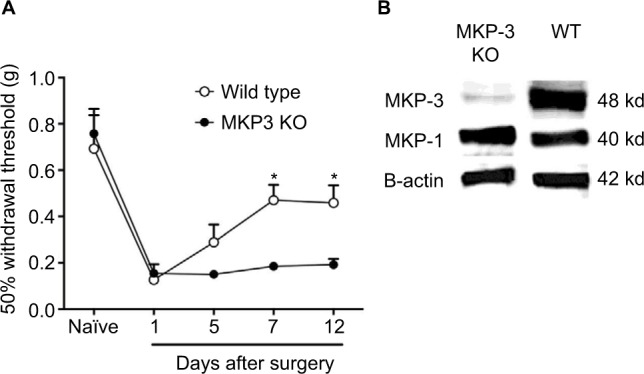
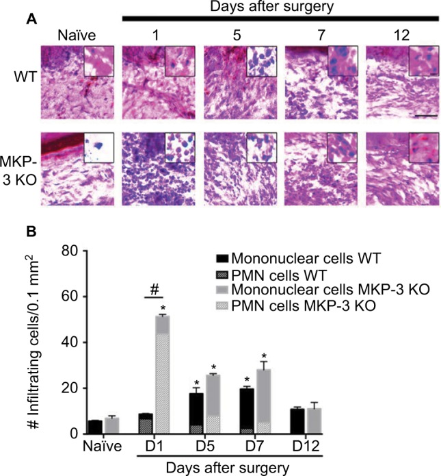
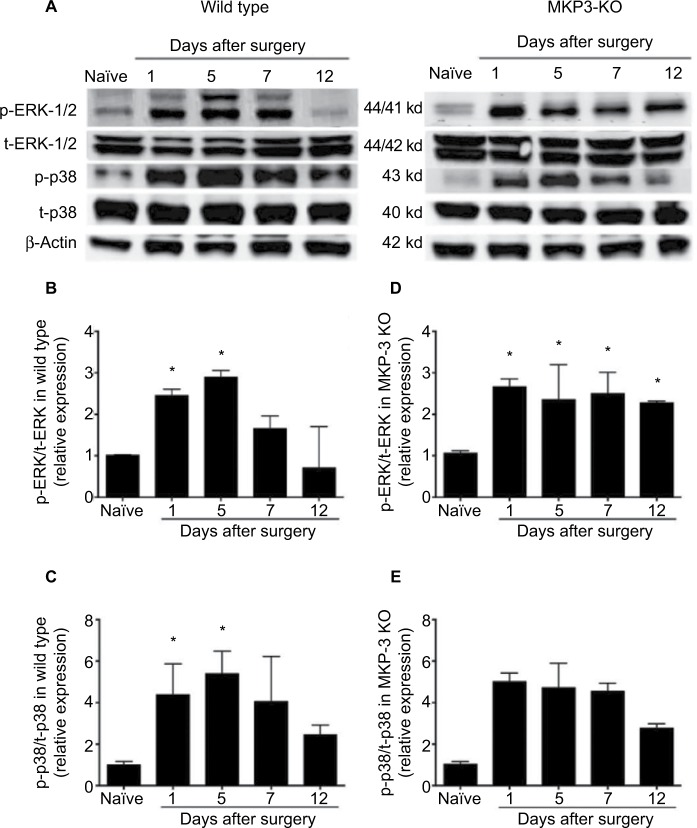
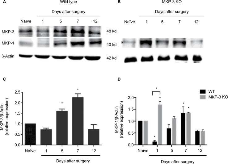
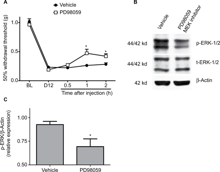
Mitogen-activated protein kinase (MAPK) phosphatase-3 (MKP-3) and its substrates (extracellular signal-regulated kinase [ERK] and p38) play an important role in pathophysiological mechanisms of acute postoperative and chronic neuropathic pain in the spinal cord. This study aimed to understand the role of MKP-3 and its target MAPKs at the site of surgical incision in nociceptive behavior. Wild-type (WT) and MKP-3 knockout (KO) mice underwent unilateral plantar hind paw incision. Mechanical allodynia was assessed by using von Frey filaments. Peripheral ERK-1/2 and p38 phosphorylation were measured by Western blot. Cell infiltration was determined using hematoxylin and eosin histological staining. Peripheral phosphorylated ERK-1/2 (p-ERK-1/2) inhibition was performed in MKP-3 KO mice. In WT mice, mechanical hypersensitivity was observed on postoperative day 1 (0.69±0.17 g baseline vs 0.13±0.08 g day 1), which resolved normally by postoperative day 12 (0.46±0.08 g, N=6). In MKP-3 KO mice, this hypersensitivity persisted at least 12 days after surgery (0.19±0.06 g; N=6). KO mice displayed higher numbers of infiltrating cells (51.4±6 cells/0.1 mm2) than WT mice (8.7±1.2 cells/0.1 mm2) on postoperative day 1 (vs 5-6 cells/0.1 mm2 at baseline) that returned to baseline 12 days after surgery (10-12 cells/0.1 mm2). In WT mice, peripheral p-p38 and p-ERK-1/2 expression increased (5- and 3-fold, respectively) on postoperative days 1 and 5, and returned to basal levels 7-12 days after surgery (N=3 per group). Peripheral p-p38 levels in MKP-3 KO mice followed a similar expression pattern as WT mice. Peripheral p-ERK-1/2 levels in MKP-3 KO mice remained elevated 12 days after surgery (2.5-fold, N=3 per group). Administration of PD98059 (MEK inhibitor, N=8, vehicle N=9) reduced p-ERK-1/2 expression in the incised tissue and blocked hypersensitivity in MKP-3 KO mice (N=6). The findings of this study suggest that MKP-3 is pivotal for normal resolution of acute postoperative allodynia, through the regulation of peripheral p-ERK-1/2.

Keywords: DUSP-6; ERK; p38.


Conflict of interest statement

Disclosure The authors report no conflicts of interest in this work.

Figures

Figure 1
Figure 1
50% paw withdrawal threshold (A) in naïve mice and mice that underwent paw incision surgery, measured on days 1, 5, 7, and 12 in wild-type (WT, open circles [○] n=6) and in MKP-3 KO mice (closed circles [●] n=6). Data were analyzed using two-way ANOVA + Bonferroni post hoc analysis. *P<0.05, WT vs MKP-3 KO. Representative Western blot images (B) of hind paw plantar tissue MKP-3 and MKP-1 (β-actin was used as the loading control) in WT and MKP-3 KO naïve mice. Data are expressed as mean ± SEM. Abbreviations: WT, wild type; KO, knockout; ANOVA, analysis of variance; MKP-3, Mitogen-activated protein kinase phosphatase-3; SEM, standard error of mean.
Figure 2
Figure 2
Representative images of H&E staining (A) (scale bar =100 µm) of paw tissue ipsilateral to incision of wild-type (WT) and MKP-3 KO mice, in naïve mice (left hind paw plantar tissue) and on days 1, 5, 7, and 12 after paw incision surgery. Cellular and nuclear morphology of some cells are shown in magnified inserted images (scale bar =20 µm). Quantification of the number of infiltrated cells (B) in paw tissue ipsilateral to incision of wild-type (WT) and MKP-3 KO mice, in naïve mice (left hind paw plantar tissue) and on days 1, 5, 7, and 12 after paw incision surgery (n=3/group). The number of polymorphonuclear cells (line pattern) and mononuclear cells (solid color) is represented within each bar. *P<0.05 vs naïve, Kruskal–Wallis tests followed by a Dunn test; #P<0.05 WT vs MKP-3 KO, Mann–Whitney U test. Data are expressed as mean ± SEM. Abbreviations: H&E, hematoxylin and eosin; WT, wild type; KO, knockout; MKP-3, Mitogen-activated protein kinase phosphatase-3; PMN, polymorphonuclear cells; SEM, standard error of mean.
Figure 3
Figure 3
Representative Western blot images of p-ERK-1/2, total-ERK-1/2, p-p38, total-p38 and β-actin (loading control) from paw tissue of wild-type and MKP-3 KO mice, in naïve condition and on days 1, 5, 7, and 12 after paw incision surgery (A). Quantification of p-ERK-1/2 (B) and p-p38 (C) expression in the paw tissue of wild-type mice, and p-ERK-1/2 (D) and p-p38 (E) expression in the paw tissue of MKP-3 KO mice, in naïve condition and on days 1, 5, 7, and 12 after paw incision surgery (n=3/group). Data were normalized to naïve condition and analyzed using one-way ANOVA + Dunnett’s post-test, *P<0.05 vs naïve. Data are expressed as mean ± SEM. Abbreviations: ERK, extracellular signal-regulated kinase; KO, knockout; MKP-3, Mitogen-activated protein kinase phosphatase-3; ANOVA, analysis of variance; SEM, standard error of mean.
Figure 4
Figure 4
Representative Western blot images of MKP-1, MKP-3, and β-actin (loading control) from paw tissue of WT mice (A), in naïve condition and on days 1, 5, 7, and 12 after paw incision surgery, and representative Western blot images of MKP-1 and β-actin (loading control) from paw tissue of MKP-3 KO mice (B), in naïve condition and on days 1, 5, 7, and 12 after paw incision surgery. Quantification of MKP-3 expression in the paw tissue of WT (C) and of MKP-1 expression in the paw tissue of MKP-3 KO mice (D), in naïve condition and on days 1, 5, 7, and 12 after paw incision surgery (n=3/group). Data were analyzed using one-way ANOVA + Dunnett’s post-test (C) or two-way ANOVA + Bonferroni post hoc (D); *P<0.05 vs naïve. Data are expressed as mean ± SEM. Abbreviations: WT, wild type; KO, knockout; MKP-3, mitogen-activated protein kinase phosphatase-3; ANOVA, analysis of variance; SEM, standard error of mean.
Figure 5
Figure 5
50% paw withdrawal threshold (A) in MKP-3 KO mice before (baseline, BL) and 12 days following paw incision (D12), and 0.5, 1, and 2 h following intraplantar administration of 25 nmol PD98059 (MEK-1/2 inhibitor, n=8) or vehicle (n=9) on postoperative day 12. Data were analyzed using two-way ANOVA + Bonferroni’s post hoc analysis. *P<0.05 vs vehicle. Representative Western blot images of p-ERK-1/2, total-ERK-1/2, and β-actin (loading control) (B) from paw tissue of MKP-3 KO mice (postoperative day 12) 2 h following intraplantar administration of PD98059 or vehicle. Quantification of p-ERK-1/2 expression (C) in paw tissue of MKP-3 KO mice (postoperative day 12) 2 h following intraplantar administration of PD98059 or vehicle (n=6 per group, five male mice and one female mouse, from each treatment group). Data were analyzed using one-way ANOVA + Bonferroni’s post hoc analysis. *P<0.05 versus vehicle. Data are expressed as mean ± SEM. Abbreviations: WT, wild type; KO, knockout; ERK, extracellular signal-regulated kinase; MKP-3, mitogen-activated protein kinase phosphatase-3; ANOVA, analysis of variance; SEM, standard error of mean.

Similar articles

Cited by

References

    1. Macrae WA. Chronic post-surgical pain: 10 years on. Br J Anaesth. 2008;101(1):77–86. - PubMed
    1. Wu CL, Raja SN. Treatment of acute postoperative pain. Lancet. 25. 2011;377(9784):2215–2225. - PubMed
    1. Alkaitis MS, Solorzano C, Landry RP, Piomelli D, DeLeo JA, Romero-Sandoval EA. Evidence for a role of endocannabinoids, astrocytes and p38 phosphorylation in the resolution of postoperative pain. PLoS One. 2010;5(5):e10891. - PMC - PubMed
    1. Wen YR, Suter MR, Ji RR, et al. Activation of p38 mitogen-activated protein kinase in spinal microglia contributes to incision-induced mechanical allodynia. Anesthesiology. 2009;110(1):155–165. - PubMed
    1. Tong SE, Daniels SE, Black P, Chang S, Protter A, Desjardins PJ. Novel p38alpha mitogen-activated protein kinase inhibitor shows analgesic efficacy in acute postsurgical dental pain. J Clin Pharmacol. 2012;52(5):717–728. - PubMed

LinkOut - more resources