. 2017 Mar 29:8:521.
doi: 10.3389/fmicb.2017.00521. eCollection 2017.

Induction of Osteopontin by Dengue Virus-3 Infection in THP-1 Cells: Inhibition of the Synthesis by Brefelamide and Its Derivative

Affiliations

Induction of Osteopontin by Dengue Virus-3 Infection in THP-1 Cells: Inhibition of the Synthesis by Brefelamide and Its Derivative

Dyshelly N Pascapurnama et al. Front Microbiol. .

Abstract

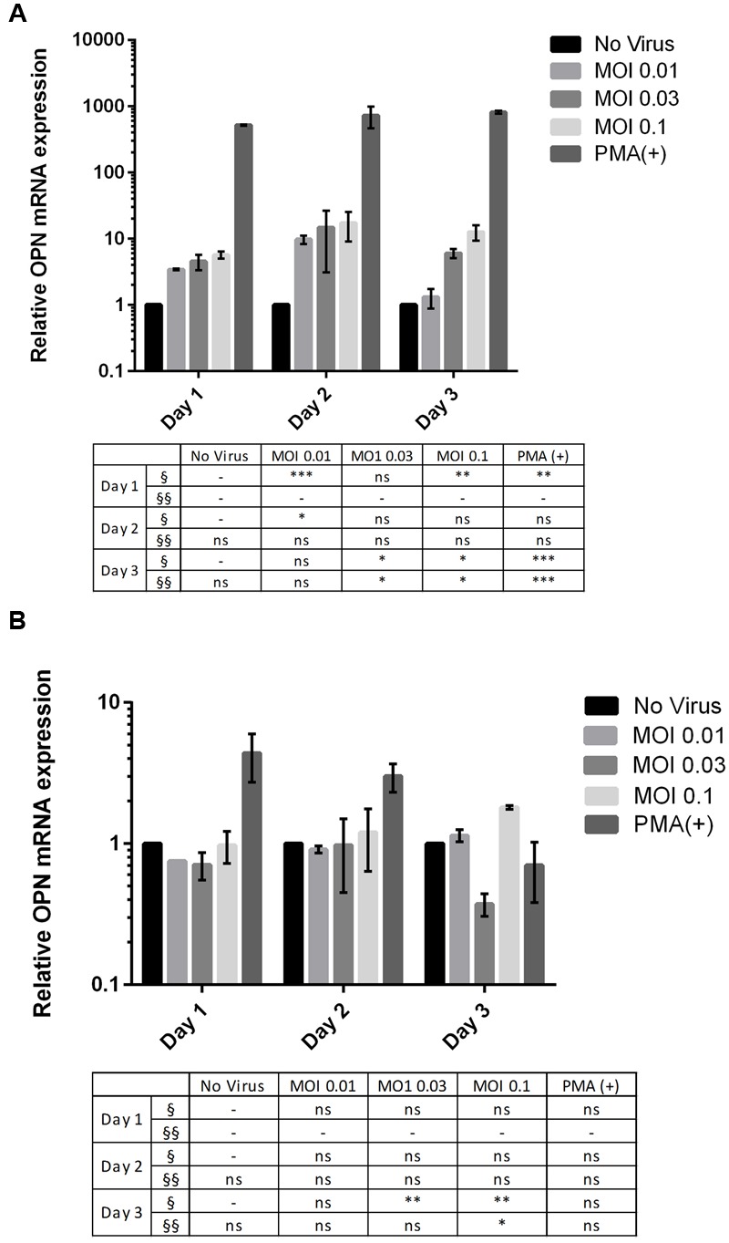
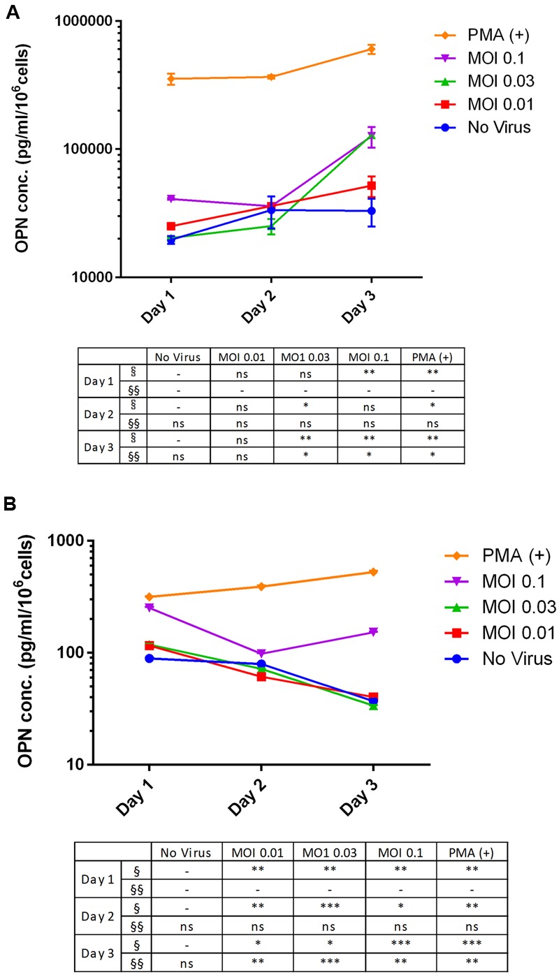
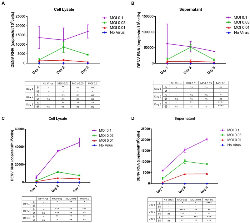
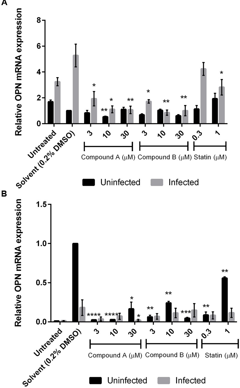
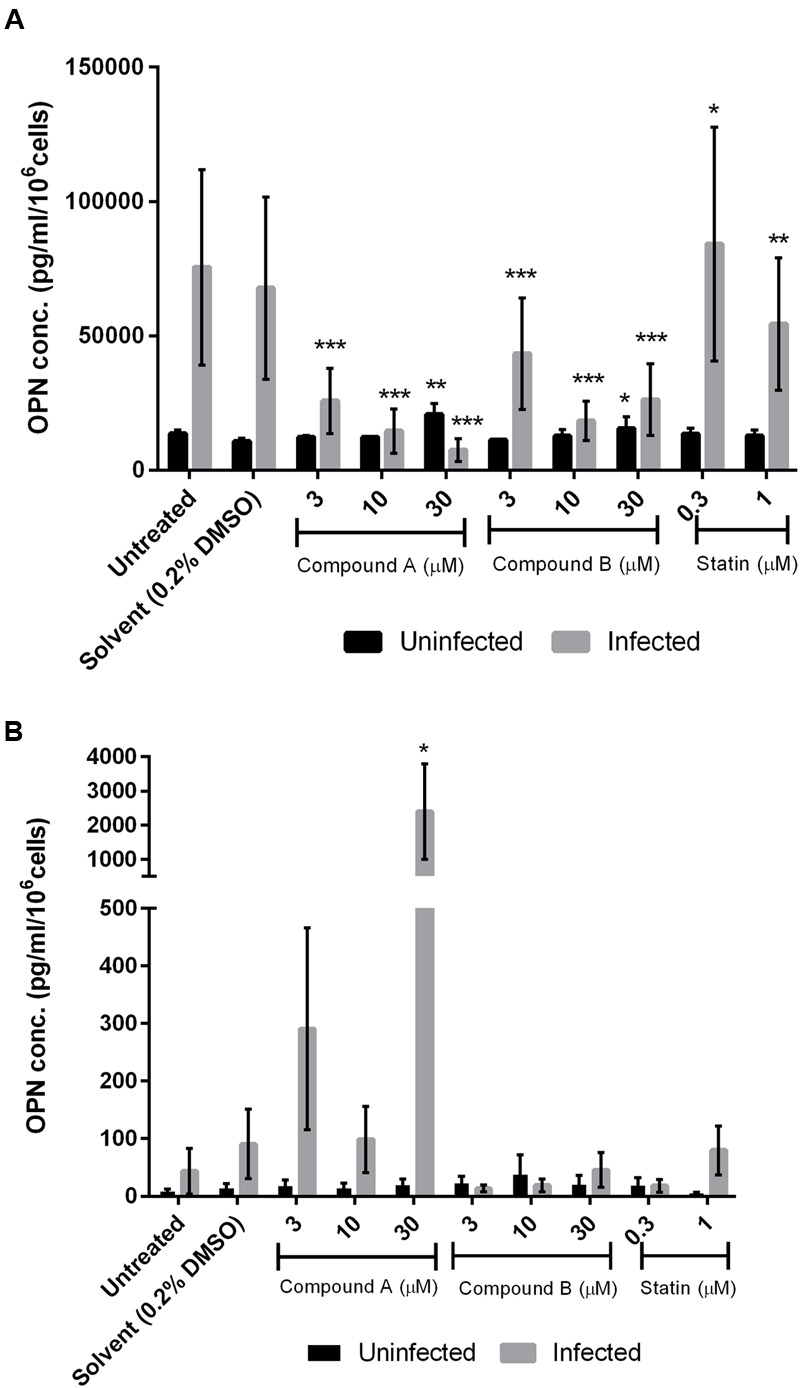
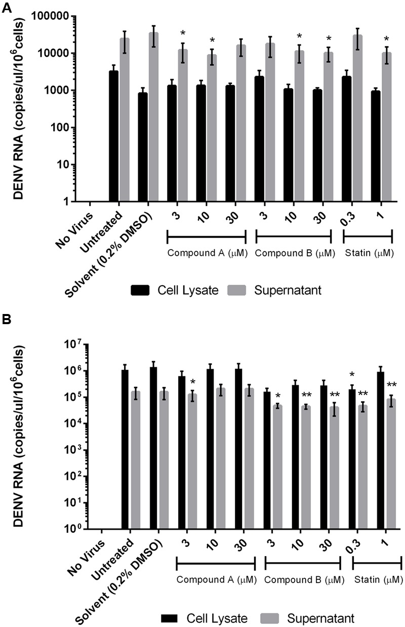
Osteopontin (OPN) is a multifunctional matricellular protein produced by a broad range of cells including osteoclasts, macrophages, T cells, endothelial cells, and vascular smooth muscle cells. OPN modulates various physiological and pathological events such as inflammation, wound healing, and bone formation and remodeling. Dengue virus (DENV) infection causes an increase in plasma OPN levels, which is correlated with the severity of symptoms and coagulation abnormalities. DENV infection also induces OPN gene expression in human macrophages. This study investigated the inhibitory effects of brefelamide and its methyl ether derivative on DENV-3 by measuring changes in OPN levels in human THP-1 and 293T cell lines infected at different multiplicities of infection and post-infection time points. OPN mRNA expression and viral RNA were detected by reverse transcriptase quantitative real-time PCR, whereas protein level was determined by enzyme-linked immunosorbent assay. We found that viral copy number was higher in 293T than in THP-1 cells. However, THP-1 constitutively expressed higher levels of OPN mRNA and protein, which were enhanced by DENV-3 infection. Brefelamide and its derivative suppressed OPN production in DENV-3 infected THP-1 cells; the effective doses of these compounds had no effect on uninfected cells, indicating low cytotoxicity. These results suggest that brefelamide and its methyl ether derivative have therapeutic effects in preventing inflammation, coagulopathy, and fibrinolysis caused by OPN upregulation induced by DENV-3 infection.

Keywords: 293T cell; THP-1 cell; brefelamide; dengue virus-3; osteopontin.


Figures

FIGURE 1
FIGURE 1
Multiplicity of infection (MOI)- and time-dependent change in OPN mRNA expression upon dengue virus (DENV) infection. (A) THP-1 and (B) 293T cells were infected with DENV-3 at various MOIs (range: 0.01–0.1). Phorbol 12-myristate 13-acetate (PMA) was used as a positive control. Cells were harvested daily (1–3 days). Total RNA was prepared from cell lysates and OPN levels were determined by RT-qPCR. GAPDH was used as reference gene to normalize the expression level. The table summarizes statistical analysis, P < 0.05, ∗∗P < 0.01, ∗∗∗P < 0.001 vs. control (uninfected cells; unpaired two-tailed t-test). Data represent mean ± SEM. § and §§ represent MOI- and time-dependent variables, respectively.
FIGURE 2
FIGURE 2
Multiplicity of infection- and time-dependent change in OPN protein expression upon DENV infection. (A) THP-1 and (B) 293T cells were infected with DENV-3 at various MOIs (range: 0.01–0.1). PMA was used as a positive control. Cells were harvested daily (1–3 days) and OPN protein levels in the supernatant were measured by ELISA. Viability was simultaneously evaluated to normalize the results. Normalized OPN levels in each sample are expressed as pg/ml/106 cells. The table summarizes statistical analysis, P < 0.05, ∗∗P < 0.01, ∗∗∗P < 0.001 vs. control (uninfected cells; unpaired two-tailed t-test). Data represent mean ± SEM. § and §§ represent MOI- and time-dependent variables, respectively.
FIGURE 3
FIGURE 3
Dengue virus RNA copy number in infected cell lines. (A,B) THP-1 and (C,D) 293T cells were left uninfected or infected with DENV-3 at various MOIs (range: 0.01–0.1). Cells were harvested at various post-infection time points (days 1, 2, and 3). Total RNA was extracted from cell lysates and the culture supernatant and DENV genome copy number was determined by RT-qPCR. The table summarizes statistical analysis, P < 0.05, ∗∗P < 0.01, ∗∗∗P < 0.001, ∗∗∗∗P < 0.0001 vs. control (uninfected cells; unpaired two-tailed t-test). Data represent mean ± SEM. § and §§ represent MOI- and time-dependent variables, respectively.
FIGURE 4
FIGURE 4
Changes in OPN mRNA expression by treatment with compounds A and B in DENV-infected cells. Uninfected and DENV-infected THP-1 (A) or 293T (B) cells were left untreated or treated with compound A or compound B (range: 3–30 μM), or statin (range: 0.3–1 μM) for 72 h. Total RNA was extracted from cell lysates and analyzed by RT-qPCR. GAPDH was used as reference gene to normalize expression levels. Results show the mean of two independent experiments. P < 0.05, ∗∗P < 0.01, ∗∗∗P < 0.001, ∗∗∗∗P < 0.0001 vs. control (0.2% DMSO; unpaired two-tailed t-test). Data represent mean ± SEM.
FIGURE 5
FIGURE 5
Effect of compounds A and B on OPN protein level in DENV-infected cells. Uninfected and DENV-infected THP-1 (A) or 293T (B) cells were left untreated or treated with compound A or compound B (range: 3–30 μM), or statin (range: 0.3–1 μM) for 72 h. Cells were then harvested and OPN levels in the culture supernatant were determined by ELISA. Viability was simultaneously measured to normalize the results. Normalized OPN levels in each sample are expressed as pg/ml/106 cells. Results show the mean of two independent experiments. P < 0.05, ∗∗P < 0.01, ∗∗∗P < 0.001 vs. control (0.2% DMSO; unpaired two-tailed t-test). Data represent mean ± SEM.
FIGURE 6
FIGURE 6
Effect of compounds A and B on DENV RNA copy number in infected cells. Uninfected and DENV-infected THP-1 (A) or 293T (B) cells were left untreated or treated with compound A or compound B (range: 3–30 μM), or statin (range: 0.3–1 μM) for 72 h. Total RNA was extracted from cell lysates and DENV RNA was detected by RT-qPCR. Results show the mean of two independent experiments. P < 0.05, ∗∗P < 0.01 vs. control (0.2% DMSO; unpaired two-tailed t-test). Data represent mean ± SEM.

Similar articles

Cited by

References

    1. Abel S., Liautaud B., Cabie A. (2012). Dengue. N. Engl. J. Med 367 180–181. 10.1056/NEJMc1205584 - DOI - PubMed
    1. Afroz S., Giddaluru J., Abbas M. M., Khan N. (2016). Transcriptome meta-analysis reveals a dysregulation in extra cellular matrix and cell junction associated gene signatures during Dengue virus infection. Sci. Rep. 6:33752 10.1038/srep33752 - DOI - PMC - PubMed
    1. Avila-Aguero M. L., Avila-Aguero C. R., Um S. L., Soriano-Fallas A., Canas-Coto A., Yan S. B. (2004). Systemic host inflammatory and coagulation response in the Dengue virus primo-infection. Cytokine 27 173–179. 10.1016/j.cyto.2004.05.007 - DOI - PubMed
    1. Avirutnan P., Malasit P., Seliger B., Bhakdi S., Husmann M. (1998). Dengue virus infection of human endothelial cells leads to chemokine production, complement activation, and apoptosis. J. Immunol. 161 6338–6346. - PubMed
    1. Bethell D. B., Flobbe K., Cao X. T., Day N. P., Pham T. P., Buurman W. A., et al. (1998). Pathophysiologic and prognostic role of cytokines in dengue hemorrhagic fever. J. Infect. Dis. 177 778–782. 10.1086/517807 - DOI - PubMed